Abstract
Central to the bone-sparing effect of estrogen (E2) is its ability to block the monocytic production of the osteoclastogenic cytokine TNF-α (TNF). However, the mechanism by which E2 downregulates TNF production is presently unknown. Transient transfection studies in HeLa cells, an E2 receptor–negative line, suggest that E2 inhibits TNF gene expression through an effect mediated by estrogen receptor β (ERβ). We also report that in RAW 264.7 cells, an E2 receptor–positive murine monocytic line, E2 downregulates cytokine-induced TNF gene expression by decreasing the activity of the Jun NH2-terminal kinase (JNK). The resulting diminished phosphorylation of c-Jun and JunD at their NH2-termini decreases the ability of these nuclear proteins to autostimulate the expression of the c-Jun and JunD genes, thus leading to lower production of c-Jun and JunD. The consequent decrease in the nuclear levels of c-Jun and JunD leads to diminished binding of c-Jun/c-Fos and JunD/c-Fos heterodimers to the AP-1 consensus sequence in the TNF promoter and, thus, to decreased transactivation of the TNF gene.
J. Clin. Invest. 104:503-513 (1999).
Introduction
It is now recognized that one of the main mechanisms by which estrogen (E2) deficiency causes bone loss is by stimulating osteoclast formation (1), a process enhanced by several inflammatory cytokines. Among them is TNF-α (2, 3), a protein produced mainly by cells of the monocyte/macrophage lineage (4). Once released in the bone microenvironment, TNF stimulates osteoclast formation, in part by inducing the production of M-CSF by bone marrow stromal cells (5, 6). M-CSF, along with osteoprotegerin ligand (OPGL; also known as ODF/TRANCE/RANKL) (7), is an essential stimulator of osteoclast precursor proliferation and differentiation (8–10).
Studies in ovariectomized mice, an established experimental model of postmenopausal osteoporosis (1), have provided compelling evidence that increased production of TNF plays a critical causal role in ovariectomy-induced bone loss. This evidence includes the finding that treatment with the TNF inhibitor TNF-binding protein completely prevents ovariectomy-induced bone loss (11) and the report that transgenic mice insensitive to TNF owing to the overexpression of a soluble TNF receptor are also protected against ovariectomy-induced bone loss (12).
Evidence has also accumulated that demonstrates that E2 downregulates the production and/or the release of TNF by several cell lineages, including bone marrow monocytes and bone cells. Thus, in monocytes and osteoblasts, TNF protein and TNF mRNA levels are increased after natural or surgical menopause and decrease with E2 replacement (13–15). E2 inhibits TNF production by direct effects on target cells, as demonstrated by the fact that in vitro E2 treatment decreases TNF levels in cultures of murine bone marrow monocytes (16) and peripheral blood monocytes (17) and decreases TNF mRNA expression in a murine monocytic cell line (18). Taken together, these studies demonstrate that the bone-sparing effect of E2 is, at least in part, a result of its ability to downregulate the bone marrow levels of TNF. Although the role of TNF as key enhancer of bone resorption in E2-deficient rodents and humans has been defined, the mechanism by which E2 downregulates TNF production remains to be determined.
The TNF gene is transcriptionally silent in unstimulated monocytes and is rapidly transcribed in response to a variety of signals, such as endotoxin (LPS), phorbol esters, and cytokines including IL-1 and TNF itself (19–22). Although LPS is the major inducer of TNF in infectious disease, in physiological conditions the production of TNF in the bone marrow is mostly induced by inflammatory cytokines. Low concentrations of IL-1 and TNF are, in fact, constitutively produced in the bone marrow (23). Monocyte stimulation with either IL-1 or TNF leads to the activation of the AP-1 family of nuclear proteins (24). The AP-1 family comprises a group of inducible proteins that bind to a common cis-acting element known as the TPA response element (TRE) (25). Factors that bind to the TRE site include members of the Jun, Fos, and ATF (CREB) families of nuclear proteins (26). The Jun family consists of c-Jun, JunB, and JunD (26), and the Fos family includes c-Fos, Fra-1, Fra-2, FosB, and FosB2 (26).
The presence of TRE sites in the TNF promoter, and the fact that AP-1 is known to be a critical inducer of human and murine TNF gene expression (27), suggests that E2 could downregulate the production of TNF by decreasing the ability of AP-1 to transactivate the TNF promoter. In this study, we have investigated the effects of E2 on cytokine-induced monocytic production of TNF. We report that E2 downregulates cytokine-induced TNF gene expression by decreasing the activity of the Jun NH2-terminal kinase (JNK) through an effect that is likely mediated by ERβ. Decreased JNK activity results in blunted production of c-Jun and JunD, thus leading to decreased binding of c-Jun/Fos and JunD/Fos heterodimers to the AP-1 binding site in the TNF promoter.
Methods
Unless otherwise specified, all reagents and media were from Sigma Chemical Co. (St. Louis, Missouri, USA). RAW 264.7 and HeLa cells were obtained from the American Type Culture Collection (Rockville, Maryland, USA) and were maintained in phenol red–free media (GIBCO BRL, Grand Island, New York, USA) and 10% charcoal-stripped serum.
RAW 264.7 and HeLa cell transfection and CAT assay.
RAW 264.7 and HeLa cells were seeded at a density of 2.5 × 10⁵ and 2 × 10⁵ cells per well, respectively. Cells were pretreated with E2 (10–8 M) or control vehicle (ethanol) for 24 hours and then transfected with 4 μg of the appropriate vectors and 1 μg of β-galactosidase. All transfections were carried out using 12 μL of Lipofectamine (GIBCO BRL) for 5 hours without serum. Once transfected, the cells were treated with IL-1 and TNF and either E2 or control vehicle for 12 hours.
To evaluate the effect of E2 on TNF transcription, either a TNF/CAT construct containing the 2.2-kb 5′ promoter region plus 1 kb of the 3′ UTR, or a TNF/CAT construct containing the 2.2-kb 5′ promoter region but lacking the 3′ UTR region, was used. Both constructs were developed (28, 29) and provided by B. Beutler (Southwestern University, Dallas, Texas, USA). For some experiments, cells were transfected with a TNF/CAT construct containing the 2.2-kb 5′ promoter region plus 1 kb of the 3′ UTR carrying a mutation previously shown to silence the TRE site in the TNF promoter (27). This mutation was generated by site-directed mutagenesis (30) and consisted of the substitution of the TG nucleotides at positions –700 to –699 with GT nucleotides. Briefly, nonoverlapping mutagenesis primers binding to different template strands were synthesized with a primer carrying the TG→GT substitutions. Introduction of the substituted bases was confirmed by DNA sequencing. After transfection, cells were stimulated with IL-1 and TNF (5 ng/mL each) for 24 hours.
To determine whether RAW 264.7 cells express functional ERs, cells were transfected with an estrogen-responsive element–driven (ERE–driven) and CAT reporter construct and then treated with either E2 (10–8 M) or control vehicle, as described previously (31).
To investigate whether the repressive effects of E2 on TNF gene expression are mediated by estrogen receptor α (ERα) or ERβ, HeLa cells were cotransfected with 2.5 μg of TNF/CAT reporter plasmid, 1 μg of β-galactosidase reporter plasmid, and 0.5 μg of either ERα expression vector (32) or ERβ expression vector (a gift of T. Brown, Pfizer, Groton, Connecticut, USA).
To verify the expression of functional ERs in transfected HeLa cells, parallel experiments were conducted by cotransfecting HeLa cells with either ERα or ERβ expression vector and the Vit2-P36L ERE/luciferase reporter plasmid (33) and then treating with either E2 (10–8 M) or control vehicle.
In all cases, cells were harvested at the end of the treatment period and lysed by 3 freeze-thaw cycles in 200 μL of 250 mM Tris-HCl (pH 7.5), as described previously (34). CAT, β-galactosidase, and luciferase activities were measured as described by Godambe et al. (35), Hall et al. (36), and Meyers et al. (33), respectively. Results were expressed as normalized CAT or luciferase activity after correction for transfection efficiency using the cotransfected β-galactosidase control for each well.
TNF ELISA.
To determine the levels of TNF secreted by RAW 264.7 cells into the culture medium, cells were stimulated for 24 hours with 5 ng/mL each of murine IL-1 and human TNF. Levels of TNF were then determined using an ELISA specific for murine TNF as described previously (37). The sensitivity of this assay was 25 pg/mL.
RT-PCR for ERα and ERβ mRNA.
RT-PCR reactions were performed as described by Lu et al. (38) with minor modifications. A total of 2.5 μg of total RNA was reverse transcribed using a commercial kit (GIBCO BRL). One microliter of first-strand reactions was amplified by PCR in a final volume of 50 μL using 30 cycles of 1 minute at 94°C, 1 minute at 54°C, and 2 minutes at 72°C. Forward and reverse primers for ERα were 5′-CTA CCT GGA GAA CGA GCC CA-3′ and 5′-AAG GCA CTG ACC ATC TGG TC-5′, respectively, and for ERβ, 5′-CTG AAC AAA GCC AAG AGA-3′ and GCT CTT ACT GTC CTC TGT CG-3′, respectively. PCR products (10 μL) were resolved by electrophoresis on 1% agarose gels, and bands were viewed by ethidium bromide staining.
RNA purification and Northern blot analysis.
Total RNA from RAW 264.7 cells was extracted using Trizol (GIBCO BRL) and was quantified by spectrophotometry. Equal amounts of total RNA (10 μg per lane) were electrophoresed on 1% Seakem agarose/formaldehyde (0.23%) gels (39) and transferred onto nitrocellulose membranes. The membranes were hybridized in Hybrizol (Oncor Inc., Gaithersburg, Maryland, USA) with 32P-labeled probes for 16 hours at 42°C. The membranes were washed 3 times in 2× SSC/0.1% SDS at room temperature, and once in 0.2× SSC/0.02% SDS at 56°C for 1 hour. For autoradiography, the blots were exposed to Kodak X-OMAT film (Eastman Kodak Co., Rochester, New York, USA) at –80°C for the appropriate duration. As a TNF probe, we used a full-length cDNA specific for murine TNF (American Type Culture Collection), labeled with [32P]dCTP using the random priming method (Boehringer Mannheim Biochemicals Inc., Indianapolis, Indiana, USA). An end-labeled oligonucleotide specific for 18S mRNA was used to control for variations in loading, as described (40).
Extraction of nuclear protein and electrophoretic mobility shift assays.
To investigate which member of AP-1 protein binds to the TRE sequence in the TNF promoter, nuclear extracts from control- and E2-treated RAW 264.7 cells were prepared from cells either stimulated or unstimulated with IL-1 and TNF (5 ng/mL each) for 1 hour, using a method described previously (41). Electrophoretic mobility shift assays (EMSAs) were performed with a double-stranded probe (5′-AAAGCAGCAGCCTGAGCTCATGATCA-3′) synthesized to represent the TRE sequence (underlined) in the murine TNF promoter (26 mer –686 to –712 nucleotide). The probes were end labeled with T4 polynucleotide kinase. The annealed probe was incubated with nuclear extract on ice for 30 minutes before separation of the DNA/protein complexes on 4% nondenaturing PAGE gels, prerun for 1 hour in 0.25× TBE running buffer. The gels were dried and exposed to Kodak XAR-5 film for the appropriate length of time. For band supershifting, 200 ng of the appropriate antibody was added to the reaction mixture on ice 40 minutes before addition of the oligonucleotide probe.
Immunoprecipitations and Western blot analysis.
Immunoprecipitation reactions were performed as described by Jain et al. (42), with modifications (41) using polyclonal antibodies directed against all members of either the Jun or Fos families of nuclear proteins. For some experiments, the supernatants were incubated with mAb’s specific for c-Jun, JunD, or JunB.
Western blots were performed as described by Towbin et al. (43) using antibodies specific for Jun proteins, Fos proteins, c-Jun, JunB, or JunD, as appropriate.
To determine the effects of E2 on the levels of total (dephosphorylated + phosphorylated) JNK and active (phosphorylated) JNK, whole-cell lysates were prepared from control- and E2-treated RAW 264.7 cells, either unstimulated or stimulated with IL-1 and TNF for 15 minutes. Cell lysates (50 μg) were resolved on 12% SDS-PAGE gels and transblotted onto nitrocellulose. Total and phosphorylated JNK were detected using antibodies specific for either total JNK or phosphorylated JNK (Santa Cruz Biotechnology Inc., Santa Cruz, California, USA), using an enhanced chemiluminescent detection system (Amersham Pharmacia Biotech, Piscataway, New Jersey, USA).
JNK activity assay.
Cells were treated with E2 or control substances for 24 hours and stimulated with IL-1 and TNF for 1 hour. Cells were washed twice with ice-cold PBS and then lysed in ice-cold lysis buffer (50 mM Tris-HCl [pH 8], 137 mM NaCl, 10% glycerol, 1% Nonidet P-40 (vol/vol), 1 mM NaF, 10 μg/mL leupeptin, 2 mM Na3VO4, and 1 mM PMSF) as described (44). JNK activity was determined in cell extracts precleared with 10–15 μL protein A-Sepharose beads. The resin was then removed by centrifugation, and the supernatant was incubated with 1 μg of anti-JNK antibody (Santa Cruz Biotechnology Inc.) for 4 hours on ice. The immunocomplexes were then precipitated using protein A-Sepharose beads. The beads were washed with lysis buffer and finally washed twice with kinase buffer (25 mM HEPES [pH 7.4], 25mM β-glycerophosphate, 25 mM MgCl2, 2 mM DTT, and 0.1 mM Na3VO4). Beads were resuspended in 25 μL of kinase buffer containing 50 μM ATP and 5 μCi [γ-32P]ATP, and were incubated for 25 minutes at 30°C with 1 μg of recombinant Jun (44, 45). The reactions were boiled in Laemmli loading dye and resolved by 12% SDS-PAGE. Phosphorylated c-Jun-GST protein was detected by autoradiography as described previously (46).
Statistical analysis.
Group mean values were compared by 2-tailed Student’s t test or 1-way ANOVA, as appropriate. Subsequent mean comparison tests were performed by the Fisher protected least significant difference test.
Results
RAW 264.7 cells express functional ERs and mRNA for both ERα and ERβ.
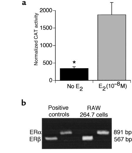
To investigate whether RAW 264.7 cells, a murine monocytic line known to produce TNF in response to a variety of stimuli (47), express functional ERs, transient transfection experiments were conducted using a reporter plasmid that contained a CAT gene under the transcriptional control of an ERE driven by an SV-40 promoter. Three replicate experiments revealed that 24-hour E2 (10–8 M) treatment increases CAT activity 6-fold (Figure 1a), thus demonstrating that RAW 264.7 cells express functional ER.
Figure 1.
RAW 264.7 cells express functional ERs and mRNA for ERα and ERβ. (a) Cells were transfected with a reporter plasmid that contained a CAT gene under the transcriptional control of an ERE driven by an SV-40 promoter, and were treated with either E2 or control vehicle. CAT activity was normalized to β-galactosidase activity to correct for variability in transfection efficiency and expressed as normalized CAT activity. Mean ± SEM of 3 experiments. *P < 0.05 vs. untreated cells. (b) RT-PCR amplification products of ERα and ERβ. Total RNA was extracted and reverse transcribed. The resulting cDNA was amplified using primers specific for ERα and ERβ. Two amplification products corresponding to ERα and ERβ were detected by ethidium bromide staining of agarose gels. Amplification of purified mRNA with ERα and ERβ primers in the absence of RT generated no detectable bands (data not shown).
Because this assay does not provide information as to whether RAW 264.7 cells express ERα, ERβ, or both, total RNA was extracted and reverse transcribed. The resulting cDNA was then amplified using primers specific for ERα or ERβ. Two amplification products corresponding to ERα and ERβ were detected by ethidium bromide staining of an agarose gel (Figure 1b).
E2 decreases the IL-1– and TNF-induced production of TNF in RAW 264.7 cells.
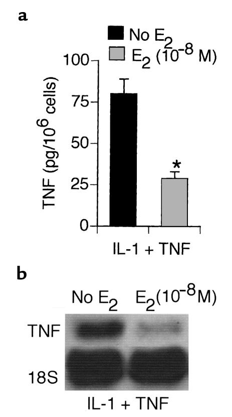
Having determined that RAW 264.7 cells respond to E2 and express functional ERs and mRNA for ERα and ERβ, we sought to investigate the effects of E2 on the production of TNF in response to cytokine stimulation. Preliminary studies (data not shown) revealed that combined stimulation with IL-1 and TNF (5 ng/mL of each) induces the secretion of 2.2-fold–higher levels of TNF into the culture medium than does treatment with either IL-1 or TNF alone. Therefore, to investigate the inhibitory effects of E2 in conditions of maximal TNF production, RAW 264.7 cells were stimulated with IL-1 and TNF (5 ng/mL of each) and treated with either ethanol (control vehicle) or 10–8 M E2 for 24 hours. The culture medium was then assayed by an ELISA specific for murine TNF. Four replicate experiments revealed that the level of murine TNF was approximately 3-fold lower in medium from E2-treated cells than in medium from controls (Figure 2a).
Figure 2.
In vitro E2 treatment of RAW 264.7 cells decreases IL-1– and TNF-induced production of TNF. (a) Levels (mean ± SEM of 4 experiments) of TNF in the culture medium as assayed by an ELISA specific for murine TNF. Equal numbers of cells were stimulated with murine IL-1 and human TNF (5 ng/mL of each) and then treated with either control vehicle or E2 (10–8 M) for 24 hours. *P < 0.05 vs. untreated cells. (b) TNF mRNA expression, as assessed by Northern blot analysis. Cells were treated with either control vehicle or E2 (10–8 M) for 24 hours and stimulated with IL-1 and TNF (5 ng/mL of each) during the last 4 hours of incubation. Representative data from 1 of 3 replicate experiments.
To investigate whether E2 regulates TNF mRNA expression, RAW 264.7 cells were stimulated with IL-1 and TNF and treated with E2 (10–8 M) as already described here. The steady-state expression of TNF mRNA was then assessed by Northern blot analysis. Data from 1 representative experiment are shown in Figure 2b. Hybridized TNF mRNA and 18S mRNA were quantified by densitometry. The relative density of the bands was expressed as TNF/18S mRNA density ratio. Triplicate experiments revealed that E2 decreases TNF mRNA levels by approximately 3-fold in cytokine-stimulated RAW 264.7 cells. Taken together, these data demonstrate that E2 regulates the signaling pathway that leads to TNF production in response to cytokine stimulation.
E2 decreases TNF gene expression in RAW 264.7 cells.
To investigate the effects of E2 on TNF gene expression, RAW 264.7 cells were pretreated with E2 (10–7 to 10–12 M) or control vehicle for 24 hours and transiently transfected with a TNF/CAT construct containing the 2.2-kb 5′ promoter region plus 1 kb of the 3′UTR (a message stability and translation regulating sequence) (28, 29). The cells were then treated with IL-1 and TNF and either E2 (10–7 to 10–12 M) or control vehicle for 12 hours. Cytosolic extracts were prepared and used for CAT assays.
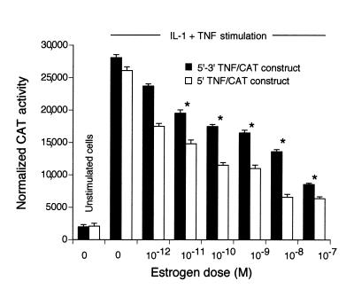
CAT activity was weak in basal conditions, but it was increased by about 14-fold after stimulation with IL-1 and TNF (Figure 3). In vitro treatment with E2 decreased IL-1– and TNF-induced TNF promoter activity in a dose-dependent manner (Figure 3, filled bars). The largest inhibition of CAT activity (∼3.5-fold) was obtained with an E2 dose equal to 10–7 M. Time course experiments (data not shown) demonstrated no inhibitory effects of E2 for treatment periods up to 2 hours. Maximal inhibitory activity was observed at 24 hours. Treatment with E2 for 48 hours resulted in no additional inhibition of CAT activity.
Figure 3.
E2 decreases TNF gene expression in a dose-dependent manner. Raw 264.7 cells were pretreated with E2 (10–7 to 10–12 M) for 24 hours before transfection and then transiently transfected with either a TNF/CAT construct containing the 2.2-kb 5′ promoter region plus 1 kb of the 3′ UTR (filled bars) or a TNF/CAT construct containing the 2.2-kb 5′ promoter region lacking the 3′ UTR region (open bars). The cells were then treated again with E2 (10–7–10–12 M) and 5 ng of TNF and IL-1 for 12 hours. Mean ± SEM of 4 experiments. *P < 0.05 vs. untreated cells.
When RAW 264.7 cells were stimulated with IL-1 and TNF and cotransfected with a TNF/CAT construct containing the 2.2-kb 5′ promoter region lacking the 3′ UTR region, an identical inhibitory effect of E2 on CAT activity was demonstrated (Figure 3, open bars). Because the 3′ UTR region of the TNF gene regulates message stability and its translation (29), the data suggest that E2 modulates cytokine-induced TNF gene transcription.
The inhibitory effects of E2 on TNF gene expression are mediated by ERβ.
Because RAW 264.7 cells express endogenous ERs, they are not suitable for investigating the contribution of a specific ER phenotype to the inhibitory effects of E2 on TNF gene expression. Therefore, to determine whether the inhibitory effects of E2 on TNF gene expression are mediated by ERα, ERβ, or both, experiments were conducted using HeLa cells, an ER-negative line that, once transfected with ERs, exhibits a response to E2 similar to the responses of natural E2 target cells (48). HeLa cells were transiently cotransfected with the TNF/CAT construct containing the 2.2-kb 5′ promoter region plus 1 kb of the 3′UTR already described here (28, 29) and either ERα or ERβ expression vectors. Cells were then treated with either E2 (10–8 M) or control vehicle for 24 hours and stimulated with 5 ng each of IL-1 and TNF for 12 hours. To verify that transfected cells express functional ERs, parallel control experiments were conducted in which HeLa cells were cotransfected with ERα or ERβ expression vector and an ERE/luciferase reporter construct driven by a minimal promoter.
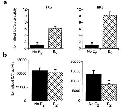
E2 significantly (P < 0.05) increased ERE luciferase activity, both in cells transfected with ERα and in those transfected with ERβ expression vector (Figure 4a), thus demonstrating that transfected HeLa cells express functional ER. Moreover, E2 decreased TNF/CAT activity in cells expressing ERβ, whereas it had no effect in those expressing ERα (Figure 4b), thus demonstrating that in HeLa cells, the inhibitory effect of E2 on TNF gene expression is specifically mediated by ERβ.
Figure 4.
ERβ specifically mediates an inhibitory effect of E2 on TNF gene expression. (a) HeLa cells were transfected with a reporter plasmid that contained a luciferase gene under the transcriptional control of an ERE driven by a minimal promoter, and treated with either E2 or control vehicle as described for the RAW 264.7 cells. Luciferase activity was normalized to β-galactosidase activity to correct for variability in transfection efficiency and expressed as normalized luciferase activity. Mean ± SEM from 1 representative experiment. *P < 0.05 vs. untreated cells. (b) HeLa cells were pretreated with E2 for 24 hours, and cotransfected with TNF/CAT construct containing the 2.2-kb 5′ promoter region plus 1 kb of the 3′UTR and either ERα or ERβ expression vectors. Cells were then treated with E2 and IL-1 and TNF for 12 hours, as described in the text. The cells were subsequently lysed and assayed for CAT and β-galactosidase activities as described in Methods. Mean ± SEM of 3 experiments. *P < 0.05 vs. untreated cells.
E2 downregulates TNF gene expression by decreasing the binding of AP-1 proteins to the TNF promoter.
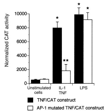
The inhibitory effects of E2 on TNF promoter activity are likely to be indirect, because there is no typical ERE sequence in the TNF promoter. We reasoned that because AP-1 is activated by IL-1 and TNF, E2 may repress TNF gene expression by blocking AP-1 binding to TNF promoter. To investigate this hypothesis, we first analyzed the functional role of the TRE site in the TNF promoter. Thus, RAW 264.7 cells were transiently transfected with a plasmid corresponding to the full TNF promoter (2.2-kb 5′ promoter region) plus 1 kb of the 3′UTR, containing point mutations of the TRE site that were reported previously to reduce TNF promoter activity (27). As shown in Figure 5, mutation of the TRE site reduced IL-1– and TNF-induced CAT activity 4-fold, whereas it did not alter the response to LPS (1 μg/mL). These data demonstrate that the TRE site is required for cytokines to induce TNF gene expression.
Figure 5.
IL-1 and TNF induce TNF gene expression via the TRE site in the TNF promoter. Raw 264.7 cells were transiently transfected with either a wild-type TNF/CAT construct containing the 2.2-kb 5′ promoter region plus 1 kb of the 3′UTR or the corresponding plasmid with a site-directed mutation of the TRE site. Cells were then stimulated with either IL-1 + TNF or LPS for 12 hours. CAT activity was normalized to β-galactosidase activity to correct for variability in transfection efficiency and expressed as normalized CAT activity. Mean ± SEM of 3 experiments. *P < 0.05 vs. unstimulated cells. **P < 0.01 vs. cells transfected with the wild-type TNF/CAT reporter gene construct.
To determine whether mutation of the AP-1 site abolishes the ability of E2 to block IL-1– and TNF-induced TNF gene expression, additional experiments were carried out in control- and E2-treated RAW 264.7 cells. As expected, the expression of the reporter gene was low in both untreated and E2-treated IL-1– and TNF-stimulated cells transfected with the AP-1 mutant (data not shown). Therefore, these experiments did not allow us to determine conclusively that mutation of the AP-1 site abrogates the repressive effects of E2 on TNF gene expression.
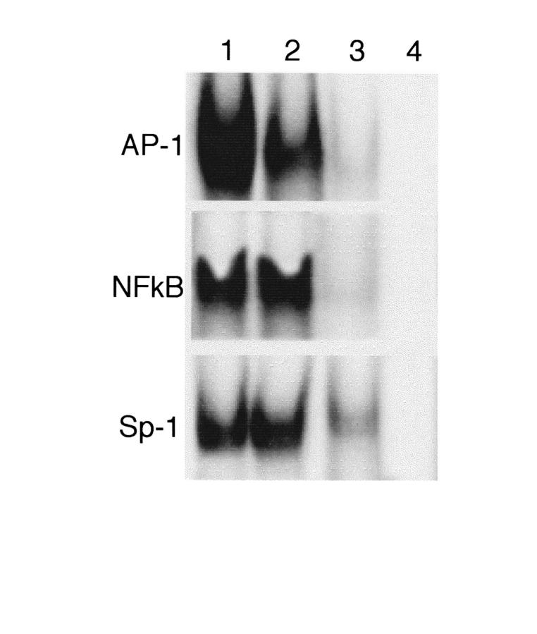
To investigate further the hypothesis that E2 specifically inhibits AP-1–induced TNF gene expression, RAW 267.4 cells were treated with and without E2 (10–8 M) for 24 hours, followed by a 1-hour stimulation with IL-1 and TNF. EMSAs were then conducted by incubating nuclear extracts from control- and E2-treated cells with a labeled oligonucleotide corresponding to the region of the murine TNF promoter containing the TRE site. As shown in Figure 6, these studies revealed that samples from both control- (lane 1) and E2-treated (lane 2) cells generate a single complex, which was confirmed as AP-1 by the ability of 50-fold M excess TRE consensus oligonucleotide to displace the complex (lane 3). Three such replicate experiments revealed that E2 decreases the binding of AP-1 to the TRE motif by approximately 3-fold.
Figure 6.
E2 decreases AP-1 binding to the TRE site in the TNF promoter, whereas it does not affect the binding of NF-κB and Sp-1 to their consensus sequences. EMSAs were performed by incubating nuclear extracts of untreated and E2-treated RAW 264.7 cells stimulated with IL-1 and TNF for 1 hour with 32P-labeled probes containing the TRE, the NF-κB, or the Sp-1 binding sites in the TNF promoter. Gel shift complexes were resolved by electrophoresis and viewed by autoradiography. Specificity of the induced complexes was determined by competition with a 50-fold M excess of unlabeled specific probe. Lane 1: cells treated with control vehicle; lane 2: cells treated with E2 (10–8 M); lane 3: 50× cold probe; lane 4: free probe. Representative data from 1 of 3 replicate experiments. Densitometric analysis of the data from all experiments revealed that AP-1 binding to the TRE site was approximately 3-fold lower in E2-treated samples than in vehicle-treated samples.
To demonstrate specificity, additional EMSAs were carried out using as probes sequences corresponding to the 4 binding sites for nuclear factor-κB (NF-κB) and the 1 binding site for Sp-1 in the TNF promoter. In accordance with the inability of E2 to repress LPS-induced TNF gene expression, we found that E2 does not downregulate the binding of NF-κB to the NF-κB1 (Figure 6) or to the NF-κB2, NF-κB3, and NF-κB4 (data not shown) consensus sequences. Further attesting to specificity, E2 did not block Sp-1 binding to its binding site (Figure 6).
Together, these observations indicate that E2 downregulates TNF gene expression by decreasing AP-1 binding to the TRE site in the TNF promoter. Because E2 decreases, but does not completely block, TNF gene expression, it is likely that TNF transcription enhancers such as NF-κB account for the residual TNF gene expression observed in E2-treated cells.
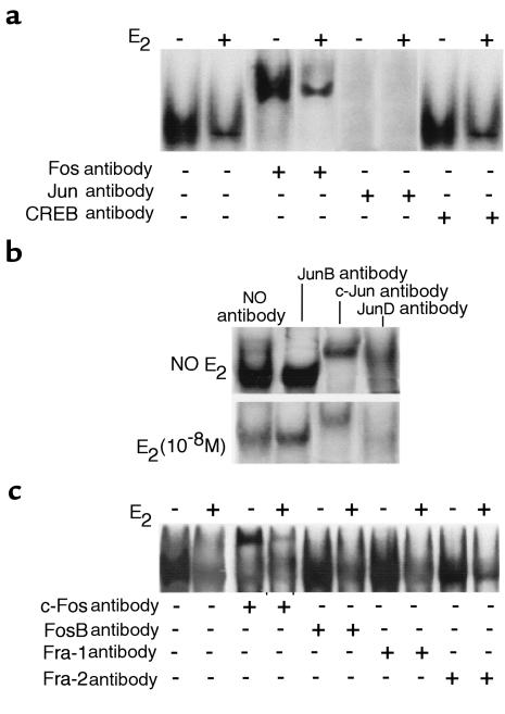
To determine which proteins form the AP-1 complex binding to the TNF promoter of cytokine-stimulated RAW 267.4 cells, EMSAs were conducted in the presence of polyclonal antibodies directed against the Jun and Fos families of proteins. An anti-Fos antibody that cross-reacts with all members of the Fos family of proteins supershifted the band, as viewed with 32P-labeled AP-1 probe in both control- and E2-treated cells (Figure 7a). Similarly, an anti-Jun antibody that cross-reacts with all members of the Jun family of proteins caused the disappearance of the complex in control- and E2-treated cells. In contrast, the AP-1 complex was not supershifted by anti-CREB antibody. Taken together, these findings demonstrate that E2 decreases the binding of Fos/Jun heterodimers to the TRE site of the TNF promoter.
Figure 7.
(a) E2 decreases the binding of Fos/Jun heterodimers to the TRE site in the TNF promoter. EMSAs were performed by incubating nuclear extracts — from untreated and E2-treated RAW 264.7 cells stimulated with IL-1 and TNF for 1 hour — with 32P-labeled probes containing an exact copy of the TRE binding site found in the TNF promoter. Polyclonal antibodies directed against all members of the Jun, Fos, and CREB families of nuclear proteins were used to supershift the relevant proteins. The figure shows representative data from 1 of 3 replicate experiments. (b) E2 treatment of RAW 264.7 cells decreases the binding of c-Jun and JunD to a probe containing an exact copy of the TRE site found in the TNF promoter. EMSAs were performed as described in the text in the presence or the absence of mAb’s directed against c-Jun, JunB, and JunD. Representative data from 1 of 3 replicate experiments. (c) E2 decreases the binding of c-Fos to a probe containing an exact copy of the TRE site in the TNF promoter. EMSAs were performed as described in the text in the presence or the absence of mAb’s directed against c-Fos, FosB, Fra-1, and Fra-2. Representative data from 1 of 3 replicate experiments.
To determine which members of the Jun and Fos families form the Jun/Fos heterodimers binding to the TNF promoter, additional EMSAs were conducted using mAb’s against specific Jun and Fos proteins. In untreated RAW 264.7 cells, the addition of a specific anti–c-Jun antibody supershifted the Jun band (Figure 7b). The addition of anti-JunD antiserum led to the almost complete disappearance of the Jun band, whereas anti-JunB antibody had no effect. As expected, in samples from E2-treated cells, AP-1 binding to the TRE site was less intense than in the corresponding samples from control cells. The addition of anti–c-Jun antibody supershifted the AP-1 band, whereas the addition of anti-JunD caused its complete disappearance. As in the case of untreated cells, antibody against JunB had no effects.
In both untreated and E2-treated cells, the AP-1 complex was also supershifted by anti–c-Fos antibody (Figure 7c), whereas antibodies against Fos-B, Fra-1, and Fra-2 had no effects, indicating that in both untreated and E2-treated cells, the AP-1 complex is formed by c-Jun/c-Fos and JunD/c-Fos heterodimers.
E2 decreases the production of c-Jun and JunD.
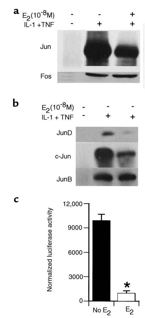
To investigate the mechanism by which E2 decreases binding of Jun and Fos to the TRE site, their nuclear levels were measured by Western blot analysis. Studies using polyclonal antibodies that recognize all members of the Fos and Jun family of proteins revealed that neither Fos nor Jun is detectable in the nuclear extracts of unstimulated cells. Stimulation with IL-1 and TNF resulted in the production of both proteins. Densitometric analysis of the data from 3 experiments revealed that E2 decreases the nuclear concentration of Jun but not that of Fos (Figure 8a), suggesting that E2 decreases the binding of Fos/Jun heterodimers to the TNF promoter by regulating the levels of Jun.
Figure 8.
(a) Western blot analysis of Jun and Fos proteins carried out using nuclear extracts from untreated and E2-treated RAW 264.7 cells stimulated with IL-1 and TNF for 1 hour and polyclonal antibodies against all members of the Jun and Fos families of nuclear proteins. Representative data from 1 of 3 replicate experiments. (b) Western blot analysis conducted using antibodies against specific Jun proteins demonstrated that E2 downregulates c-Jun and JunD levels, but not JunB levels. Representative data from 1 of 3 replicate experiments. (c) E2 decreases c-Jun gene expression in IL-1– and TNF-stimulated RAW 264.7 cells. Control- and E2-treated RAW 264.7 cells stimulated with IL-1 and TNF were transiently transfected with a c-Jun/luciferase construct containing the full-length c-Jun promoter. Luciferase activity was normalized to β-galactosidase activity to correct for variability in transfection efficiency and expressed as normalized luciferase activity. Mean ± SEM of 3 experiments. *P < 0.05 vs. untreated cells.
To characterize further the effects of E2 on the nuclear levels of Jun proteins, Western blot studies were carried out using nuclear extracts from cytokine-stimulated RAW 264.7 cells and antibodies specific for c-Jun, JunB, and JunD (Figure 8b). Densitometric analysis of the data revealed that E2 decreases the nuclear concentration of JunD by approximately 4-fold and that of c-Jun by 3-fold. Conversely, E2 had no effects on JunB levels. To investigate whether E2 decreases Jun gene expression, control- and E2-treated RAW 264.7 cells stimulated with IL-1 and TNF were transiently transfected with a c-Jun/luciferase construct containing the full-length c-Jun promoter (49). These experiments revealed that E2 decreases the activity of the reporter construct by approximately 10-fold, thus establishing that E2 represses the expression of the c-Jun gene (Figure 8c). Taken together, the data demonstrate that E2 blocks TNF gene expression by decreasing the levels of c-Jun and JunD, a phenomenon that results in diminished binding of c-Jun/c-Fos and JunD/c-Fos heterodimers to the AP-1 site in the TNF promoter.
E2 decreases JNK activity in RAW 264.7 cells.
The promoter regions of both the c-Jun and JunD genes contain 2 TRE sites constitutively occupied by Jun/ATF2 heterodimers. However, the magnitude of the autoinduction of Jun genes by Jun/ATF2 heterodimers is enhanced by JNK-induced phosphorylation of c-Jun and JunD. E2 may thus downregulate the nuclear levels of c-Jun and JunD, thereby decreasing AP-1–induced TNF transcription, by blunting JNK activity and the resulting phosphorylation of c-Jun and JunD.
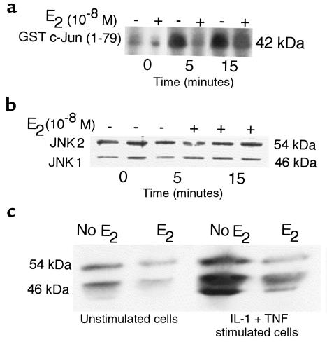
To investigate whether E2 regulates JNK activity, RAW 264.7 cells were treated with either E2 or control vehicle for 24 hours, a length of time sufficient to induce genomic effects, and then stimulated with IL-1 and TNF for 0–15 minutes. Cell extracts were then immunoprecipitated using an antibody that recognizes 2 members of the JNK family, JNK1 and JNK2. Equal amounts of proteins were recovered and incubated with recombinant GST-c-Jun (a JNK substrate) and [γ-32P]ATP. Phosphorylated material was resolved by SDS-PAGE and viewed by autoradiography. These studies revealed that E2 decreases IL-1– and TNF-induced JNK activity by approximately 3-fold (Figure 9a), with a peak effect at 5 minutes.
Figure 9.
(a) E2 decreases JNK activity in RAW 264.7 cells. RAW 264.7 cells were treated with either E2 or vehicle for 24 hours and then stimulated with IL-1 and TNF for 0–15 minutes. Cell extracts were then immunoprecipitated by anti-JNK antibody. Equal amounts of immunoprecipitation material were recovered and incubated with recombinant GST-c-Jun and [γ-32P]ATP. Phosphorylated material was resolved by SDS-PAGE and viewed by autoradiography. Representative data from 1 experiment. (b) E2 has no effects on total JNK levels. Western blot studies were conducted as described in the text using an antibody that recognizes the dephosphorylated and the phosphorylated species of both JNK1 and JNK2. Representative data from 1 of 3 experiments. (c) E2 decreases the level of phosphorylated (active JNK). Western blot analysis was conducted using cell lysates from control- and E2-treated RAW 264.7 cells stimulated with IL-1β and TNF for 5 minutes, and an antibody specific for phospho JNK. Representative data from 1 of 3 experiments.
Decreased JNK activity may result from either lower cell levels of the kinase or from decreased enzyme activation. To investigate this matter, Western blot studies were conducted using an antibody directed against both the dephosphorylated and phosphorylated species of JNK1 and JNK2. As shown in Figure 9b, experiments revealed that E2 does not regulate the levels of total (active and inactive) JNK1 and JNK2.
Because JNK is activated by dual phosphorylation in its NH2-terminal region, we measured the levels of phosphorylated (active) JNK in untreated and E2-treated RAW 264.7 cell extracts by Western blot analysis, using an antibody specific for phosphorylated JNK1 and JNK2. As shown in Figure 9c, we found that E2 decreases the levels of phosphorylated JNK by about 3-fold. Together, these data demonstrate that one of the mechanisms by which E2 decreases the production of Jun proteins is by downregulating JNK activity, a phenomenon that results in decreased c-Jun and JunD transactivation activity and, thus, diminished autoinduction of the c-Jun and JunD genes.
Discussion
The genomic effects of E2 are mediated by a ligand-inducible transcription factor known as ER. After the discovery of a second ER phenotype (ERβ), the first identified ER is now known as ERα (50). However, no published information is available on the expression of either ERα or ERβ in cells of the monocytic lineage. In this study, we have found that the murine monocytic cell line RAW 264.7 is responsive to E2 and expresses both ERα and ERβ mRNAs.
Although ERα and ERβ have similar DNA-binding domains, their A/B domains and activation-function region 1 (AF-1) are quite different, suggesting that ERα and ERβ may differentially regulate ER-responsive genes (51). Recent studies have, indeed, demonstrated that ERα and ERβ respond differently to ligand, leading to opposite effects on AP-1–induced gene expression (52). Specifically, although the ERα-mediated effects of E2 lead to stimulation of AP-1–induced gene expression, E2 acts as a repressor of AP-1–induced transcription when bound to ERβ (52). In accordance with these data, we have found that in HeLa cells, the inhibitory effects of E2 on the activity of a TNF/CAT reporter construct are mediated by ERβ.
Mutational analysis of the murine TNF promoter revealed that silencing of the AP-1 binding site in the full-length TNF promoter abrogates the ability of IL-1 and TNF to induce TNF gene expression. These findings extend previous observations demonstrating that the AP-1 binding site is critical for PMA induction of TNF gene expression (27). In contrast, mutation of the AP-1 site does not block the ability of LPS to induce TNF gene expression, a finding consistent with the notion that LPS induces the expression of the murine TNF gene primarily by activating the NF-κB family of nuclear proteins (53).
Cytokine stimulation often leads to the activation of both the NF-κB and AP-1 families of transcription factors (24, 54). Thus, it could be argued that mutation of the AP-1 site should not repress TNF gene expression due to because of NF-κB stimulation of the promoter. Because mutation of the AP-1 site decreased TNF gene expression by approximately 4-fold, our findings suggest the possibility that AP-1 and NF-κB cooperate to activate the murine TNF promoter. Regardless of whether AP-1 interacts with NF-κB, the data demonstrate the critical role of the AP-1 site in the mechanism by which inflammatory cytokines induce the TNF gene.
A surprising finding of our study was that in RAW 264.7 cells, E2 does not inhibit NF-κB binding to the TNF promoter, suggesting that E2 does not regulate NF-κB–induced TNF gene expression. These findings are in apparent disagreement with reports indicating that in human osteoblasts, E2 represses the IL-6 promoter by blunting NF-κB–induced transcription (55). The finding that E2 represses NF-κB–induced IL-6 production in human osteoblasts, whereas it has no effects on NF-κB–induced TNF production in murine monocytes, indicates that the effects of E2 on NF-κB–induced gene expression are promoter and cell specific.
In accordance with the key role of AP-1 as mediator of the stimulatory effects of inflammatory cytokines on the transcriptional activity of the TNF gene, we found that E2 downregulates cytokine-induced TNF production by decreasing the binding of c-Jun/c-Fos and JunD/c-Fos heterodimers to the AP-1 consensus sequence in the TNF promoter, a phenomenon resulting from decreased nuclear levels of c-Jun and JunD.
Attesting to specificity, our data also demonstrate that E2 does not regulate the nuclear levels of Fos proteins. Because Fos DNA binding is proportional to the nuclear levels of Fos proteins, our findings argue against the possibility that an inhibitory effect of E2 on Fos DNA-binding activity contributes to the downregulation of AP-1–induced transcription observed in E2-treated cells.
Our study has revealed that long-term (24-hour) E2 treatment decreases the levels of c-Jun by decreasing the expression of the c-Jun gene. Although we have not directly assessed the effects of E2 on JunD gene expression, as the level of all Jun proteins is regulated primarily at the transcriptional level (26), we hypothesize that E2 regulates the transcription of the c-Jun and the JunD genes.
The transcription of c-Jun and JunD is regulated by 2 TRE sites present in the promoter regions of the c-Jun and JunD genes (26, 56). The 2 TRE sites of each gene are constitutively occupied by c-Jun/ATF2 and JunD/ATF2 heterodimers (26, 56), which generate a basal rate of gene autostimulation. However, the magnitude of the autoinduction of c-Jun and JunD genes by Jun/ATF2 heterodimers is enhanced by phosphorylation of 2 sites within the NH2-terminal activation domain of c-Jun and JunD, Ser 63, and Ser 73 (56). The responsible kinase is JNK (also known as SAPK), a member of the mitogen-activated protein kinase (MAPK) family (56, 57).
We found that E2 downregulates JNK activity by blocking the activation of this kinase, an event leading to decreased NH2-terminal phosphorylation of c-Jun and JunD and, hence, diminished autoinduction of the c-Jun and JunD promoters. Because JNK activation plays a key role in stimulating the production of c-Jun and JunD, JNK activation is also critical for enhancing the expression of distal genes activated by AP-1 (58), such as TNF (27). Thus our data demonstrate that a key mechanism by which E2 downregulates AP-1–induced TNF gene expression is by inhibiting JNK activity.
Contrary to the glucocorticoid receptor, ER modulates AP-1–dependent gene expression without binding to either DNA or AP-1 proteins (48, 59, 60). The finding that E2 downregulates JNK activity, and the resulting AP-1–induced activation of the c-Jun, JunD, and TNF gene transcription, provide a novel explanation of how the E2/ER complex represses AP-1–induced transcription without interacting with DNA, Jun, or Fos.
The mechanism by which E2 blunts JNK activation remains to be determined. E2 may downregulate one of the upstream MAPKs that phosphorylate and activate JNK (57), or enhance the activity of phosphatases that deactivate JNK. Among these are the ubiquitously expressed mouse MAPK phosphatases 1 and 2 (61). Because E2 inhibits JNK activity, and the resulting production of Jun and TNF, only when cells are exposed to the sex steroid for at least 12 hours, the data suggest that a genomic effect of E2 inhibits the production of a protein involved in the activation of JNK. According to this model, the time course of JNK inhibition would parallel the decay of the regulated protein.
The finding that E2 downregulates JNK activity does not exclude the possibility that additional mechanisms may contribute to the inhibitory effects of E2 on the production of c-Jun and JunD. For example, E2 could increase the COOH-terminal phosphorylation of these proteins, thus impairing their binding to DNA (62). Similarly, E2 could repress p38, the kinase that, by phosphorylating ATF-2, contributes to the autoinduction of the c-Jun and JunD promoters (63). However, it should be noted that compounds that block p38 do not diminish TNF mRNA accumulation, but instead diminish TNF protein production (64). Given that we have found that E2 decreases TNF mRNA levels, it is unlikely that E2 regulates the activation of p38.
In summary, we found that in the murine myeloid cell line RAW 264.7, E2 downregulates TNF production by decreasing the binding of c-Jun/c-Fos and JunD/c-Fos heterodimers to the AP-1 consensus sequence in the TNF promoter. Decreased binding of these complexes to the TNF promoter results from diminished levels of c-Jun and JunD, whose production is reduced by an inhibitory effect of E2 on the activity of JNK, an enzyme that phosphorylates c-Jun and JunD in their NH2-termini. NH2 terminally phosphorylated c-Jun and JunD interact with AP-1 binding sites in the c-Jun and JunD promoters and autostimulate the expression of the c-Jun and JunD genes (65, 66). Thus, E2-mediated inhibition of JNK activity downregulates the autostimulated expression of c-Jun and JunD genes, resulting in decreased AP-1–induced TNF gene expression.
Inhibition of TNF production is one of the mechanisms by which E2 prevents osteoclast formation and bone loss (67). Thus, the results of this study are relevant for the pathogenesis and the treatment of postmenopausal osteoporosis, as identification of the molecular targets of E2 in monocytes may lead to the development of new anti-osteoclastogenic therapeutic agents.
Acknowledgments
This study was supported in part by grants from the Lilly Center for Women’s Health, the National Institutes of Health (AR-41412 and AR-45829), and the Barnes-Jewish Hospital Research Foundation.
References
- 1.Manolagas SC, Jilka RL. Bone marrow, cytokines, and bone remodeling. N Engl J Med. 1995;332:305–311. doi: 10.1056/NEJM199502023320506. [DOI] [PubMed] [Google Scholar]
- 2.Gowen M, Wood DD, Ihrie EJ, McGuire MKB, Russell RGG. An interleukin-1–like factor stimulates bone resorption in vitro. Nature. 1983;306:378–380. doi: 10.1038/306378a0. [DOI] [PubMed] [Google Scholar]
- 3.Horowitz M. Cytokines and estrogen in bone: anti-osteoporotic effects. Science. 1993;260:626–627. doi: 10.1126/science.8480174. [DOI] [PubMed] [Google Scholar]
- 4.Dinarello CA. Interleukin-1 and interleukin-1 antagonism. Blood. 1991;77:1627–1652. [PubMed] [Google Scholar]
- 5.Dexter TM, Allen TD, Lajyha LG. Conditions controlling the proliferation of hematopoietic cells in vitro. J Cell Physiol. 1977;91:335–344. doi: 10.1002/jcp.1040910303. [DOI] [PubMed] [Google Scholar]
- 6.Dexter, T.M., et al. 1990. Stromal cells in haemopoiesis. In Molecular control of haemopoiesis. G. Bock and J. Marsh, editors. John Wiley & Sons Ltd. West Sussex, United Kingdom. 86–95.
- 7.Lacey DL, et al. Osteoprotegerin ligand is a cytokine that regulates osteoclast differentiation and activation. Cell. 1998;93:165–176. doi: 10.1016/s0092-8674(00)81569-x. [DOI] [PubMed] [Google Scholar]
- 8.Quinn JM, Elliott J, Gillespie MT, Martin TJ. A combination of osteoclast differentiation factor and macrophage-colony stimulating factor is sufficient for both human and mouse osteoclast formation in vitro. Endocrinology. 1998;139:4424–4427. doi: 10.1210/endo.139.10.6331. [DOI] [PubMed] [Google Scholar]
- 9.Suda T, Takahashi N, Martin TJ. Modulation of osteoclast differentiation. Endocr Rev. 1992;13:66–80. doi: 10.1210/edrv-13-1-66. [DOI] [PubMed] [Google Scholar]
- 10.Yoshida HS, et al. The murine mutation osteopetrosis is in the coding region of macrophage colony stimulating factor gene. Nature. 1990;345:442–444. doi: 10.1038/345442a0. [DOI] [PubMed] [Google Scholar]
- 11.Kimble R, Bain S, Pacifici R. The functional block of TNF but not of IL-6 prevents bone loss in ovariectomized mice. J Bone Miner Res. 1997;12:935–941. doi: 10.1359/jbmr.1997.12.6.935. [DOI] [PubMed] [Google Scholar]
- 12.Ammann P, et al. Transgenic mice expressing soluble tumor necrosis factor-receptor are protected against bone loss caused by estrogen deficiency. J Clin Invest. 1997;99:1699–1703. doi: 10.1172/JCI119333. [DOI] [PMC free article] [PubMed] [Google Scholar]
- 13.Pacifici R, et al. Effect of surgical menopause and estrogen replacement on cytokine release from human blood mononuclear cells. Proc Natl Acad Sci USA. 1991;88:5134–5138. doi: 10.1073/pnas.88.12.5134. [DOI] [PMC free article] [PubMed] [Google Scholar]
- 14.Pacifici R, et al. Ovarian steroid treatment blocks a postmenopausal increase in blood monocyte interleukin 1 release. Proc Natl Acad Sci USA. 1989;86:2398–2402. doi: 10.1073/pnas.86.7.2398. [DOI] [PMC free article] [PubMed] [Google Scholar]
- 15.Ralston SH. Analysis of gene expression in human bone biopsies by polymerase chain reaction: evidence for enhanced cytokine expression in postmenopausal osteoporosis. J Bone Miner Res. 1994;9:883–890. doi: 10.1002/jbmr.5650090614. [DOI] [PubMed] [Google Scholar]
- 16.Kimble RB, Srivastava S, Ross FP, Matayoshi A, Pacifici R. Estrogen deficiency increases the ability of stromal cells to support osteoclastogenesis via an IL-1 and TNF mediated stimulation of M-CSF production. J Biol Chem. 1996;271:28890–28897. doi: 10.1074/jbc.271.46.28890. [DOI] [PubMed] [Google Scholar]
- 17.Ralston SH, Russell RGG, Gowen M. Estrogen inhibits release of tumor necrosis factor from peripheral blood mononuclear cells in postmenopausal women. J Bone Miner Res. 1990;5:983–988. doi: 10.1002/jbmr.5650050912. [DOI] [PubMed] [Google Scholar]
- 18.Shanker G, Sorci-Thomas M, Adams MR. Estrogen modulates the expression of tumor necrosis factor alpha mRNA in phorbol ester-stimulated human monocytic THP-1 cells. Lymphokine Cytokine Res. 1994;13:377–382. [PubMed] [Google Scholar]
- 19.Sung SS, Bjorndahl JM, Wang CY, Kao HT, Fu SM. Production of tumor necrosis factor/cachectin by human T cell lines and peripheral blood T lymphocytes stimulated by phorbol myristate acetate and anti-CD3 antibody. J Exp Med. 1988;167:937–953. doi: 10.1084/jem.167.3.937. [DOI] [PMC free article] [PubMed] [Google Scholar]
- 20.Yao J, Mackman N, Edgington TS, Fan ST. Lipopolysaccharide induction of the tumor necrosis factor-alpha promoter in human monocytic cells. Regulation by Egr-1, c-Jun, and NF-kappaB transcription factors. J Biol Chem. 1997;272:17795–17801. doi: 10.1074/jbc.272.28.17795. [DOI] [PubMed] [Google Scholar]
- 21.Philip R, Epstein LB. Tumour necrosis factor as immunomodulator and mediator of monocyte cytotoxicity induced by itself, γ-interferon and interleukin-1. Nature. 1986;323:86–89. doi: 10.1038/323086a0. [DOI] [PubMed] [Google Scholar]
- 22.Chaplin, D.D., and Hogqist, K. 1992. Interactions between TNF and interleukin-1. In Tumor necrosis factors: the molecules and their emerging role in medicine. B. Beutler, editor. Raven Press. New York, NY. 197–220.
- 23.Dinarello CA. The pathophysiology of the pro-inflammatory cytokines. Biotherapy. 1990;2:189–191. doi: 10.1007/BF02173518. [DOI] [PubMed] [Google Scholar]
- 24.Brenner DA, O’Hara M, Angel P, Chojkier M, Karin M. Prolonged activation of jun and collagenase genes by tumour necrosis factor-alpha. Nature. 1989;337:661–663. doi: 10.1038/337661a0. [DOI] [PubMed] [Google Scholar]
- 25.Angel P, Karin M. The role of Jun, Fos and the AP-1 complex in cell-proliferation and transformation. Biochim Biophys Acta. 1991;1072:129–157. doi: 10.1016/0304-419x(91)90011-9. [DOI] [PubMed] [Google Scholar]
- 26.Foletta VC, Segal DH, Cohen DR. Transcriptional regulation in the immune system: all roads lead to AP-1. J Leukoc Biol. 1998;63:139–152. doi: 10.1002/jlb.63.2.139. [DOI] [PubMed] [Google Scholar]
- 27.Rhoades KL, Golub SH, Economou JS. The regulation of the human tumor necrosis factor alpha promoter region in macrophage, T cell, and B cell lines. J Biol Chem. 1992;267:22102–22107. [PubMed] [Google Scholar]
- 28.Beutler B, Brown T. A CAT reporter construct allows ultrasensitive estimation of TNF synthesis, and suggests that the TNF-gene has been silenced in non-macrophage cell lines. J Clin Invest. 1991;87:1336–1344. doi: 10.1172/JCI115137. [DOI] [PMC free article] [PubMed] [Google Scholar]
- 29.Han J, Brown T, Beutler B. Endotoxin-responsive sequences control cachectin/tumor necrosis factor biosynthesis at the translational level. J Exp Med. 1990;171:465–475. doi: 10.1084/jem.171.2.465. [DOI] [PMC free article] [PubMed] [Google Scholar]
- 30.Hemsley A, Arnheim N, Toney MD, Cortopassi G, Galas DJ. A simple method for site-directed mutagenesis using the polymerase chain reaction. Nucleic Acids Res. 1989;17:6545–6551. doi: 10.1093/nar/17.16.6545. [DOI] [PMC free article] [PubMed] [Google Scholar]
- 31.Rifas L, et al. Production of interleukin-6 in human osteoblasts and human bone marrow stromal cells: evidence that induction by interleukin-1 and tumor necrosis factor-α is not regulated by ovarian steroids. Endocrinology. 1995;136:4056–4067. doi: 10.1210/endo.136.9.7649114. [DOI] [PubMed] [Google Scholar]
- 32.Tora L, et al. The cloned human oestrogen receptor contains a mutation which alters its hormone binding properties. EMBO J. 1989;8:1981–1986. doi: 10.1002/j.1460-2075.1989.tb03604.x. [DOI] [PMC free article] [PubMed] [Google Scholar]
- 33.Meyers CY, Lutfi HG, Adler S. Transcriptional regulation of estrogen-responsive genes by non-steroidal estrogens: doisynolic and allenolic acids. J Steroid Biochem Mol Biol. 1997;62:477–489. doi: 10.1016/s0960-0760(97)00063-0. [DOI] [PubMed] [Google Scholar]
- 34.Sambrook, J., Fritsch, E.F., and Maniatis, T. 1989. Molecular cloning: a laboratory manual. Cold Spring Harbor Laboratory Press. Cold Spring Harbor, NY. 16.60.
- 35.Godambe SA, Chaplin DD, Takora T, Read LM, Bellone CJ. A novel cis-acting element for lipopolysaccharide-induced transcription of the murine interleukin-1 β gene. Mol Cell Biol. 1995;15:112–119. doi: 10.1128/mcb.15.1.112. [DOI] [PMC free article] [PubMed] [Google Scholar]
- 36.Hall CV, Jacob PE, Ringold GM, Lee F. Expression and regulation of Escherichia coli Lac Z fusion in mammalian cells. J Mol Appl Genet. 1983;2:101–109. [PubMed] [Google Scholar]
- 37.Kitazawa R, Kimble RB, Vannice JL, Kung VT, Pacifici R. Interleukin-1 receptor antagonist and tumor necrosis factor binding protein decrease osteoclast formation and bone resorption in ovariectomized mice. J Clin Invest. 1994;94:2397–2406. doi: 10.1172/JCI117606. [DOI] [PMC free article] [PubMed] [Google Scholar]
- 38.Lu B, et al. Estrogen receptor-beta mRNA variants in human and murine tissues. Mol Cell Endocrinol. 1998;138:199–203. doi: 10.1016/s0303-7207(98)00050-1. [DOI] [PubMed] [Google Scholar]
- 39.Chaplin DD, et al. Molecular map of the murine S region. Proc Natl Acad Sci USA. 1983;80:6947–6951. doi: 10.1073/pnas.80.22.6947. [DOI] [PMC free article] [PubMed] [Google Scholar]
- 40.Abu-Amer Y, Ross FP, Edwards J, Teitelbaum SL. Lipopolysaccharide-stimulated osteoclastogenesis is mediated by tumor necrosis factor via its P55 receptor. J Clin Invest. 1997;100:1557–1565. doi: 10.1172/JCI119679. [DOI] [PMC free article] [PubMed] [Google Scholar]
- 41.Srivastava S, et al. Estrogen blocks M-CSF gene expression and osteoclast formation by regulating CKII-induced phosphorylation of Egr-1 and its interaction with Sp-1. J Clin Invest. 1998;102:1850–1859. doi: 10.1172/JCI4561. [DOI] [PMC free article] [PubMed] [Google Scholar]
- 42.Jain N, et al. Casein kinase II associates with Egr-1 and acts as a negative modulator of its DNA binding and transcription activities in NIH 3T3 cells. J Biol Chem. 1996;271:13530–13536. doi: 10.1074/jbc.271.23.13530. [DOI] [PubMed] [Google Scholar]
- 43.Towbin H, Staehelin T, Gordon J. Electrophoretic transfer of proteins from polyacrylamide gels to nitrocellulose sheets: procedure and some applications. Proc Natl Acad Sci USA. 1979;76:4350–4354. doi: 10.1073/pnas.76.9.4350. [DOI] [PMC free article] [PubMed] [Google Scholar]
- 44.Hibi M, Lin A, Smeal T, Minden A, Karin M. Identification of an oncoprotein- and UV-responsive protein kinase that binds and potentiates the c-Jun activation domain. Genes Dev. 1993;7:2135–2148. doi: 10.1101/gad.7.11.2135. [DOI] [PubMed] [Google Scholar]
- 45.Chan ED, Winston BW, Jarpe MB, Wynes MW, Riches DW. Preferential activation of the p46 isoform of JNK/SAPK in mouse macrophages by TNF alpha. Proc Natl Acad Sci USA. 1997;94:13169–13174. doi: 10.1073/pnas.94.24.13169. [DOI] [PMC free article] [PubMed] [Google Scholar]
- 46.Reinhard C, Shamoon B, Shyamala V, Williams LT. Tumor necrosis factor alpha-induced activation of c-jun N-terminal kinase is mediated by TRAF2. EMBO J. 1997;16:1080–1092. doi: 10.1093/emboj/16.5.1080. [DOI] [PMC free article] [PubMed] [Google Scholar]
- 47.Beutler, B. 1992. Tumor necrosis factors: the molecules and their emerging role in medicine. Raven Press. New York, NY. 485–577.
- 48.Webb P, Lopez GN, Uht RM, Kusher PJ. Tamoxifen activation of the estrogen receptor/AP-1 pathway: potential origin for the cell-specific estrogen-like effects of antiestrogens. Mol Endocrinol. 1995;9:443–456. doi: 10.1210/mend.9.4.7659088. [DOI] [PubMed] [Google Scholar]
- 49.Wei P, Inamdar N, Vedeckis WV. Transrepression of c-jun gene expression by the glucocorticoid receptor requires both AP-1 sites in the c-jun promoter. Mol Endocrinol. 1998;12:1322–1333. doi: 10.1210/mend.12.9.0158. [DOI] [PubMed] [Google Scholar]
- 50.Kuiper GG, Enmark E, Pelto-Huikko M, Nilsson S, Gustafsson JA. Cloning of a novel receptor expressed in rat prostate and ovary. Proc Natl Acad Sci USA. 1996;93:5925–5930. doi: 10.1073/pnas.93.12.5925. [DOI] [PMC free article] [PubMed] [Google Scholar]
- 51.Couse JF, Lindzey J, Grandien K, Gustafsson JA, Korach KS. Tissue distribution and quantitative analysis of estrogen receptor-alpha (ERalpha) and estrogen receptor-beta (ERbeta) messenger ribonucleic acid in the wild-type and ERalpha-knockout mouse. Endocrinology. 1997;138:4613–4621. doi: 10.1210/endo.138.11.5496. [DOI] [PubMed] [Google Scholar]
- 52.Paech K, et al. Differential ligand activation of estrogen receptors ERalpha and ERbeta at AP1 sites. Science. 1997;277:1508–1510. doi: 10.1126/science.277.5331.1508. [DOI] [PubMed] [Google Scholar]
- 53.Shakhov AN, Collart MA, Vassalli P, Nedospasov SA, Jongeneel CV. kB enhancers are involved in liposaccharide-mediated transcriptional activation of the TNF α gene in primary macrophages. J Exp Med. 1991;171:35–30. doi: 10.1084/jem.171.1.35. [DOI] [PMC free article] [PubMed] [Google Scholar]
- 54.Muegge K, Durum S. Cytokines and transcription factors [review] Cytokine. 1990;2:1–8. doi: 10.1016/1043-4666(90)90036-s. [DOI] [PubMed] [Google Scholar]
- 55.Stein B, Yang MX. Repression of the interleukin-6 promoter by estrogen receptor is mediated by NF-kappa-B and C/EBP-BETA. Mol Cell Biol. 1995;15:4971–4979. doi: 10.1128/mcb.15.9.4971. [DOI] [PMC free article] [PubMed] [Google Scholar]
- 56.Minden A, Karin M. Regulation and function of the JNK subgroup of MAP kinases. Biochim Biophys Acta. 1997;1333:F85–F104. doi: 10.1016/s0304-419x(97)00018-8. [DOI] [PubMed] [Google Scholar]
- 57.Ip YT, Davis RJ. Signal transduction by the c-Jun N-terminal kinase (JNK): from inflammation to development. Curr Opin Cell Biol. 1998;10:205–219. doi: 10.1016/s0955-0674(98)80143-9. [DOI] [PubMed] [Google Scholar]
- 58.Westwick JK, Weitzel C, Minden A, Karin M, Brenner DA. Tumor necrosis factor alpha stimulates AP-1 activity through prolonged activation of the c-Jun kinase. J Biol Chem. 1994;269:26396–26401. [PubMed] [Google Scholar]
- 59.Schmitt M, et al. Positive and negative effects of nuclear receptors on transcription activation by AP-1 of the human choline acetyltransferase proximal promoter. J Neurosci Res. 1995;40:152–164. doi: 10.1002/jnr.490400203. [DOI] [PubMed] [Google Scholar]
- 60.Umayahara Y, et al. Estrogen regulation of the insulin-like growth factor I gene transcription involves an AP-1 enhancer. J Biol Chem. 1994;269:16433–16442. [PubMed] [Google Scholar]
- 61.Liu Y, Gorospe M, Yang C, Holbrook NJ. Role of mitogen-activated protein kinase phosphatase during the cellular response to genotoxic stress. Inhibition of c-Jun N-terminal kinase activity and AP-1-dependent gene activation. J Biol Chem. 1995;270:8377–8380. doi: 10.1074/jbc.270.15.8377. [DOI] [PubMed] [Google Scholar]
- 62.Hunter T, Karin M. The regulation of transcription by phosphorylation. Cell. 1992;70:375–387. doi: 10.1016/0092-8674(92)90162-6. [DOI] [PubMed] [Google Scholar]
- 63.van Dam H, et al. Heterodimer formation of cJun and ATF-2 is responsible for induction of c-jun by the 243 amino acid adenovirus E1A protein. EMBO J. 1993;12:479–487. doi: 10.1002/j.1460-2075.1993.tb05680.x. [DOI] [PMC free article] [PubMed] [Google Scholar]
- 64.Bradbeer JN, Stroup SJ, Hoffman JC, Gowen M. An orally active inhibitor of cytokine synthesis prevents bone loss in the ovariectomized rat. J Bone Miner Res. 1996;11(Suppl. 1):S125–(Abstr.). [Google Scholar]
- 65.Gupta S, et al. Selective interaction of JNK protein kinase isoforms with transcription factors. EMBO J. 1996;15:2760–2770. [PMC free article] [PubMed] [Google Scholar]
- 66.Whitmarsh AJ, Davis RJ. Transcription factor AP-1 regulation by mitogen-activated protein kinase signal transduction pathways. J Mol Med. 1996;74:589–607. doi: 10.1007/s001090050063. [DOI] [PubMed] [Google Scholar]
- 67.Pacifici R. Estrogen, cytokines and pathogenesis of postmenopausal osteoporosis. J Bone Miner Res. 1996;11:1043–1051. doi: 10.1002/jbmr.5650110802. [DOI] [PubMed] [Google Scholar]


