Abstract
A 51-year-old woman with relapsing–remitting multiple sclerosis (RRMS) and 3-year history of natalizumab use developed expressive aphasia. A brain magnetic resonance image (MRI) showed left frontotemporal and right parietal lesion with mild contrast enhancement and cerebrospinal fluid (CSF) was positive for John Cunningham virus (JCV) by polymerase chain reaction (PCR). The patient received five cycles of plasmapheresis followed by intravenous immunoglobulin. Despite this intervention, her speech deteriorated and she developed right hemiparesis. Upon referral to our institution, CSF quantitative JCV PCR was notable for 834 copies/ml. The patient was given an initial dose of 50,000 units of interleukin-2 (IL-2) subcutaneously (SQ) followed by 1 million units IL-2 SQ daily. Due to concern for immune reconstitution inflammatory syndrome (IRIS), the patient also received intravenous methylprednisone weekly. The regimen was tolerated well by the patient with no severe adverse effects. Clinically, the patient showed some improvement, and became more responsive and regained right lower extremity antigravity strength. After 12 weeks of IL-2 therapy, JCV quantitative PCR was notable for 31 copies/ml and the patient was more responsive. Due to persistence of JCV, IL-2 therapy was changed to mefloquine. At follow up after 6 months, the patient showed no clinical deterioration.
Keywords: Progressive multifocal leukoencephalopathy, Interleukin-2, Natalizumab, Multiple sclerosis, Immune Reconstitution Inflammatory Syndrome
Introduction
Progressive multifocal leukoencephalopathy (PML) is an opportunistic infection caused by John Cunningham virus (JCV) in immunocompromised patients [Aksamit, 2012]. PML has been described in acquired immune deficiency syndrome (AIDS) patients, hematological malignancies, rheumatoid arthritis, systemic lupus erythematosous and sarcoidosis, and in patients receiving immunomodulatory therapies such as rituximab, natalizumab and efalizumab [Aksamit, 2012]. So far, more than 400 cases of PML have been reported among patients on natalizumab. Among these patients, progression of neuropathology due to JCV infection is gradual, but many of these cases are complicated by development of immune reconstitution inflammatory syndrome (IRIS) which requires administration of high dose corticosteroids [Calabrese, 2011].
We report a case of a patient who received natalizumab for prolonged duration, even after JCV serology was positive. Unfortunately she developed PML and was transferred to our facility for a higher level of care. We attributed her clinical deterioration to extension of PML rather than IRIS based on high copy number JCV. We attempted interleukin-2 (IL-2) as a therapeutic agent for PML based on the pathophysiologic principle of IL-2 promoting reconstitution of T lymphocytes to fight the infection.
Case presentation
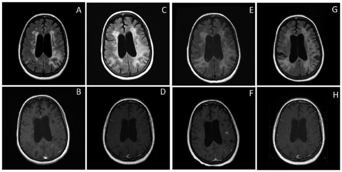
A 51-year-old woman with relapsing–remitting multiple sclerosis (RRMS) and a 3-year history of natalizumab use developed progressive expressive aphasia in March 2013. A brain magnetic resonance image (MRI) revealed new left frontoparietal and right parietal lesions with mild contrast enhancement (Figure 1a and b), and natalizumab was discontinued. The patient’s cerebrospinal fluid (CSF) was positive for the JCV via polymerase chain reaction (PCR) testing and she received five cycles of plasmapheresis in June followed by intravenous (IV) immunoglobulin (2 g/kg body weight, divided over 5 days). Despite this intervention her speech deteriorated and she developed right hemiparesis. Upon referral to our institution in August, she had global aphasia, right sided neglect and right sided hemiparesis. Another brain MRI was obtained which showed subcortical, periventricular, left frontoparietal and right parietal lesions (increased in size compared with the previous MRI) with no contrast enhancement (Figure 1c and d). Lumbar puncture was performed, which was consistent with mild lymphocytic pleocytosis (total nucleated cells 20 cells/dl, 85% lymphocyte). Quantitative PCR of JCV showed 834 copies/ml. Serum flow cytometry analysis showed 319 cells/µl CD8 positive cells (normal range: 330–920 cells/µl) and 570 cells/µl CD4 positive cells (normal range: 530–1300 cells/µl).
Figure 1.
(A–H): Initial magnetic resonance imaging (MRI) fluid attenuation inversion recovery (FLAIR) and post contrast T1 images (A,B) of the patient from outside facility showing subcortical, periventricular, left frontoparietal and right parietal FLAIR hyperintensity (A) with mild contrast enhancement in left parietal lesions (B). Images obtained at our institution 16 weeks into clinical course, showing increase in size of biparietal lesions (C) but no contrast enhancement (D. Images (E,F) obtained after 4 weeks of IL-2 therapy showing area of contrast enhancement left frontal subcortical area concerning for IRIS. Images obtained following completion 12 weeks of IL-2 therapy (G,H).
Over the next few days her clinical condition worsened. She also had starring spells with automatisms concerning of complex partial seizures. She was started on levetiracetam 1 g twice a day. Additional therapies for JCV were considered. The patient was given an initial dose of 50,000 units of IL-2 subcutaneously (SQ) on 25 August followed by 1 million units IL-2 SQ daily. Due to increased risk for IRIS with use of daily IL-2, the patient also received IV methylprednisone weekly. The effect of the off-label therapy was monitored with daily clinical assessments, weekly brain MRI and quantitative CSF JCV PCR. Subsequent CSF JCV PCR showed the JCV copy number had reduced to 240 copies/ml after 1 week of therapy and to 43 copies/ml at 2 weeks. Clinically, the patient showed some improvement and became more responsive and regained right lower extremity antigravity strength. A brain MRI obtained after 4 weeks of therapy showed some contrast enhancement in the left frontoparietal region, which was concerning for development of IRIS (Figure 1e and f). The patient was given IV methylprednisone 1 g daily for 3 days and IL-2 therapy was continued. The patient’s neurological examination showed no clinical deterioration. The next JCV PCR copy number 1 week after the last test was 70 copies/ml, but the contrast enhancement was reduced on subsequent MRI (Figure 1g and h). After 12 weeks of IL-2 therapy, JCV copy number was 31 copies/ml. There was also a mild increase in CSF lymphocytic pleocytosis (total nucleated cells 32 cells/dl, 97% lymphocytes). The patient was more responsive and there was improvement in right sided weakness. Due to the persistence of JCV, IL-2 therapy was changed to mefloquine. At follow up after 6 months, the patient showed no clinical deterioration.
Discussion
This case brings forth a therapeutic option for management of PML, a fatal opportunistic infection which is a complication of natalizumab infusion. Our patient was treated with daily IL-2 and weekly methylprednisone for 3 months with significant reduction in JCV PCR quantitative titers and stabilization of clinical presentation. Although there was some increase in extent of white matter involvement on MRI (Figure 1a–h), no adverse effects were reported from IL-2 administration.
Various therapies have been tried so far for management of PML, largely without success. These therapies have been reported to be effective by directly acting on viral neuronal entry or replication, or by potentiating host immune system mediated viral eradication [Aksamit, 2012]. Currently the standard approach to manage PML related to natalizumab infusion is to discontinue the monoclonal antibody and institute plasmapheresis to remove the integrin α-4/β-1 antibody from the circulation, subsequently increasing central nervous system (CNS) immunosurveillance [Calabrese, 2011].
Based on the patient’s clinical deterioration following initial plasma exchange therapy, there was no definitive way to differentiate between progression of PML versus IRIS. However, lack of gadolinium enhancement on brain MRI and a high copy number of JCV despite conventional treatment suggested a lack of immune response to the infection. We decided to use IL-2 due to its role in potentiation of the anti-JCV cytotoxic T-cell response. IL-2 administration influences CD-8 count, and increases granzyme and perforin formation [Smith, 1984]. It has also been shown to enhance response to cytotoxic natural killer (NK) cells and to induce lymphokine activated killer cells [Lotze et al. 1986]. IL-2 is usually produced by antigen-activated T cells. It subsequently promotes T cells to switch from the G1 phase to the proliferative phase and increases the levels of proinflammatory cytokines leading to T-cell differentiation [Smith, 1984].
Following discontinuation of therapy, the immune system has extensive access to the CNS and patients are at a high risk of developing IRIS [Calabrese, 2011]. Therefore we also continued weekly treatment with high dose methylprednisone. The IL-2 regimen theoretically also puts the patient at increased risk of progression of multiple sclerosis, therefore extensive consenting of patient and family was performed. As the fatality and rate of progression associated with PML is significantly higher than with multiple sclerosis, we decided that the benefits of IL-2 therapy outweighed the risks.
In the past, IL-2 therapy has been utilized for treatment of PML in the patients with hematological malignancies (Hodgkin’s lymphoma, myelodysplatic syndrome) [Buckanovich et al. 2002; Kunschner and Scott, 2005; Przepiorka et al. 1997]. These patients have shown stabilization or improvement in neurological status (Table 1).
Table 1.
Case reports of use of IL-2 therapy for management of PML.
| Age (years)/sex | Clinical presentation | MRI findings | Dose | Duration of therapy | Underlying predisposing factor | JCV CSF PCR/brain biopsy | Improvement in neurological status | |
|---|---|---|---|---|---|---|---|---|
| Dubey et al.(in press) | 51/F | Global aphasia, right sided neglect, right hemiparesis, complex partial seizure | Left frontoparietal and right parietal lesions hyperintense on T2-WI and mild contrast enhancement | IL-2: 50,000 units/m2 initial dose, then 1 million units/m2 daily, SQ | 84 days | Natalizumab therapy for RRMS | + | Decrease in JCV quantitative PCR, improvement and stabilization of neurological status |
| Methylprednisone: 1 g weekly | ||||||||
| Buckanovich et al. [2002] | 29/F | Ataxic, decreased, visual acuity, bilateral inferior,visual field deficits | Irregular diffuse noncontrast enhancing lesions in bilateral parietal lobes, hyperintese on T2-WI | IL-2: 0.5 million units/m2 per day, IV | 116 days, followed reinitiation of therapy 20 days later, duration of therapy NS | Hodgkin’s lymphoma treated with NMASCT | _ | Neurological deficits completely resolved; patient able to perform all activities of daily living |
| status post radiation therapy, | ||||||||
| MOPP/ABV chemotherapy, cyclosporiene for GVHD prophylaxis | ||||||||
| Kunschner and Scott [2005] | 58/F | Cognitive deterioration, dysarthria, right hemiparesis | Irregular no contrast enhancing 3–4 cm lesion in the left centrum semiovale, hyper-intense on T2-WI | IL-2: 0.5 million units /m2 per day for 5 weeks, 1.0 million units /m2 per day for a sixth week, IV | 42 days | Myelodysplastic syndrome | + | 5-year follow up |
| Improved cognition, mild dysarthria, and moderate right hemiparesis | ||||||||
| Przepiorka et al. [1997] | 46/F | Vertigo, aphasia and right hemiparesis | Contrast nonenhancing T2 hyperintense lesion in left frontoparietal region | IL-2: 0.5 million units/m2 per day, IV | 182 days | Low-grade lymphoma status post etoposide, cyclophosphamide, total body irradiation, and autologous marrow and blood stem cell transplantation | + | Improvement in speech and motor function |
ABV, doxorubicin, bleomycin, vinblastine chemotherapy; CSF, cerebrospinal fluid; GVHD, graft-versus-host disease; IL-2, interleukin-2; IV, intravenous; JCV, John Cunningham virus; MOPP, mechlorethamine, vincristine, procarbazine; MRI, magnetic resonance imaging; PCR, polymerase chain reaction; NMASCT, nonmyeloablative allogeneic stem cell transplantation; PML, progressive multifocal leukoencephalopathy; RRMS, relapsing–remitting multiple sclerosis; SQ, subcutaneous; T2-WI, T2-weighted images.
So far, there are no published cases of the management of natalizumab-related PML with a regimen of daily IL-2 and weekly high dose corticosteroids. Based on our experience, this novel regimen not only was well tolerated by the patient with no severe adverse effects, but it may also have contributed to the reduction in CSF JCV copy number. Further prospective studies and randomized controlled trials are required to study IL-2 as a therapeutic agent for PML following natalizumab administration.
Footnotes
Funding: The author(s) received no financial support for the research, authorship, and/or publication of this article.
Conflict of interest statement: The author(s) declared the following potential conflicts of interest with respect to the research, authorship, and/or publication of this article: D.G. has consulted for Teva Pharmaceuticals and Bayer, has grants or grants pending from Novartis, and has received payment for or given lectures for Teva Pharmaceuticals, Bayer, Novartis and Pfizer. A.D.D.’s fellowship funding was provided by the Transverse Myelitis Association. E.F. has received personal compensation as a speaker and consultant from Acorda Therapeutics, Genzyme, Novartis International AG and Teva Pharmaceutical Industries Ltd. B.G. owns stock or stock options and serves as a consultant for Amplimmune, Inc. and DioGenix, Inc; he has also received personal compensation for developing education presentations from MediLogix and for serving as a consultant for Novartis International AG. D.D. and Y.Z. declare no conflicts of interest in preparing this article.
Contributor Information
Divyanshu Dubey, Department of Neurology and Neurotherapeutics, UT Southwestern Medical Center, 5323 Harry Hines Blvd. Dallas, Texas-75235, USA.
Yinan Zhang, Department of Neurology and Neurotherapeutics, UT Southwestern Medical Center, Dallas, Texas, USA.
Donna Graves, Department of Neurology and Neurotherapeutics, UT Southwestern Medical Center, Dallas, Texas, USA.
Allen D. DeSena, Department of Neurology, University of Cincinnati College of Medicine, Cincinnati, Ohio, USA
Elliot Frohman, Department of Neurology and Neurotherapeutics, UT Southwestern Medical Center, Dallas, Texas, USA.
Benjamin Greenberg, Department of Neurology and Neurotherapeutics, UT Southwestern Medical Center, Dallas, Texas, USA.
References
- Aksamit A., Jr. (2012) Progressive multifocal leukoencephalopathy. Continuum 18: 1374–1391. [DOI] [PubMed] [Google Scholar]
- Buckanovich R., Liu G., Stricker C., Luger S., Stadtmauer E., Schuster S., et al. (2002) Nonmyeloablative allogeneic stem cell transplantation for refractory Hodgkin’s lymphoma complicated by interleukin-2 responsive progressive multifocal leukoencephalopathy. Ann Hematol 81: 410–413. [DOI] [PubMed] [Google Scholar]
- Calabrese L. (2011) A rational approach to PML for the clinician. Clev Clin J Med 78(Suppl. 2): S38–S41. [DOI] [PubMed] [Google Scholar]
- Dubey D., Zhang Y., Graves D., DeSena A. D., Frohman E., Greenberg B. (in press) Use of interleukin-2 for management of natalizumab-associated progressive multifocal leukoencephalopathy: case report and review of literature. Therapeutic Advances in Neurological Disorders DOI: 10.1177/1756285615621029. [DOI] [PMC free article] [PubMed] [Google Scholar]
- Kunschner L., Scott T. (2005) Sustained recovery of progressive multifocal leukoencephalopathy after treatment with IL-2. Neurology 65: 1510. [DOI] [PubMed] [Google Scholar]
- Lotze M., Matory Y., Rayner A., Ettinghausen S., Vetto J., Seipp C., et al. (1986) Clinical effects and toxicity of interleukin-2 in patients with cancer. Cancer 58: 2764–2772. [DOI] [PubMed] [Google Scholar]
- Przepiorka D., Jaeckle K., Birdwell R., Fuller G., Kumar A., Huh Y., et al. (1997) Successful treatment of progressive multifocal leukoencephalopathy with low-dose interleukin-2. Bone Marrow Transplant 20: 983–987. [DOI] [PubMed] [Google Scholar]
- Smith K. (1984) Interleukin 2. Annu Rev Immunol 2: 319–333. [DOI] [PubMed] [Google Scholar]


