Abstract
Stem cells hold remarkable promise for applications in tissue engineering and disease modeling. During the past decade, significant progress has been made in developing soluble factors (e.g., small molecules and growth factors) to direct stem cells into a desired phenotype. However, the current lack of suitable synthetic materials to regulate stem cell activity has limited the realization of the enormous potential of stem cells. This can be attributed to a large number of materials properties (e.g., chemical structures and physical properties of materials) that can affect stem cell fate. This makes it challenging to design biomaterials to direct stem cell behavior. To address this, polymer microarray technology has been developed to rapidly identify materials for a variety of stem cell applications. In this article, we summarize recent developments in polymer array technology and their applications in stem cell engineering.
Statement of significance
Stem cells hold remarkable promise for applications in tissue engineering and disease modeling. In the last decade, significant progress has been made in developing chemically defined media to direct stem cells into a desired phenotype. However, the current lack of the suitable synthetic materials to regulate stem cell activities has been limiting the realization of the potential of stem cells. This can be attributed to the number of variables in material properties (e.g., chemical structures and physical properties) that can affect stem cells. Polymer microarray technology has shown to be a powerful tool to rapidly identify materials for a variety of stem cell applications. Here we summarize recent developments in polymer array technology and their applications in stem cell engineering.
Keywords: Polymer microarray, Stem cell, Surface chemistry, Surface topography, Elastic modulus
1. Introduction
Stem cells hold remarkable potential for applications in regenerative medicine and disease modeling. They have the unique ability to undergo self-renewal in an undifferentiated state and have the potential to differentiate into multiple cell types [4–7]. Adult stem cells, such as hematopoietic stem cells (HSCs) and mesenchymal stem cells (MSCs), have been thoroughly investigated and their clinical benefits have been well established [8–10]. The recently derived human pluripotent stem cells (hPSCs), including both human embryonic stem cells (hESCs) and human induced pluripotent stem cells (hiPSCs), have the ability to self-renew indefinitely and differentiate into all of the human cell types in in vitro culture [11–14]. With these characteristics, hPSCs provide an ideal source for the large number of cells (>109 cells/patient) needed for cell replacement therapies [15–17].
To direct differentiation of hPSCs, many of the early advances were accomplished through the study of embryology, with the intent of replicating embryonic development [18–20]. By using soluble inductive factors (e.g., growth factors and small molecules) to recapitulate embryonic stage cell signaling, hPSCs can be differentiated into a desired cell phenotype. One textbook example is to modulate Wnt signaling in a temporally defined manner to produce functional cardiomyocytes from hPSCs [21]. In addition to this rational design-based strategy, high throughput approaches have been utilized to screen small molecules, growth factors and their combinations to direct hPSC differentiations [22]. For example, Borowiak and coworker screened 4000 small molecules and identified two molecules that can direct hESCs into endothermal cells [23]. With these advances, soluble factors have been extensively utilized to differentiate hPSCs into various functional cells.
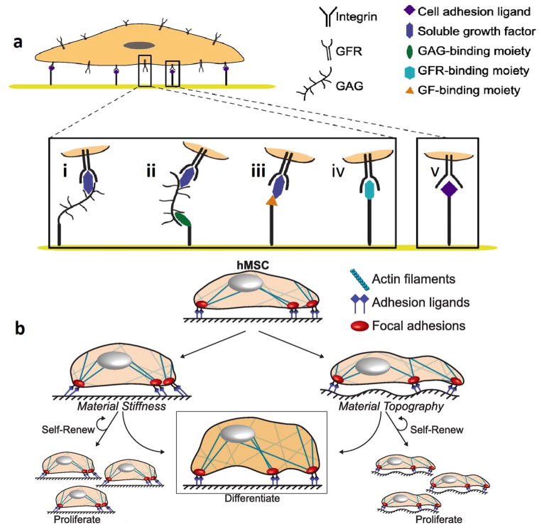
In addition to soluble factors, insoluble factors (e.g., cell culture substrates and 3D scaffolds) have been shown to exhibit controlling effects on stem cells [19,24,25]. While soluble factors can modulate specific target(s) in signaling pathways to influence stem cells via chemical interactions, insoluble factors can provide both chemical and physical cues to direct stem cell fate [19,26–29]. As shown in Fig. 1, signals provided by the materials can be separated into two categories: surface bound chemical structures and material physical properties [30]. Surface bound chemical structures can engage a variety of cell membrane-bound proteins and receptors to initiate various cellular signaling cascades and influence stem cell activities [31]. These surface bound bioactive molecules can be derived from a variety of sources. Some studies have utilized naturally derived ECM proteins (e.g., fibronectin and laminin) due to their biological functions and abundant presence in the extracellular space within the human body [32]. Other research has suggested that it is advantageous to utilize the effective groups of these proteins in order to increase efficiency [33]. This has led to the popularity of peptide-mediated stem cell adhesion and fate determination. One example is the RGD peptide sequence that is known for its ability to induce cell adhesion [34,35]. Though certain integrin has a high affinity for RGD, the resulting interaction alone is not sufficient to control cell fate. As a result, it is inadequate to simply use RGD, requiring a combination of different ligands to elicit an optimized response from the cell membrane [36,37].
Fig. 1.
Stem cell interactions with chemical and physical cues. (a) Chemical interactions on materials can regulate growth factor signaling. Engineered materials may incorporate (i) covalently bound glycosaminoglycans (GAGs) or proteoglycans (PGs) or (ii) moieties that bind GAGs/PGs, which can in turn sequester growth factors from the stem cell microenvironment. Alternatively, materials may be functionalized with (iii) moieties that bind growth factors or (iv) moieties that directly interact with growth factor receptors (GFRs), to upregulate or downregulate GFR signaling. Finally, GFRs and their associated signaling pathways may synergize with (v) integrin-mediated adhesion and signaling downstream of adhesion. (b) Mechanical properties of the microenvironment. Resistance to deformation on stiff materials increases cytoskeletal tension of human mesenchymal stem cells (hMSC) through focal adhesion kinase (FAK) and Rho-associated kinase (ROCK) activity, leading to differentiation. (left) Introduction of topography results in rearrangement of integrins at the cell–material interface, promoting topography-dependent hMSC proliferation, self-renewal, or differentiation. (right) (Figure from Ref. [30] with permission).
Within the last decade, studies assessing the effects of material physical properties on stem cells have received significant attention. One facet is the use of nanoscale topographical features to modulate cell adhesion and direct stem cell fate [38,39]. Due to the high-binding affinity of integrin, and its ability to produce focal adhesion points through cell-surface interaction, cells can bind surfaces of varying roughness and patterned geometries. It has been shown that nanotopographic features of materials surface can influence the key steps in cell adhesion (e.g., integrin clustering) and influence stem cell fate determination through self-renewal and directed differentiation [40–42]. In addition to nanotopographic features, it has been shown that cells are sensitive to the microscale features and stiffness of the materials [43,44]. Both the microscale features and materials stiffness can alter cellular cytoskeletal structures, affecting stem cell fate [44–46]. Due to the large number of variables of chemical and physical properties of materials that can influence cell behavior, it is necessary to develop a highly tunable platform that can be used to direct stem cells.
The traditional model for discovering effective factors for materials-directed cell activity and fate determination utilizes a methodical approach, where only a few conditions can be thoroughly investigated at a time [47]. Given the complexity of the interactions between stem cells and materials, there is a growing need for the development of high throughput approaches for the quick and efficient determination of optimal materials properties. One such strategy is the use of surface gradients, where the material is spatially arranged into regions of gradually varied chemical concentrations or physical properties [48–51]. These methods can allow for rapid establishment of the relationship between one or two materials properties and (stem) cell behaviors. Other studies leveraged the recent development in robotic liquid-dispensing technology and microfabrication to develop polymer microarray systems for the rapid examination of a variety of tunable testing conditions [47,52–56]. These polymer arrays can facilitate the simultaneous screening of a variety of factors, allowing for side-by-side analysis and small-scale experiments that require relatively few resources.
In this review, we will firstly give a brief overview of the technologies used to fabricate polymer microarrays. Secondly, we will discuss the recent development of polymer microarrays to screen the effects of material chemical structures on stem cells. Thirdly, we will describe the development of polymer microarrays to examine the effects of materials physical properties on stem cells with a focus on surface topography and elastic modulus. Lastly, we will review microarrays capable of examining combinatorial effects of materials chemical structures and physical properties as well as recent developments in 3D hydrogel microarray fabrication. We will conclude with an outlook on the field.
2. Polymer microarray fabrication
Polymer microarrays have been fabricated using a variety of patterning technologies. In general, these technologies can be categorized into two groups: (robotic) liquid-dispensing systems and microfabrication technologies [52,57]. The (robotic) liquid-dispensing systems include both contact printing and non-contact printing (i.e., ink-jet printing) [58]. After liquid dispensing, sometimes a crosslinking step (e.g., UV crosslinking of monomers) is needed [54,59]. The contact printing often uses a pin to load the “ink” (e.g., monomer and polymer solutions) and then deposits the “ink” onto a substrate by direct or close contact. The non-contact printing can directly eject “ink” droplets through a nozzle onto substrates [60]. Liquid-dispensing technologies can allow for accurate deposition of a tiny amount of monomer/polymer solutions onto substrates in a spatially defined manner to form microarrays. By changing compositions of the deposited monomer/polymer solutions, this approach has been widely used to create microarrays comprised of polymers with different chemistries.
While certain physical properties of materials (e.g., elasticity) can be controlled through their chemical composition, other physical properties (e.g., topography) are less dependent on the material chemistry. To prepare materials with defined nano-/microscale topography, numerous microfabrication-based techniques including electron beam lithography and nanoimprinting have been employed [52]. These techniques have been used to fabricate material arrays with defined yet varied physical properties to rapidly identify the influence of material physical cues on stem cell behavior.
3. Screening the effects of materials chemical structures on stem cells
Surface chemistry of biomaterials can significantly affect cell behavior through cell surface receptor mediated signaling cascades. Based on the materials chemistry, we have separated the existing polymer microarrays into four different categories: synthetic polymer microarrays, ECM protein microarrays, peptide microarrays, and small molecule microarrays.
3.1. Synthetic polymer microarrays
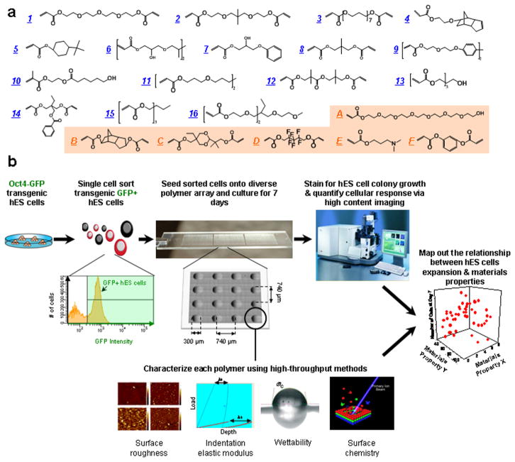
Due to the high polymerization rate of photopolymerization and high solubility of (meth)-acrylates in high boiling point organic solvents (e.g., DMF), light-assisted polymerizations of (meth)-acrylates have been extensively used to prepare synthetic polymer microarrays. Although polymers within these microarrays usually do not contain biological ligands, they can adsorb serum/ECM proteins from pre-conditioning solution and/or cell culture media to effectively bind to cell surface receptors. One example of this strategy is to rapidly identify synthetic substrates for hPSC culture through the screening of polyacrylate-based microarrays. To this end, we have developed a library of 496 different polymers constructed by 22 acrylate monomers, accomplished using a robotic liquid-dispensing system and UV photopolymerization. Subsequently, physical characteristics including surface wettability, surface roughness, and indentation elastic modulus of each polymer in the library were quantified using high throughput methods. This allowed for the establishment of a structure–function relationship between the biological performance of hESC culture and the material properties [54]. The results showed that the substrate properties affected the undifferentiated growth of hESCs to different degrees; with the surface chemistry having showed the most pronounced effects (Fig. 2). The surface chemistry, especially esters and hydrocarbons, were recognized as key cues on materials surface to promote hES cell self-renewal, as they can effectively enhance adsorption of vitronectin to improve self-renewal of hPSCs. Enlightened by this information, we have developed an ultraviolet/ozone radiation modified polystyrene (PS) culture system in the hopes of producing a favorable cell culture environment. It is possible to introduce certain chemical cues that have been shown to promote hESC self-renewal, such as hydrocarbons and ester/carboxylic acid, by treating PS with an optimized dose of short wavelength UV [61]. UV exposed PS (UVPS) was able to produce three-fold more cells per area when compared to the more commonly used mouse embryonic fibroblast (mEF) feeder cells. Further investigation has revealed that UVPS can promote vitronectin adsorption, improving surface interaction with integrin αvβ3 and αvβ5 to better support hPSCs self-renewal.
Fig. 2.
High-throughput screening of biomaterials for clonal growth. (a) Monomers used for array synthesis were classified into two categories: “major” monomers that constitute >50% of the reactant mixture and “minor” monomers that constitute <50% of the mixture. Sixteen major monomers were named numerically (blue), and six minor monomers were labeled alphabetically (orange). (b) Schematic diagram of the screen. First, transgenic Oct4-GFP hES cells were maintained on mEFs. Then flow cytometry enabled the isolation of high purity undifferentiated hES cells from the completely dissociated coculture of hES cells and mEFs. A flow cytometry histogram during a representative cell sort is shown. GFP + cells (right of the black gate) were seeded onto the arrays, whereas the differentiated cells and mEFs (GFP−, left of the black gate) were not used. A photograph of the polymer microarray with 16 polymer spots is shown to illustrate dimensions and separation. Each polymer was also characterized using high-throughput methods to characterize its surface roughness, indentation elastic modulus, wettability (water contact angle, °C) and surface chemistry. Finally, the cellular response on the polymer array was quantified using laser-scanning cytometry, and structure–function relationships were determined by numerical analysis of both the cellular response and materials characterization data. (Figure from Ref. [54] with permission). (For interpretation of the references to color in this figure legend, the reader is referred to the web version of this article.)
Recently, Celiz and coworkers expanded this system to use 141 different (meth)-acrylate monomers to prepare combinatorial polymer microarrays to screen for culture substrates that are capable of supporting hPSCs adhesion and expansion. This system has allowed for the identification of several new chemical moieties that can promote hPSC adhesion [62]. In addition, the current methods to passage hESCs and hiPSCs (e.g., proteolytic enzymatic digestion) are very inefficient and tend to result in cellular abnormality. To improve this, Zhang and coworkers prepared a 2-(diethylamino)ethyl acrylate-based thermoresponsive hydrogel array to support passaging hESCs in a reagent-free manner. From the library, the authors identified a hydrogel that can support long-term expansion (2–6 months) of hESCs and allow for reagent-free cell passaging by simply changing the culture temperature from 37 to 15 °C for 30 min [63].
In addition to dispensing monomer solutions onto glass slides and polymerizing them in situ, this technology can be extended to printing existing polymers and their combinations onto a microarray. To this end, Anderson and coworkers directly printed a number of biodegradable polymers onto glass slides to fabricate polymer microarrays and explored their effects on human mesenchymal stem cells (hMSCs), neural stem cells (NSCs) and primary articular chondrocytes [53]. Using a comparable technology, Hay and coworkers were able to screen potential substrates for hESC-derived hepatocyte (hESC-HE) culture by spotting 380 presynthesized polyurethanes and polyacrylates onto agarose coated glass slides [64]. The system revealed six polymers that could support hESC-HE attachment, but neither chemical nor physical cues were found to elicit biological responses. This highlights the need for unbiased screening to discover suitable substrates for the culturing of stem cells.
3.2. ECM protein microarrays
In addition to the synthetic polymers, ECM proteins provide another source for the construction of a combinatorial library. Though relatively expensive and possibly containing contaminants of animal origin, they hold great potential in having more potent signaling moieties capable of initiating cellular signaling cascades to regulate stem cell growth and differentiation. As early as 2000, MacBeth and coworkers developed protein microarray technology [65]. Then, Flaim and coworkers printed ECM proteins onto dehydrated polyacrylamide substrates to fabricate ECM protein microarrays. They used these arrays to examine mouse embryonic stem (mES) cells and hepatocytes, revealing the effects of different protein combinations on hepatocyte function and mES cell differentiation. Through the use of “factorial analysis methods”, the role of each ECM protein was identified from the combinations. The results showed that collagen III and laminin caused down regulation of albumin (a liver-specific function marker), while collagen IV and fibronectin could lead to an increase of intracellular albumin expression [66]. Further, the group studied the combinatorial effects of ECM proteins and growth factors with this technology [67]. For this study, the authors designed microarrays composed of 20 combinations of 5 ECM proteins (fibronectin, laminin, collagen I, collagen III, collagen IV) and arranged them into a multiwell system. 12 different growth factors were mixed and added into microwells, allowing for the simultaneous study of 1200 experiments on 240 unique signaling environments. This assay offered a method for the rapid identification of co-signaling between ECM protein, growth factors, and combinations of the two.
Rather than using growth factors in a soluble form, Soen and coworkers printed the premixed combinations of growth factors, ECM components, cell adhesion molecules, and morphogens onto a microarray to study the proliferation and differentiation of primary bipotent human neural precursor cells [68]. With a similar strategy, Brafman and coworkers also developed an extended arrayed cellular microenvironment (ACMEs) [69]. Besides growth factors and ECM proteins, ACMEs also contains proteoglycans and glycans, carbohydrates that occupy a large portion of extracellular space. The authors then applied this technology to examine the effects of extracellular components on multiple hESC and hiPSC lines.
One of the potential limitations of the previously described microarrays lies in the possible delamination of the coating during the long-term culture. To address this problem, Ghaemi and coworkers robotically printed ECM proteins onto freshly prepared epoxy-silane-coated slides to covalently immobilize ECM proteins. Subsequently, the surface was passivated by poly(ethylene glycol) bisamine to improve long-term cell pattern integrity. With this system, they successfully studied hMSC osteogenic differentiation over a 3-week span [70].
3.3. Peptide microarray
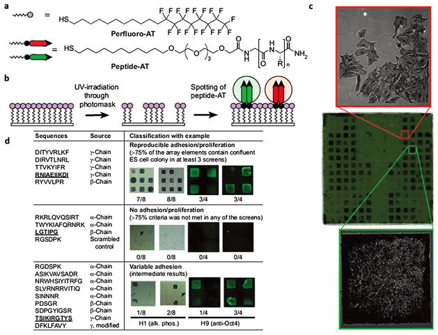
ECM proteins and growth factors hold great potential as source materials to fabricate polymer microarrays due to their high biological activities. However, their applications could be potentially limited due to their high costs, batch-to-batch variations and contaminants of animal origin. In contrast, synthetic materials containing biological ligands can be made at low cost and with high purity. A recent trend is to conjugate peptides (i.e., functional segments of ECM proteins) onto substrates in a microarray format. To this end, Kiessling and coworkers prepared thiolated peptides and spotted them onto gold-coated glass slides to prepare self-assembling monolayer (SAM) peptide microarrays [71]. They then applied this technology to examine the functions of peptides derived from laminin on maintaining hESC pluripotence, as laminin is well known for its ability to promote hESC pluripotency (Fig. 3) [72]. Next, this group synthesized the thiolated heparin-binding peptides that target cell-membrane glycosaminoglycans (GAGs) and included them in the SAM peptide microarray. They then applied this array system to screen for peptides that can maintain long-term self-renewal of hPSC lines [37]. Recently, they applied the heparin-binding peptides discovered by the SAM peptide microarray to hPSC differentiation and elucidated the signal pathways switch between Akt and Smad for different germ layer cell differentiation [55].
Fig. 3.
SAM surface array screen for ES cell growth and self-renewal. (a) Structure of alkanethiols used for the assembly of the array. “R” denotes amino acid side chains. (b) A two-step process was used to generate the arrays. A uniform SAM composed of perfluoro-AT was photopatterned and solutions of peptide-ATs were spotted onto the exposed gold areas to form peptide-terminated SAM array elements. (c) A representative array containing 18 different peptide-ATs was screened to identify surfaces that promote proliferation of ES cell line H9. The peptide-displaying SAMs are arranged in a mirror-symmetric pattern (bottom and top halves of chip have identical array elements). Each half of the array contains 18 laminin-derived peptide-ATs spotted in groups of four (2 × 2 elements) arranged in a left-to-right horizontal comb pattern. The remaining array elements are filled with nonadhesive glucamine-AT. The array was incubated with media containing 20% FBS and thoroughly washed with serum-free media. Cells were grown on the array for 6 d in culture media, fixed, and stained for alkaline phosphatase (AP). The positions of cell growth are symmetric and emphasize the reproducibility of ES cell responses to the array elements. The array was mounted onto a glass slide and scanned using a flatbed scanner. The phase-contrast 10× images reveal morphological differences between AP-positive undifferentiated (left) and AP-negative differentiated ES cells (right). Array dimensions: 22 × 22 mm; each element is 0.8 mm. (d) Summary of results from multiple screens for 18 laminin-derived peptides and ES cell lines H1 and H9. Laminin chain origin is shown for each peptide. Following 5–7 d of growth, substrates were categorized according to their ability to accommodate confluent (square-shaped) and undifferentiated colonies, as judged by staining for alkaline phosphatase (purple) or Oct4 (green). Each synthetic substrate was tested four to eight times in each screen; therefore, the fraction of array elements presenting square ES cell colonies serves as a convenient measure of substrate efficiency. Representative results for one peptide in each category are shown, and the corresponding peptide sequences are underlined. The apparent higher intensity of Oct4 around the edges of the array element can be attributed to differences in thicknesses of ES cell colony around the edges or differentiation of overcrowded cells in the middle of the array element. The latter is not observed when cells are cultured on large area substrates that do not restrict colony growth. (Figure from Ref. [72] with permission).
Instead of using thiol-gold interactions to immobilize peptides, Kilian’s group has taken advantage of recent advances in “click” chemistry [73]. They synthesized the alkyne group functionalized peptides and robotically spotted them with azide-terminated alkanethiolate to prepare peptide microarrays. They used these arrays to study the human adipose-derived stem cell (hADSCs) attachment abilities on four ECM adhesion peptides (YIGSR, GRGDS, KPSSAPTQLN, and KRSR) and their various combinations. They demonstrated enhanced osteogenic expression of ADSCs when using KPPSAPTQLN peptides, which provided evidence of the peptide’s role in osteogenic differentiation [74]. In addition to directly spotting peptide solution onto glass slides, Murphy and coworkers developed a straightforward yet robust approach to prepare SAM peptide microarrays. They put an elastomeric stencil, containing an array of holes, onto gold substrates to produce arrayed microwells, which can facilitate local formation of SAMs and carbodiimide catalyzed conjugation reactions between n-terminal primary amine of peptides and carboxylic acid terminated SAMs. They used this array system to study the effects of multiple peptides on human mesenchymal stem cell (hMSC) activities by engaging multiple cellular receptors including integrin, bone morphogenic protein (BMP)-2 receptor and growth factor receptors [75]. This technology can be extended to fabricate 3D peptide-functionalized synthetic hydrogel microarrays, with further details discussed in Section 6 (3D hydrogel microarray fabrication) [56].
3.4. Small molecule microarrays
Another potential method to direct stem cell differentiation is to employ small molecules that mimic chemical aspects of the native ECM. Utilizing this strategy, Benoit and coworkers robotically spotted methacrylates functionalized with small molecules to develop a hydrogel microarray that could emulate the functional groups of various extracellular microenvironments to induce hMSCs differentiation [76]. hMSCs are well known to possess the capacity to differentiate into multiple cell types, including cartilage, bone, and fat. The researchers chose each of the following small molecules for their roles in connective tissue development: pedant carboxyl groups that resemble glycosaminoglycans in cartilage, phosphate groups that induce bone mineralization, and tert-butyl groups that mimic the lipid rich environment of adipose tissue. In line with their hypothesis, these functionalized hydrogels were able to induce differentiation of hMSCs into chondrogenic, osteogenic, or adipogenic cells.
4. Assessing the effects of materials physical properties on stem cells
Physical properties of materials can affect a variety of stem cell activities. Here, we describe the application of microarray strategies to study the effects of topography and stiffness of materials on stem cell activities.
4.1. Topographical biomaterials
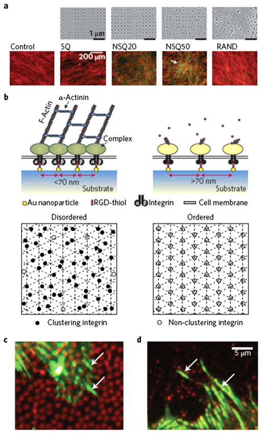
One important facet of research investigating the effects of materials’ physical properties is the study of material topographical features. It has been shown that cells are able to respond to the nanotopographical features of different materials using integrin receptors through the process of developing focal adhesion points [42]. In order to utilize these cell-surface interactions to direct stem cells, significant research has been devoted to investigating the governing mechanisms behind the interplay between integrin-ligand binding and nanotopography. To this end, Dalby and coworkers showed that the interactions between cells and nanoscale substrates are to a large extent mediated by cellular constructs called nanopodia, smaller and more sensitive versions of cellular filopodia [77]. In addition, they have utilized electron beam lithography to develop an array system to investigate the effects of nanotopography on hMSC self-renewal and differentiation. Using a PMMA nanoarray with a controlled disorder (i.e., neither highly ordered nor random), they were able to direct osteogenesis in hMSCs with comparable efficiency to osteogenic media (Fig 4) [78]. Through further investigation, the authors showed that the highly ordered nanostructures reduce hMSCs adhesion yet promote their self-renewal [79]. The altered integrin clustering on the nanoscale substrates has been identified as having a major influence on the changes in stem cell fate [42]. In addition, the authors applied meta-bolomics methodology to help understand the effects of the nanoscale substrates on hMSC multipotency and to systematically identify useful pluripotent factors [80].
Fig. 4.
Nanoscale disorder and adhesion bridging. (a) Electron-beam lithography was used to demonstrate that neither order nor randomness successfully led to osteoinduction of MSCs. SQ, square (within the square array the individual pits are 120 nm in diameter, 100-nm deep and have a 300-nm centre–centre spacing); RAND, random. However, controlled disorder (NSQ20 and NSQ50, same as SQ but with ±20 nm and ±50 nm offset from the 300 nm centre–centre position) produced abundant, spontaneous, osteogenesis in basal media. Cells are shown in red (actin) and osteogenesis is shown in green (osteopontin). (b) At the adhesion level, adding a level of disorder to RGDs placed 70 nm apart allowed much greater integrin clustering in MSCs. Bottom panels show an ordered lattice (right) with RGDs placed >70 nm apart with little integrin clustering possible (open circles). However, if a level of disorder (left) is added while there are areas with gaps where adhesion does not occur, more RGDs are moved within gathering distance (closed circles). (c and d) Fluorescence microscopy images showing 800-nm-diameter fibronectin circles (red, c) and 200-nm-diameter vitronectin circles (red, d) with adhesions (vinculin in green) seen bridging between the circles (arrows). (Figure from Ref. [42] with permission). (For interpretation of the references to color in this figure legend, the reader is referred to the web version of this article.)
An interesting application of nanotopographic substrates for stem cell engineering is cell sorting. Chen and coworkers utilized the established etching methodology to produce nanorough patterns on an otherwise smooth glass surface. They have demonstrated that the undifferentiated hESCs prefer smooth surfaces while the differentiated hESCs favorably attach to nanorough surfaces. When a mixed cell population of hESCs and NIH 3T3 fibroblasts were applied to the glass slides, the cells sorted themselves according to the rough and smooth regions, with the majority of hESCs attaching to the smooth surface and the fibroblasts attaching to the nanorough surface [81].
In addition to nanotopography, stem cells can be readily manipulated using microscale substrates. It is important to make note of the difference in scale between features found on the nano- and microscale patterning. The nanoscale patterning operates on the same scale as the individual cell receptors, which allows for the nanotopographical features to directly modulate the binding activities of integrin and other adhesion protein receptors [41]. In contrast, microscale topography deals on the scale of the cell as a whole. As a result, the microscale substrates can control stem cell size and shape that in turn can affect cellular cytoskeletal contractility and determine stem cell phenotype [44,46,82]. Feature size of nano-/microscale substrates can significantly affect cellular cytoskeletal contraction and stem cell differentiation. To rapidly identify the relationship between stem cell differentiation and feature size of micro-/nano-patterned substrates, Cabezas and coworkers leveraged the high precision 3D spatial control of scanning probe lithography and the robustness of soft lithography to develop polymer pen lithography (PPL) technology. This technology allows for the rapid fabrication of as many as 25 million features with sizes ranging from the nano- to micrometer scale. The authors then used this technology to demonstrate that controlling the feature size of the substrate can effectively induce osteogenic differentiation of MSCs in the basal medium [83].
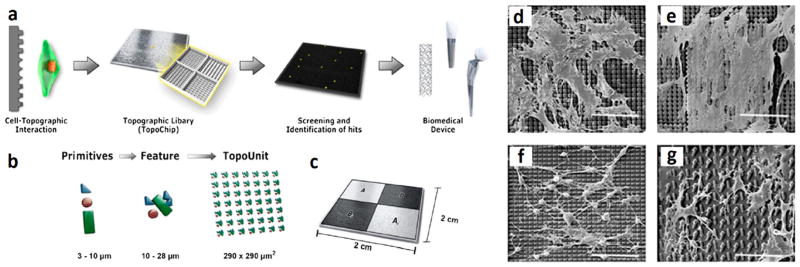
Given the nearly unlimited number of possible topographical features and their combinations, it is necessary to develop high throughput screening systems to identify the best possible combination of topographical features that can determine stem cell fate. Unadkat and coworkers used three types of primitive shapes (i.e., circles, triangles and lines) as building blocks to construct random microscale topographic features. The circles can provide large smooth surfaces, triangles can create angles and rectangles can lead to stretched elements. By using an unbiased algorithm, the authors selected and fabricated 2176 random topographies to form “TopoChip”, upon which hMSCs were seeded and assessed for expression of differentiation markers [84] (Fig. 5). The chips were made from poly(DL-lactic acid) (PLA), with individual features being 5 μm and the walls being 20 μm. Using this system, the researchers were able to screen for patterns that improved osteogenic differentiation and cell proliferation. They have since improved upon this model by developing a chip carrier system that allows for a controlled culturing environment, eliminating many variables that may otherwise influence cell adhesion and viability [85].
Fig. 5.
TopoChip design. (a) A schematic representation of a sequence of events that is proposed to be followed for high-throughput screening of biomedical materials starting from initial design to clinical application. (b) Design of the TopoChip is based on the use of primitives. Three types of primitives, namely circles, triangles, and lines were used to construct features. Repeated features constitute a TopoUnit and two times 2176 = 4352 TopoUnits constitute a TopoChip (size ranges are indicated). In addition, four flat control surfaces are included. (c) TopoChip is divided into four quadrants. TopoUnits in quadrant A are repeated in quadrant Ai and similarly TopoUnits in quadrant B are repeated in quadrant Bi in order to exclude site specific or localized effects. (d–g) SEM images of cells showing diverse cellular morphologies. Scale bar, 90 μm. (Figure from Ref. [84] with permission).
4.2. Stiffness of substrates
In addition to topography, one tunable physical characteristic that affects stem cell fate is the elastic modulus (i.e., stiffness) of substrates. The influence of substrate stiffness on stem cells is mediated through the cellular cytoskeleton. It is easy to envision that soft substrates are relatively easy for a cell to deform, and stiff surfaces force the cells to spread. This will directly influence cellular cytoskeletal contractility and determine stem cell fate [86]. The very recent results from Swift and coworkers further suggests that outside mechanical forces are transducted as far as the nucleus, where nucleoskeletal protein lamin-a expression changes relative to outside tissue stiffness [87].
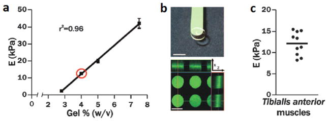
As a pioneer in the field, Engler and coworkers demonstrated that hydrogel stiffness effectively influences the differentiation process of hMSCs by replicating in vivo tissue elasticity. Their results showed that gels similar to muscle elasticity led to myogenic differentiation, while gels similar to calcified bone elasticity led to osteogenic differentiation [88]. In addition, Musah and coworkers showed neurons can be derived from hESCs and hiPSCs by using compliant hydrogel substrates [89]. Due to the limited knowledge of the adult stem cell niche in vivo, it is very challenging to expand adult stem cells in vitro [90]. To address this, Blau and coworkers showed through a microarray system that it was possible to maintain muscle stem cells (MuSCs) for longer periods of time using laminin modified PEG hydrogels with comparable elasticity to that of skeletal muscle (Fig. 6) [91]. They further developed this technology to improve transplantation efficacy of aged stem cells through the use of hydrogels with appropriate elasticity [92].
Fig. 6.
Pliant hydrogel promotes MuSC survival and prevents differentiation in culture. (a) PEG hydrogels with tunable mechanical properties. Young’s modulus (E) is linearly correlated with precursor polymer concentration (n = 4); red circle indicates muscle elasticity. (b) Image of a pliant PEG hydrogel on a green spatula. Scale bar, 7 mm (top). Confocal immunofluorescence image of hydrogel microcontact printed with laminin specifically at the bottom of hydrogel microwells (i.e., from the “tips” of the micropillars). Scale bar, 125 mm (bottom). (c) Dissected tibialis anterior muscles (n = 5 mice, 10 muscles total) were analyzed by rheometry (horizontal line indicates the mean). (Figure from Ref. [91] with permission). (For interpretation of the references to color in this figure legend, the reader is referred to the web version of this article.)
5. Simultaneously testing the effects of chemical and physical cues
While the array systems described above provide insight on cell activity, the singular control of physical or chemical parameters may not be sufficient to elicit the desired stem cell phenotypic responses for tissue engineering purposes. The combined testing of chemical and physical cues in an array format has emerged as a powerful strategy to screen stem cells. For example, some of the previously mentioned studies investigated not only the effects of materials chemistry mediated cell adhesion, but also attempted varying the stiffness of the materials [54].
A textbook example to study biochemical and biophysical effectors of stem cells using a high throughput microarray system was developed by Gobaa and coworkers. They utilized a robotic spotting system to examine different combinations of adhesive proteins on increasingly stiff hydrogel surfaces [93]. The authors further modified this technology for individual cell sorting, which allowed for the building of custom live mammalian cell arrays for the culture and interrogation of cell libraries [94]. Through the careful integration of hydrogels of different stiffness, roughness, or hydrophobicity, it may be possible to induce a wider variety of responses that would otherwise be difficult to access through one parameter alone.
In another example, Junmin and coworkers developed a novel microarray to investigate the combined effects of cell geometry, hydrogel substrate mechanics and cell adhesive ligand composition on hMSC differentiation [95]. In addition to hydrogel systems, Weng and coworkers developed a micropost array system, which contains ECM protein-coated PDMS microposts with varied length, diameter, spacing, and stiffness, to understand the synergistic effects of matrix stiffness and surface-bound cell adhesive ligands. This PDMS micropost array can allow the authors to independently control and completely decouple substrates’ chemical and physical properties, including cell adhesive ligand density and patterning, and bulk and nanoscale mechanics of the substrates [96].
5.1. 3D hydrogel microarray fabrication
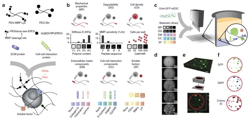
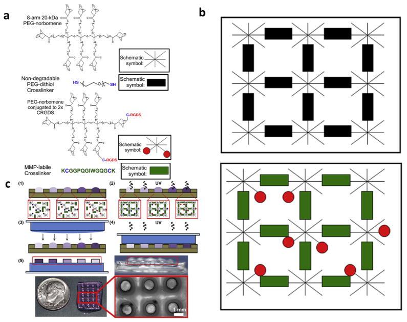
While most of the above-described microarray systems are limited to 2D substrates, recent research has shifted to preparing 3D culture environments in a microarray format to better understand stem cell behavior in an in vivo-like environment. To this end, Ranga and coworker utilized a liquid-dispensing robot to prepare 3D hydrogel microarrays that were used to test the combined effects of matrix elasticity, proteolytic degradability, and signaling proteins on the self-renewal of mESCs (Fig. 7) [97]. Using a similar technology, Khadmhosseini and coworkers spotted miniaturized cell-laden 3D hydrogel microarrays. They evaluated the osteogenic differentiation of hMSCs using different combinations of ECM proteins including gelatin, fibronectin, laminin, and osteocalcin in 8 kinds of media (totaling 96 conditions). Their results revealed that the combination of multiple ECM proteins significantly enhanced osteogenic differentiation of hMSCs when compared to the individual protein. They further validated the results obtained from the microarray experiments by using macroscale hydrogels [98]. In addition to liquid-dispensing systems, microfabrication based technologies can facilitate 3D hydrogel array fabrication. To this end, Murphy and coworkers have extended their microwell array technology to prepare 3D hydrogel microarrays (Fig. 8) [76]. They used standard soft lithography techniques to fabricate a PDMS stencil containing arrays of microwells. Then, hydrogel precursor solutions containing different ratios of RGD cell adhesion peptides were spotted into wells and cross-linked using UV light to form 3D hydrogel arrays. Another potentially powerful strategy to fabricate 3D hydrogel microarrays is to utilize 3D photopatterning techniques. For example, Mosiewicz and coworkers applied photocage chemistry to prepare a hydrogel that can manipulate stem cells in 3D environments in a spatiotemporally controlled manner [99].
Fig. 7.
3D combinatorial screening from a modular materials library. (a) Enzymatically mediated cross-linking scheme, where xxxx xxxx represents specific peptide sequence. (b) Components of the combinatorial toolbox are assembled from biologically relevant factors in categorized form. Stiffness and MMP sensitivity of the matrix are set within the experimentally measured ranges shown (n = 3 replicates). (c) Experimental process consists of combining the components library with reporter cells using robotic mixing and dispensing technology into 1536-well plates (n = 3 replicates). (d–f) Automated microscopy and image processing to determine colony size and GFP intensity. Average cell density per well is set by the initial cell concentration used in the experiment, and exact initial cell density for each well is determined retrospectively by imaging. Examples of a set of images tracking colony growth in a single well over the course of a 5-day experiment (d) 3D confocal reconstruction (e) and image segmentation are shown (f). Scale bar, 200 mm. (Figure from Ref. [97] with permission).
Fig. 8.
Molecules included in PEG hydrogels. (a) The hydrogels are composed of (i) 8-arm PEG molecules, with each arm functionalized with a norbornene molecule; (ii) Di-thiolated PEG crosslinking molecules bridge multiple 8-arm PEG molecules together into an ordered polymer network. A di-thiolated PEG molecule acts as an inert crosslinking molecule that is not cell-degradable; (iii) In bioactive hydrogels, PEG molecules are decorated with CRGDS adhesion peptide or CRDGS scrambled peptide to modulate cell adhesion to the hydrogel; (iv) Di-thiolated matrix metalloproteinase (MMP) labile crosslinking peptides enable cell-driven hydrogel degradation. (b) “Background” hydrogels are void of cell adhesion molecules and are not subject to cell-driven degradation (top). “Hydrogel spots” modulate cell behavior through covalently attached adhesion molecules and are biodegradable via MMP activity (bottom). (c) Schematic representation of hydrogel array fabrication. (1) Separate hydrogel spot solutions containing various ratios of CRGDS adhesion peptide (red circles) and a scrambled CRDGS non-functional peptide (blue circles) are pipetted into wells of a PDMS stencil. Total pendant peptide concentration is fixed at 2 mM in all solutions. (2) The hydrogel spots are crosslinked in the stencil using UV light. (3) A crosslinked 1-mm thick “background” hydrogel slab is laid on top of the crosslinked bioactive hydrogel spots. A thin layer of background hydrogel solution is added to the slab to anchor the cured spots to the background. (4) The hydrogel spots are anchored to the background after treatment with UV light. (5) The completed hydrogel array is removed from the stencil. Red boxes highlight the raised spots in the schematic and side view images of the arrays. (Figure from Ref. [56] with permission.). (For interpretation of the references to color in this figure legend, the reader is referred to the web version of this article.)
6. Perspective and conclusion
Polymer array technology has emerged as a powerful tool to rapidly examine the relationship between material properties (i.e., chemical and physical) and stem cell responses. To further accelerate its applications in stem cell engineering, several key factors need to be carefully considered. For example, it is unrealistic to screen for every possible combination of cell-material interactions and the resulting downstream effects. The polymer array-based research would benefit tremendously from careful experimental design. Celiz and coworkers have attempted to improve upon this through a stepwise approach, which includes high throughput materials discovery, “hit” optimization, and the identification of “lead” compounds [100]. Mathematical modeling can play a critical role in synergistically improving the power of polymer microarray technology. Kohn and coworkers have developed a quantitative structure property relationship (QSPR) modeling method for predicting cellular responses within large combinatorial libraries [101]. Through the development of more advanced modeling methods, it will be possible to narrow down the pool of polymer candidates that more likely elicit a desired stem cell response, allowing for a more direct investigation without the need to examine every possible combination.
There is a growing need for the polymer array technology in stem cell research. One of the largest problems faced by hPSC technology in tissue engineering applications is the difficulty in producing large numbers of patient-specific, functional cells. Despite the progress made in optimizing soluble factors (e.g., small molecules and growth factors) for hPSC self-renewal and differentiation, Matrigel has remained the gold standard substrate for hPSC culture and differentiation. Due to the complexity of the chemical/biological composition (>1800 proteins) of Matrigel, it is expected that high throughput microarray technology will play a vital role in the discovery of chemically defined culture substrates for hPSC culture. Given the large variation between different hESCs and hiPSC lines [102,103], it is anticipated that each hESC and hiPSC line may need its own synthetic substrates for optimal expansion and differentiation. This will necessitate the further development and wide application of polymer microarray technology in personalized/precision regenerative medicine and disease modeling.
In addition to hPSCs, very recent research clearly demonstrated the power of adult stem cells in regenerative medicine [104,105]. Due to the limited understanding of adult stem cell niche in vivo, it is very difficult to isolate and expand most human adult stem cells in vitro. The emerging microarray technology can dramatically accelerate the discovery of innovative materials to facilitate the development of adult stem cell mediated regenerative medicine.
While most research has been focused on the optimization of materials for a specific stem cell application, there is growing interest in studying the fundamental mechanisms of signaling pathways involved in the stem cell-materials interactions. To this end, Kiessling and coworkers have identified heparin-binding peptides by using microarray technology and elucidated how the binding interactions between substrates and cell surface receptors can switch signal pathways between Akt and Smad for different germ layer cell differentiation [55].
Finally, while most research to date has been limited to developing culture substrates for stem cells, the paradigm in stem cell research is gradually shifting from cell production to cell delivery and integration of these delivered (stem) cells with host tissues. For example, human cardiomyocytes can be routinely derived from hESCs and hiPSCs. However, only ~10% hiPSC-derived cardiomyocytes have been found to be viable after they were injected into infarcted myocardium [106,107]. This clearly demonstrates an unmet need to rapidly develop three-dimensional, semi-vivo systems to better understand and engineer stem cell behaviors in physiological and pathological environments. Further advances in robotic liquid handling technology, hydrogel chemistry, and photolithography would greatly accelerate the development of three-dimensional, matrix-like scaffolds for stem cell engineering [38,108,109].
Acknowledgments
The authors would like to thank the financial support from the National Institutes of Health (8P20 GM103444, U54 GM104941), the startup funds from Clemson University, and the National Science Foundation (NSF - EPS-0903795).
Glossary
- Click Chemistry
A set of conjugation reactions that fulfill the following prerequisites: (i) high yield, nearly quantitative conversion; (ii) biologically benign conditions (aqueous solution, ambient temperature, and near physiologic pH); (iii) limited or no residual byproduct [1]
- Combinatorial Array
Array design that produces large numbers of testing conditions through the combination/mixing of (bio)molecules
- Factorial Analysis
Statistical method used to describe variability among observed, correlated variables in terms of a smaller number of defined “factors”
- Soft-Lithography
A set of related techniques uses stamps or channels fabricated in an elastomeric (’soft’) material for pattern generation [2]
- Self-Assembled Monolayers (SAM)
Ordered molecular assemblies formed by the adsorption of an active surfactant on a solid surface [3]
References
- 1.Tang W, Becker ML. “Click” reactions: a versatile toolbox for the synthesis of peptide-conjugates. Chem Soc Rev. 2014;43:7013–7039. doi: 10.1039/c4cs00139g. [DOI] [PubMed] [Google Scholar]
- 2.Kane RS, Takayama S, Ostuni E, Ingber DE, Whitesides GM. Patterning proteins and cells using soft lithography. Biomaterials. 1999;20:2363–2376. doi: 10.1016/s0142-9612(99)00165-9. [DOI] [PubMed] [Google Scholar]
- 3.Ulman A. Formation and structure of self-assembled monolayers. Chem Rev. 1996;96:1533–1554. doi: 10.1021/cr9502357. [DOI] [PubMed] [Google Scholar]
- 4.Weissman I. Stem cell therapies could change medicine... if they get the chance. Cell Stem Cell. 2012;10:663–665. doi: 10.1016/j.stem.2012.05.014. [DOI] [PubMed] [Google Scholar]
- 5.Daley GQ. The promise and perils of stem cell therapeutics. Cell Stem Cell. 2012;10:740–749. doi: 10.1016/j.stem.2012.05.010. [DOI] [PMC free article] [PubMed] [Google Scholar]
- 6.Melton DA. Using stem cells to study and possibly treat type 1 diabetes. Philos Trans R Soc Lond B Biol Sci. 2011;366:2307–2311. doi: 10.1098/rstb.2011.0019. [DOI] [PMC free article] [PubMed] [Google Scholar]
- 7.Saha K, Jaenisch R. Technical challenges in using human induced pluripotent stem cells to model disease. Cell Stem Cell. 2009;5:584–595. doi: 10.1016/j.stem.2009.11.009. [DOI] [PMC free article] [PubMed] [Google Scholar]
- 8.Martinez EC, Kofidis T. Adult stem cells for cardiac tissue engineering. J Mol Cell Cardiol. 2011;50:312–319. doi: 10.1016/j.yjmcc.2010.08.009. [DOI] [PubMed] [Google Scholar]
- 9.Pittenger MF, Mackay AM, Beck SC, Jaiswal RK, Douglas R, Mosca JD, et al. Multilineage potential of adult human mesenchymal stem cells. Science. 1999;284:143–147. doi: 10.1126/science.284.5411.143. [DOI] [PubMed] [Google Scholar]
- 10.Weissman IL, Anderson DJ, Gage F. Stem and progenitor cells: origins, phenotypes, lineage commitments, and transdifferentiations. Annu Rev Cell Dev Biol. 2001;17:387–403. doi: 10.1146/annurev.cellbio.17.1.387. [DOI] [PubMed] [Google Scholar]
- 11.Takahashi K, Tanabe K, Ohnuki M, Narita M, Ichisaka T, Tomoda K, et al. Induction of pluripotent stem cells from adult human fibroblasts by defined factors. Cell. 2007;131:861–872. doi: 10.1016/j.cell.2007.11.019. [DOI] [PubMed] [Google Scholar]
- 12.Takahashi K, Yamanaka S. Induction of pluripotent stem cells from mouse embryonic and adult fibroblast cultures by defined factors. Cell. 2006;126:663–676. doi: 10.1016/j.cell.2006.07.024. [DOI] [PubMed] [Google Scholar]
- 13.Thomson JA, Itskovitz-Eldor J, Shapiro SS, Waknitz MA, Swiergiel JJ, Marshall VS, et al. Embryonic stem cell lines derived from human blastocysts. Science. 1998;282:1145–1147. doi: 10.1126/science.282.5391.1145. [DOI] [PubMed] [Google Scholar]
- 14.Yu J, Vodyanik MA, Smuga-Otto K, Antosiewicz-Bourget J, Frane JL, Tian S, et al. Induced pluripotent stem cell lines derived from human somatic cells. Science. 2007;318:1917–1920. doi: 10.1126/science.1151526. [DOI] [PubMed] [Google Scholar]
- 15.Shiba Y, Hauch KD, Laflamme MA. Cardiac applications for human pluripotent stem cells. Curr Pharm Des. 2009;15:2791–2806. doi: 10.2174/138161209788923804. [DOI] [PMC free article] [PubMed] [Google Scholar]
- 16.Mignone JL, Kreutziger KL, Paige SL, Murry CE. Cardiogenesis from human embryonic stem cells. Circulation J: Off J Jpn Circulation Soc. 2010;74:2517–2526. doi: 10.1253/circj.cj-10-0958. [DOI] [PMC free article] [PubMed] [Google Scholar]
- 17.Laflamme MA, Zbinden S, Epstein SE, Murry CE. Cell-based therapy for myocardial ischemia and infarction: pathophysiological mechanisms. Ann Rev Pathol. 2007;2:307–339. doi: 10.1146/annurev.pathol.2.010506.092038. [DOI] [PubMed] [Google Scholar]
- 18.Murry CE, Keller G. Differentiation of embryonic stem cells to clinically relevant populations: lessons from embryonic development. Cell. 2008;132:661–680. doi: 10.1016/j.cell.2008.02.008. [DOI] [PubMed] [Google Scholar]
- 19.Discher DE, Mooney DJ, Zandstra PW. Growth factors, matrices, and forces combine and control stem cells. Science. 2009;324:1673–1677. doi: 10.1126/science.1171643. [DOI] [PMC free article] [PubMed] [Google Scholar]
- 20.Heng BC, Haider H, Sim EK, Cao T, Ng SC. Strategies for directing the differentiation of stem cells into the cardiomyogenic lineage in vitro. Cardiovasc Res. 2004;62:34–42. doi: 10.1016/j.cardiores.2003.12.022. [DOI] [PubMed] [Google Scholar]
- 21.Ueno S, Weidinger G, Osugi T, Kohn AD, Golob JL, Pabon L, et al. Biphasic role for Wnt/beta-catenin signaling in cardiac specification in zebrafish and embryonic stem cells. Proc Natl Acad Sci USA. 2007;104:9685–9690. doi: 10.1073/pnas.0702859104. [DOI] [PMC free article] [PubMed] [Google Scholar]
- 22.Ding S, Schultz PG. A role for chemistry in stem cell biology. Nat Biotechnol. 2004;22:833–840. doi: 10.1038/nbt987. [DOI] [PubMed] [Google Scholar]
- 23.Borowiak M, Maehr R, Chen S, Chen AE, Tang W, Fox JL, et al. Small molecules efficiently direct endodermal differentiation of mouse and human embryonic stem cells. Cell Stem Cell. 2009;4:348–358. doi: 10.1016/j.stem.2009.01.014. [DOI] [PMC free article] [PubMed] [Google Scholar]
- 24.Vunjak-Novakovic G, Scadden DT. Biomimetic platforms for human stem cell research. Cell Stem Cell. 2011;8:252–261. doi: 10.1016/j.stem.2011.02.014. [DOI] [PMC free article] [PubMed] [Google Scholar]
- 25.Chai C, Leong KW. Biomaterials approach to expand and direct differentiation of stem cells. Mol Ther: J Am Soc Gene Ther. 2007;15:467–480. doi: 10.1038/sj.mt.6300084. [DOI] [PMC free article] [PubMed] [Google Scholar]
- 26.Trappmann B, Gautrot JE, Connelly JT, Strange DG, Li Y, Oyen ML, et al. Extracellular-matrix tethering regulates stem-cell fate. Nat Mater. 2012;11:642–649. doi: 10.1038/nmat3339. [DOI] [PubMed] [Google Scholar]
- 27.Wen JH, Vincent LG, Fuhrmann A, Choi YS, Hribar KC, Taylor-Weiner H, et al. Interplay of matrix stiffness and protein tethering in stem cell differentiation. Nat Mater. 2014;13:979–987. doi: 10.1038/nmat4051. [DOI] [PMC free article] [PubMed] [Google Scholar]
- 28.Efe JA, Ding S. The evolving biology of small molecules: controlling cell fate and identity. Philos Trans R Soc Lond B Biol Sci. 2011;366:2208–2221. doi: 10.1098/rstb.2011.0006. [DOI] [PMC free article] [PubMed] [Google Scholar]
- 29.Li W, Jiang K, Ding S. Concise review: a chemical approach to control cell fate and function. Stem Cells. 2012;30:61–68. doi: 10.1002/stem.768. [DOI] [PubMed] [Google Scholar]
- 30.Khalil AS, Xie AW, Murphy WL. Context clues: the importance of stem cell-material interactions. ACS Chem Biol. 2014;9:45–56. doi: 10.1021/cb400801m. [DOI] [PMC free article] [PubMed] [Google Scholar]
- 31.Kong HJ, Boontheekul T, Mooney DJ. Quantifying the relation between adhesion ligand-receptor bond formation and cell phenotype. Proc Natl Acad Sci USA. 2006;103:18534–18539. doi: 10.1073/pnas.0605960103. [DOI] [PMC free article] [PubMed] [Google Scholar]
- 32.LaNasa SM, Bryant SJ. Influence of ECM proteins and their analogs on cells cultured on 2-D hydrogels for cardiac muscle tissue engineering. Acta Biomater. 2009;5:2929–2938. doi: 10.1016/j.actbio.2009.05.011. [DOI] [PubMed] [Google Scholar]
- 33.Lutolf MP, Hubbell JA. Synthetic biomaterials as instructive extracellular microenvironments for morphogenesis in tissue engineering. Nat Biotechnol. 2005;23:47–55. doi: 10.1038/nbt1055. [DOI] [PubMed] [Google Scholar]
- 34.Shachar M, Tsur-Gang O, Dvir T, Leor J, Cohen S. The effect of immobilized RGD peptide in alginate scaffolds on cardiac tissue engineering. Acta Biomater. 2011;7:152–162. doi: 10.1016/j.actbio.2010.07.034. [DOI] [PubMed] [Google Scholar]
- 35.Tugulu S, Silacci P, Stergiopulos N, Klok HA. RGD-functionalized polymer brushes as substrates for the integrin specific adhesion of human umbilical vein endothelial cells. Biomaterials. 2007;28:2536–2546. doi: 10.1016/j.biomaterials.2007.02.006. [DOI] [PubMed] [Google Scholar]
- 36.Sapir Y, Kryukov O, Cohen S. Integration of multiple cell-matrix interactions into alginate scaffolds for promoting cardiac tissue regeneration. Biomaterials. 2011;32:1838–1847. doi: 10.1016/j.biomaterials.2010.11.008. [DOI] [PubMed] [Google Scholar]
- 37.Klim JR, Li L, Wrighton PJ, Piekarczyk MS, Kiessling LL. A defined glycosaminoglycan-binding substratum for human pluripotent stem cells. Nat Methods. 2010;7:989–994. doi: 10.1038/nmeth.1532. [DOI] [PMC free article] [PubMed] [Google Scholar]
- 38.Walters NJ, Gentleman E. Evolving insights in cell-matrix interactions: elucidating how non-soluble properties of the extracellular niche direct stem cell fate. Acta Biomater. 2015;11:3–16. doi: 10.1016/j.actbio.2014.09.038. [DOI] [PMC free article] [PubMed] [Google Scholar]
- 39.Chen W, Shao Y, Li X, Zhao G, Fu J. Nanotopographical surfaces for stem cell fate control: engineering mechanobiology from the bottom. Nano Today. 2014;9:759–784. doi: 10.1016/j.nantod.2014.12.002. [DOI] [PMC free article] [PubMed] [Google Scholar]
- 40.Nikkhah M, Edalat F, Manoucheri S, Khademhosseini A. Engineering microscale topographies to control the cell-substrate interface. Biomaterials. 2012;33:5230–5246. doi: 10.1016/j.biomaterials.2012.03.079. [DOI] [PMC free article] [PubMed] [Google Scholar]
- 41.Riveline D, Zamir E, Balaban NQ, Schwarz US, Ishizaki T, Narumiya S, et al. Focal contacts as mechanosensors: externally applied local mechanical force induces growth of focal contacts by an mDia1-dependent and ROCK-independent mechanism. J Cell Biol. 2001;153:1175–1186. doi: 10.1083/jcb.153.6.1175. [DOI] [PMC free article] [PubMed] [Google Scholar]
- 42.Dalby MJ, Gadegaard N, Oreffo RO. Harnessing nanotopography and integrin-matrix interactions to influence stem cell fate. Nat Mater. 2014;13:558–569. doi: 10.1038/nmat3980. [DOI] [PubMed] [Google Scholar]
- 43.Discher DE, Janmey P, Wang YL. Tissue cells feel and respond to the stiffness of their substrate. Science. 2005;310:1139–1143. doi: 10.1126/science.1116995. [DOI] [PubMed] [Google Scholar]
- 44.Kilian KA, Bugarija B, Lahn BT, Mrksich M. Geometric cues for directing the differentiation of mesenchymal stem cells. Proc Natl Acad Sci USA. 2010;107:4872–4877. doi: 10.1073/pnas.0903269107. [DOI] [PMC free article] [PubMed] [Google Scholar]
- 45.Bhana B, Iyer RK, Chen WL, Zhao R, Sider KL, Likhitpanichkul M, et al. Influence of substrate stiffness on the phenotype of heart cells. Biotechnol Bioeng. 2010;105:1148–1160. doi: 10.1002/bit.22647. [DOI] [PubMed] [Google Scholar]
- 46.McBeath R, Pirone DM, Nelson CM, Bhadriraju K, Chen CS. Cell shape, cytoskeletal tension, and RhoA regulate stem cell lineage commitment. Dev Cell. 2004;6:483–495. doi: 10.1016/s1534-5807(04)00075-9. [DOI] [PubMed] [Google Scholar]
- 47.Anderson DG, Levenberg S, Langer R. Nanoliter-scale synthesis of arrayed biomaterials and application to human embryonic stem cells. Nat Biotechnol. 2004;22:863–866. doi: 10.1038/nbt981. [DOI] [PubMed] [Google Scholar]
- 48.Lutolf MP, Gilbert PM, Blau HM. Designing materials to direct stem-cell fate. Nature. 2009;462:433–441. doi: 10.1038/nature08602. [DOI] [PMC free article] [PubMed] [Google Scholar]
- 49.Yang F, Mei Y, Langer R, Anderson DG. High throughput optimization of stem cell microenvironments. Comb Chem High Throughput Screening. 2009;12:554–561. doi: 10.2174/138620709788681916. [DOI] [PMC free article] [PubMed] [Google Scholar]
- 50.Mei Y, Elliott JT, Smith JR, Langenbach KJ, Wu T, Xu C, et al. Gradient substrate assembly for quantifying cellular response to biomaterials. J Biomed Mater Res Part A. 2006;79:974–988. doi: 10.1002/jbm.a.30883. [DOI] [PubMed] [Google Scholar]
- 51.Mei Y, Wu T, Xu C, Langenbach KJ, Elliott JT, Vogt BD, et al. Tuning cell adhesion on gradient poly(2-hydroxyethyl methacrylate)-grafted surfaces. Langmuir: ACS J Surf Colloids. 2005;21:12309–12314. doi: 10.1021/la050668x. [DOI] [PubMed] [Google Scholar]
- 52.Mei Y. Microarrayed materials for stem cells. Mater Today. 2012;15:444–452. doi: 10.1016/S1369-7021(12)70196-7. [DOI] [PMC free article] [PubMed] [Google Scholar]
- 53.Anderson DG, Putnam D, Lavik EB, Mahmood TA, Langer R. Biomaterial microarrays: rapid, microscale screening of polymer-cell interaction. Biomaterials. 2005;26:4892–4897. doi: 10.1016/j.biomaterials.2004.11.052. [DOI] [PubMed] [Google Scholar]
- 54.Mei Y, Saha K, Bogatyrev SR, Yang J, Hook AL, Kalcioglu ZI, et al. Combinatorial development of biomaterials for clonal growth of human pluripotent stem cells. Nat Mater. 2010;9:768–778. doi: 10.1038/nmat2812. [DOI] [PMC free article] [PubMed] [Google Scholar]
- 55.Wrighton PJ, Klim JR, Hernandez BA, Koonce CH, Kamp TJ, Kiessling LL. Signals from the surface modulate differentiation of human pluripotent stem cells through glycosaminoglycans and integrins. Proc Natl Acad Sci USA. 2014;111:18126–18131. doi: 10.1073/pnas.1409525111. [DOI] [PMC free article] [PubMed] [Google Scholar]
- 56.Nguyen EH, Zanotelli MR, Schwartz MP, Murphy WL. Differential effects of cell adhesion, modulus and VEGFR-2 inhibition on capillary network formation in synthetic hydrogel arrays. Biomaterials. 2014;35:2149–2161. doi: 10.1016/j.biomaterials.2013.11.054. [DOI] [PMC free article] [PubMed] [Google Scholar]
- 57.Oliveira MB, Mano JF. High-throughput screening for integrative biomaterials design: exploring advances and new trends. Trends Biotechnol. 2014;32:627–636. doi: 10.1016/j.tibtech.2014.09.009. [DOI] [PubMed] [Google Scholar]
- 58.Algahtani MS, Scurr DJ, Hook AL, Anderson DG, Langer RS, Burley JC, et al. High throughput screening for biomaterials discovery. J Controlled Release: Off J Controlled Release Soc. 2014;190:115–126. doi: 10.1016/j.jconrel.2014.06.045. [DOI] [PubMed] [Google Scholar]
- 59.Celiz AD, Smith JG, Patel AK, Hook AL, Rajamohan D, George VT, et al. Discovery of a novel polymer for human pluripotent stem cell expansion and multilineage differentiation. Adv Mater. 2015;27:4006–4012. doi: 10.1002/adma.201501351. [DOI] [PMC free article] [PubMed] [Google Scholar]
- 60.de Gans BJ, Schubert US. Inkjet printing of well-defined polymer dots and arrays. Langmuir: the ACS journal of surfaces and colloids. 2004;20:7789–93. doi: 10.1021/la049469o. [DOI] [PubMed] [Google Scholar]
- 61.Saha K, Mei Y, Reisterer CM, Pyzocha NK, Yang J, Muffat J, et al. Surface-engineered substrates for improved human pluripotent stem cell culture under fully defined conditions. Proc Natl Acad Sci USA. 2011;108:18714–18719. doi: 10.1073/pnas.1114854108. [DOI] [PMC free article] [PubMed] [Google Scholar]
- 62.Celiz AD, Smith JG, Patel AK, Langer R, Anderson DG, Barrett DA, et al. Chemically diverse polymer microarrays and high throughput surface characterisation: a method for discovery of materials for stem cell culture. Biomater Sci. 2014;2:1604–1611. doi: 10.1039/c4bm00054d. [DOI] [PMC free article] [PubMed] [Google Scholar]
- 63.Zhang R, Mjoseng HK, Hoeve MA, Bauer NG, Pells S, Besseling R, et al. A thermoresponsive and chemically defined hydrogel for long-term culture of human embryonic stem cells. Nat Commun. 2013;4:1335. doi: 10.1038/ncomms2341. [DOI] [PMC free article] [PubMed] [Google Scholar]
- 64.Hay DC, Pernagallo S, Diaz-Mochon JJ, Medine CN, Greenhough S, Hannoun Z, et al. Unbiased screening of polymer libraries to define novel substrates for functional hepatocytes with inducible drug metabolism. Stem Cell Res. 2011;6:92–102. doi: 10.1016/j.scr.2010.12.002. [DOI] [PubMed] [Google Scholar]
- 65.MacBeath G, Schreiber SL. Printing proteins as microarrays for high-throughput function determination. Science. 2000;289:1760–1763. doi: 10.1126/science.289.5485.1760. [DOI] [PubMed] [Google Scholar]
- 66.Flaim CJ, Chien S, Bhatia SN. An extracellular matrix microarray for probing cellular differentiation. Nat Methods. 2005;2:119–125. doi: 10.1038/nmeth736. [DOI] [PubMed] [Google Scholar]
- 67.Flaim CJ, Teng D, Chien S, Bhatia SN. Combinatorial signaling microenvironments for studying stem cell fate. Stem Cells Develop. 2008;17:29–39. doi: 10.1089/scd.2007.0085. [DOI] [PubMed] [Google Scholar]
- 68.Soen Y, Mori A, Palmer TD, Brown PO. Exploring the regulation of human neural precursor cell differentiation using arrays of signaling microenvironments. Mol Syst Biol. 2006;2:37. doi: 10.1038/msb4100076. [DOI] [PMC free article] [PubMed] [Google Scholar]
- 69.Brafman DA, Chien S, Willert K. Arrayed cellular microenvironments for identifying culture and differentiation conditions for stem, primary and rare cell populations. Nat Protoc. 2012;7:703–717. doi: 10.1038/nprot.2012.017. [DOI] [PubMed] [Google Scholar]
- 70.Rasi Ghaemi S, Harding F, Delalat B, Vasani R, Voelcker NH. Surface engineering for long-term culturing of mesenchymal stem cell microarrays. Biomacromolecules. 2013;14:2675–2683. doi: 10.1021/bm400531n. [DOI] [PubMed] [Google Scholar]
- 71.Orner BP, Derda R, Lewis RL, Thomson JA, Kiessling LL. Arrays for the combinatorial exploration of cell adhesion. J Am Chem Soc. 2004;126:10808–10809. doi: 10.1021/ja0474291. [DOI] [PubMed] [Google Scholar]
- 72.Derda R, Li L, Orner BP, Lewis RL, Thomson JA, Kiessling LL. Defined substrates for human embryonic stem cell growth identified from surface arrays. ACS Chem Biol. 2007;2:347–355. doi: 10.1021/cb700032u. [DOI] [PubMed] [Google Scholar]
- 73.Rostovtsev VV, Green LG, Fokin VV, Sharpless KB. A stepwise huisgen cycloaddition process: copper(I)-catalyzed regioselective “ligation” of azides and terminal alkynes. Angew Chem. 2002;41:2596–2599. doi: 10.1002/1521-3773(20020715)41:14<2596::AID-ANIE2596>3.0.CO;2-4. [DOI] [PubMed] [Google Scholar]
- 74.Zhang D, Kilian KA. Peptide microarrays for the discovery of bioactive surfaces that guide cellular processes: a single step azide–alkyne “click” chemistry approach. J Mater Chem B. 2014;2:4280. doi: 10.1039/c4tb00375f. [DOI] [PubMed] [Google Scholar]
- 75.Koepsel JT, Brown PT, Loveland SG, Li WJ, Murphy WL. Combinatorial screening of chemically defined human mesenchymal stem cell culture substrates. J Mater Chem. 2012;22:19474–19481. doi: 10.1039/C2JM32242K. [DOI] [PMC free article] [PubMed] [Google Scholar]
- 76.Benoit DS, Schwartz MP, Durney AR, Anseth KS. Small functional groups for controlled differentiation of hydrogel-encapsulated human mesenchymal stem cells. Nat Mater. 2008;7:816–823. doi: 10.1038/nmat2269. [DOI] [PMC free article] [PubMed] [Google Scholar]
- 77.McNamara LE, Sjostrom T, Seunarine K, Meek RD, Su B, Dalby MJ. Investigation of the limits of nanoscale filopodial interactions. J Tissue Eng. 2014;5:2041731414536177. doi: 10.1177/2041731414536177. [DOI] [PMC free article] [PubMed] [Google Scholar]
- 78.Dalby MJ, Gadegaard N, Tare R, Andar A, Riehle MO, Herzyk P, et al. The control of human mesenchymal cell differentiation using nanoscale symmetry and disorder. Nat Mater. 2007;6:997–1003. doi: 10.1038/nmat2013. [DOI] [PubMed] [Google Scholar]
- 79.McMurray RJ, Gadegaard N, Tsimbouri PM, Burgess KV, McNamara LE, Tare R, et al. Nanoscale surfaces for the long-term maintenance of mesenchymal stem cell phenotype and multipotency. Nat Mater. 2011;10:637–644. doi: 10.1038/nmat3058. [DOI] [PubMed] [Google Scholar]
- 80.Tsimbouri PM, McMurray RJ, Burgess KV, Alakpa EV, Reynolds PM, Murawski K, et al. Using nanotopography and metabolomics to identify biochemical effectors of multipotency. ACS Nano. 2012;6:10239–10249. doi: 10.1021/nn304046m. [DOI] [PubMed] [Google Scholar]
- 81.Chen W, Villa-Diaz LG, Sun Y, Weng S, Kim JK, Lam RH, et al. Nanotopography influences adhesion, spreading, and self-renewal of human embryonic stem cells. ACS Nano. 2012;6:4094–4103. doi: 10.1021/nn3004923. [DOI] [PMC free article] [PubMed] [Google Scholar]
- 82.Khademhosseini A, Langer R, Borenstein J, Vacanti JP. Microscale technologies for tissue engineering and biology. Proc Natl Acad Sci USA. 2006;103:2480–2487. doi: 10.1073/pnas.0507681102. [DOI] [PMC free article] [PubMed] [Google Scholar]
- 83.Giam LR, Massich MD, Hao L, Shin Wong L, Mader CC, Mirkin CA. Scanning probe-enabled nanocombinatorics define the relationship between fibronectin feature size and stem cell fate. Proc Natl Acad Sci USA. 2012;109:4377–4382. doi: 10.1073/pnas.1201086109. [DOI] [PMC free article] [PubMed] [Google Scholar]
- 84.Unadkat HV, Hulsman M, Cornelissen K, Papenburg BJ, Truckenmuller RK, Carpenter AE, et al. An algorithm-based topographical biomaterials library to instruct cell fate. Proc Natl Acad Sci USA. 2011;108:16565–16570. doi: 10.1073/pnas.1109861108. [DOI] [PMC free article] [PubMed] [Google Scholar]
- 85.Unadkat HV, Rewagad RR, Hulsman M, Hulshof GF, Truckenmuller RK, Stamatialis DF, et al. A modular versatile chip carrier for high-throughput screening of cell-biomaterial interactions. J R Soc Interface/R Soc. 2013;10:20120753. doi: 10.1098/rsif.2012.0753. [DOI] [PMC free article] [PubMed] [Google Scholar]
- 86.Hoffman BD, Grashoff C, Schwartz MA. Dynamic molecular processes mediate cellular mechanotransduction. Nature. 2011;475:316–323. doi: 10.1038/nature10316. [DOI] [PMC free article] [PubMed] [Google Scholar]
- 87.Swift J, Ivanovska IL, Buxboim A, Harada T, Dingal PC, Pinter J, et al. Nuclear lamin-A scales with tissue stiffness and enhances matrix-directed differentiation. Science. 2013;341:1240104. doi: 10.1126/science.1240104. [DOI] [PMC free article] [PubMed] [Google Scholar]
- 88.Engler AJ, Sen S, Sweeney HL, Discher DE. Matrix elasticity directs stem cell lineage specification. Cell. 2006;126:677–689. doi: 10.1016/j.cell.2006.06.044. [DOI] [PubMed] [Google Scholar]
- 89.Musah S, Wrighton PJ, Zaltsman Y, Zhong X, Zorn S, Parlato MB, et al. Substratum-induced differentiation of human pluripotent stem cells reveals the coactivator YAP is a potent regulator of neuronal specification. Proc Natl Acad Sci USA. 2014;111:13805–13810. doi: 10.1073/pnas.1415330111. [DOI] [PMC free article] [PubMed] [Google Scholar]
- 90.Scadden DT. The stem-cell niche as an entity of action. Nature. 2006;441:1075–1079. doi: 10.1038/nature04957. [DOI] [PubMed] [Google Scholar]
- 91.Gilbert PM, Havenstrite KL, Magnusson KE, Sacco A, Leonardi NA, Kraft P, et al. Substrate elasticity regulates skeletal muscle stem cell self-renewal in culture. Science. 2010;329:1078–1081. doi: 10.1126/science.1191035. [DOI] [PMC free article] [PubMed] [Google Scholar]
- 92.Cosgrove BD, Gilbert PM, Porpiglia E, Mourkioti F, Lee SP, Corbel SY, et al. Rejuvenation of the muscle stem cell population restores strength to injured aged muscles. Nat Med. 2014;20:255–264. doi: 10.1038/nm.3464. [DOI] [PMC free article] [PubMed] [Google Scholar]
- 93.Gobaa S, Hoehnel S, Roccio M, Negro A, Kobel S, Lutolf MP. Artificial niche microarrays for probing single stem cell fate in high throughput. Nat Methods. 2011;8:949–955. doi: 10.1038/nmeth.1732. [DOI] [PubMed] [Google Scholar]
- 94.Woodruff K, Fidalgo LM, Gobaa S, Lutolf MP, Maerkl SJ. Live mammalian cell arrays. Nat Methods. 2013;10:550–552. doi: 10.1038/nmeth.2473. [DOI] [PubMed] [Google Scholar]
- 95.Lee J, Abdeen AA, Zhang D, Kilian KA. Directing stem cell fate on hydrogel substrates by controlling cell geometry, matrix mechanics and adhesion ligand composition. Biomaterials. 2013;34:8140–8148. doi: 10.1016/j.biomaterials.2013.07.074. [DOI] [PubMed] [Google Scholar]
- 96.Weng S, Fu J. Synergistic regulation of cell function by matrix rigidity and adhesive pattern. Biomaterials. 2011;32:9584–9593. doi: 10.1016/j.biomaterials.2011.09.006. [DOI] [PMC free article] [PubMed] [Google Scholar]
- 97.Ranga A, Gobaa S, Okawa Y, Mosiewicz K, Negro A, Lutolf MP. 3D niche microarrays for systems-level analyses of cell fate. Nat Commun. 2014;5:4324. doi: 10.1038/ncomms5324. [DOI] [PMC free article] [PubMed] [Google Scholar]
- 98.Dolatshahi-Pirouz A, Nikkhah M, Gaharwar AK, Hashmi B, Guermani E, Aliabadi H, et al. A combinatorial cell-laden gel microarray for inducing osteogenic differentiation of human mesenchymal stem cells. Sci Rep. 2014;4:3896. doi: 10.1038/srep03896. [DOI] [PMC free article] [PubMed] [Google Scholar]
- 99.Mosiewicz KA, Kolb L, van der Vlies AJ, Martino MM, Lienemann PS, Hubbell JA, et al. In situ cell manipulation through enzymatic hydrogel photopatterning. Nat Mater. 2013;12:1072–1078. doi: 10.1038/nmat3766. [DOI] [PubMed] [Google Scholar]
- 100.Celiz AD, Smith JG, Patel AK, Hook AL, Rajamohan D, George VT, et al. Discovery of a novel polymer for human pluripotent stem cell expansion and multilineage differentiation. Adv Mater. 2015;27:4006–4012. doi: 10.1002/adma.201501351. [DOI] [PMC free article] [PubMed] [Google Scholar]
- 101.Ghosh J, Lewitus DY, Chandra P, Joy A, Bushman J, Knight D, et al. Computational modeling of in vitro biological responses on polymethacrylate surfaces. Polymer. 2011;52:2650–2660. doi: 10.1016/j.polymer.2011.04.014. [DOI] [PMC free article] [PubMed] [Google Scholar]
- 102.Osafune K, Caron L, Borowiak M, Martinez RJ, Fitz-Gerald CS, Sato Y, et al. Marked differences in differentiation propensity among human embryonic stem cell lines. Nat Biotechnol. 2008;26:313–315. doi: 10.1038/nbt1383. [DOI] [PubMed] [Google Scholar]
- 103.Hu BY, Weick JP, Yu J, Ma LX, Zhang XQ, Thomson JA, et al. Neural differentiation of human induced pluripotent stem cells follows developmental principles but with variable potency. Proc Natl Acad Sci USA. 2010;107:4335–4340. doi: 10.1073/pnas.0910012107. [DOI] [PMC free article] [PubMed] [Google Scholar]
- 104.Lee CH, Rodeo SA, Fortier LA, Lu C, Erisken C, Mao JJ. Protein-releasing polymeric scaffolds induce fibrochondrocytic differentiation of endogenous cells for knee meniscus regeneration in sheep. Sci Transl Med. 2014;6:266ra171. doi: 10.1126/scitranslmed.3009696. [DOI] [PMC free article] [PubMed] [Google Scholar]
- 105.Lee CH, Cook JL, Mendelson A, Moioli EK, Yao H, Mao JJ. Regeneration of the articular surface of the rabbit synovial joint by cell homing: a proof of concept study. Lancet. 2010;376:440–448. doi: 10.1016/S0140-6736(10)60668-X. [DOI] [PMC free article] [PubMed] [Google Scholar]
- 106.Ye L, Chang YH, Xiong Q, Zhang P, Zhang L, Somasundaram P, et al. Cardiac repair in a porcine model of acute myocardial infarction with human induced pluripotent stem cell-derived cardiovascular cells. Cell Stem Cell. 2014;15:750–761. doi: 10.1016/j.stem.2014.11.009. [DOI] [PMC free article] [PubMed] [Google Scholar]
- 107.Chong JJ, Yang X, Don CW, Minami E, Liu YW, Weyers JJ, et al. Human embryonic-stem-cell-derived cardiomyocytes regenerate non-human primate hearts. Nature. 2014;510:273–277. doi: 10.1038/nature13233. [DOI] [PMC free article] [PubMed] [Google Scholar]
- 108.Ranga A, Lutolf MP. High-throughput approaches for the analysis of extrinsic regulators of stem cell fate. Curr Opin Cell Biol. 2012;24:236–244. doi: 10.1016/j.ceb.2012.01.006. [DOI] [PubMed] [Google Scholar]
- 109.DeForest CA, Anseth KS. Advances in bioactive hydrogels to probe and direct cell fate. Ann Rev Chem Biomol Eng. 2012;3:421–444. doi: 10.1146/annurev-chembioeng-062011-080945. [DOI] [PubMed] [Google Scholar]


