Abstract
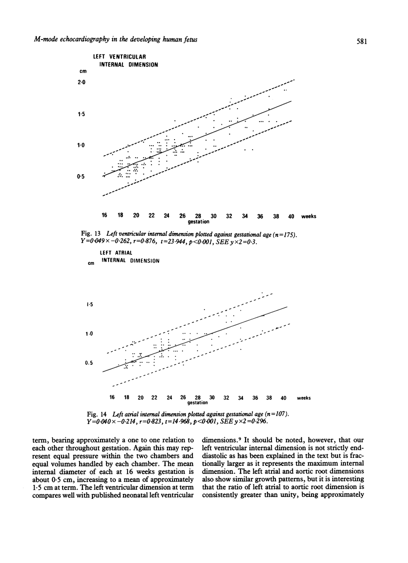
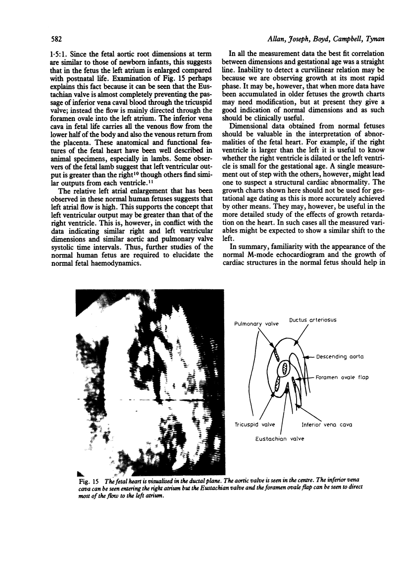
The M-mode echocardiogram can be obtained in the developing human fetus from around the 16th week of gestation until term. Errors in interpretation of the M-mode echocardiogram can be avoided by concurrent two dimensional fetal cardiac examination. The pattern of motion of the mitral, tricuspid, aortic, and pulmonary valves and ventricular wall motion can be studied. Motion of the foramen ovale flap, which is characteristic in fetal life, can also be observed. Using the M-mode tracing, measurement data for six variables were made and growth charts constructed. The variables recorded were septal and left ventricular wall thickness, right and left ventricular internal dimension in diastole, aortic root dimension, and left atrial internal dimension. The measurements were made in normal pregnancies where the fetal heart was structurally normal. Confidence limits for each measurement were derived for gestational ages between 16 and 39 weeks.
Full text
PDF
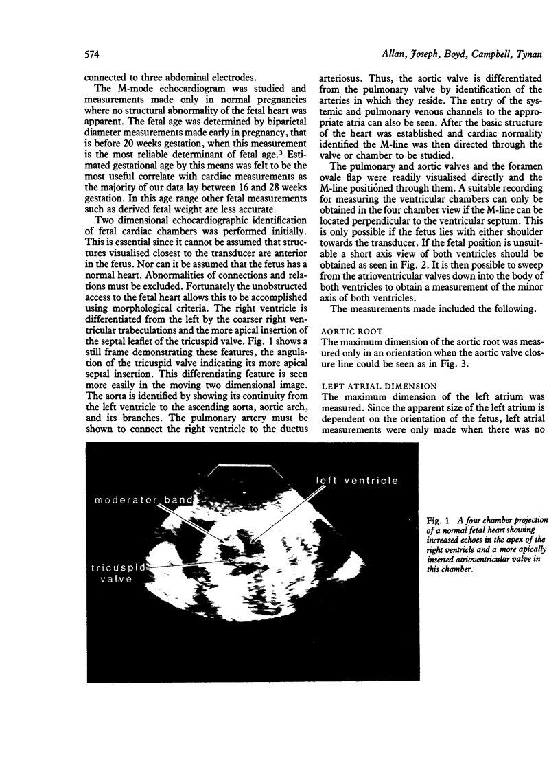
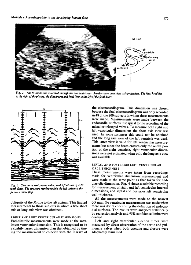
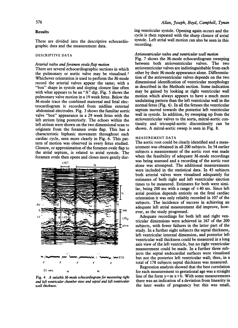
Images in this article
Selected References
These references are in PubMed. This may not be the complete list of references from this article.
- Allan L. D., Tynan M., Campbell S., Anderson R. H. Identification of congenital cardiac malformations by echocardiography in midtrimester fetus. Br Heart J. 1981 Oct;46(4):358–362. doi: 10.1136/hrt.46.4.358. [DOI] [PMC free article] [PubMed] [Google Scholar]
- Campbell S., Newman G. B. Growth of the fetal biparietal diameter during normal pregnancy. J Obstet Gynaecol Br Commonw. 1971 Jun;78(6):513–519. doi: 10.1111/j.1471-0528.1971.tb00309.x. [DOI] [PubMed] [Google Scholar]
- DAWES G. S., MOTT J. C., WIDDICOMBE J. G. The foetal circulation in the lamb. J Physiol. 1954 Dec 10;126(3):563–587. doi: 10.1113/jphysiol.1954.sp005227. [DOI] [PMC free article] [PubMed] [Google Scholar]
- Henrion R., Aubry J. P. Fetal cardiac abnormality and real-time ultrasound study:a case of Ivemark syndrome. Contrib Gynecol Obstet. 1979;6:119–122. [PubMed] [Google Scholar]
- Kleinman C. S., Hobbins J. C., Jaffe C. C., Lynch D. C., Talner N. S. Echocardiographic studies of the human fetus: prenatal diagnosis of congenital heart disease and cardiac dysrhythmias. Pediatrics. 1980 Jun;65(6):1059–1067. [PubMed] [Google Scholar]
- Sahn D. J., Lange L. W., Allen H. D., Goldberg S. J., Anderson C., Giles H., Haber K. Quantitative real-time cross-sectional echocardiography in the developing normal humam fetus and newborn. Circulation. 1980 Sep;62(3):588–597. doi: 10.1161/01.cir.62.3.588. [DOI] [PubMed] [Google Scholar]
- Solinger R., Elbl F., Minhas K. Echocardiography in the normal neonate. Circulation. 1973 Jan;47(1):108–118. doi: 10.1161/01.cir.47.1.108. [DOI] [PubMed] [Google Scholar]
- Winsberg F. Echocardiography of the fetal and newborn heart. Invest Radiol. 1972 May-Jun;7(3):152–158. doi: 10.1097/00004424-197205000-00004. [DOI] [PubMed] [Google Scholar]


