

Abstract
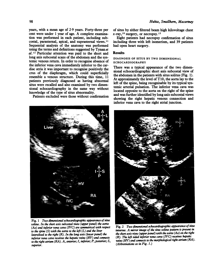
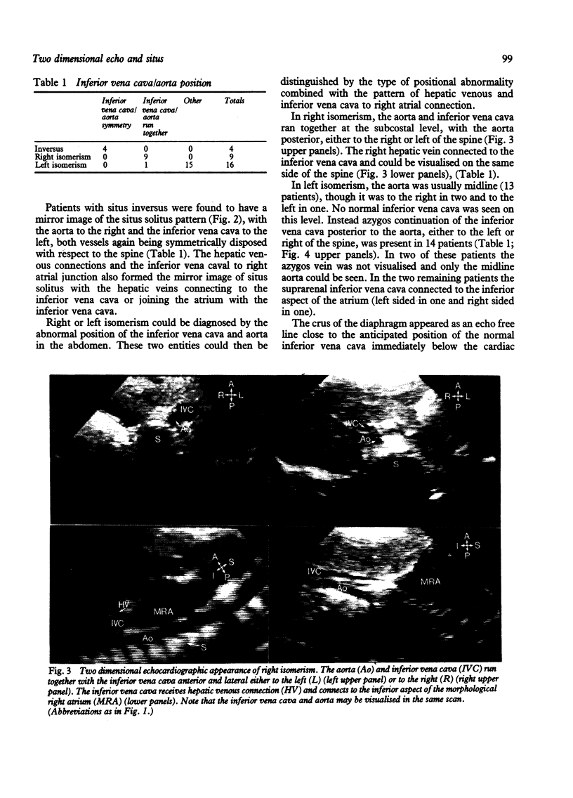
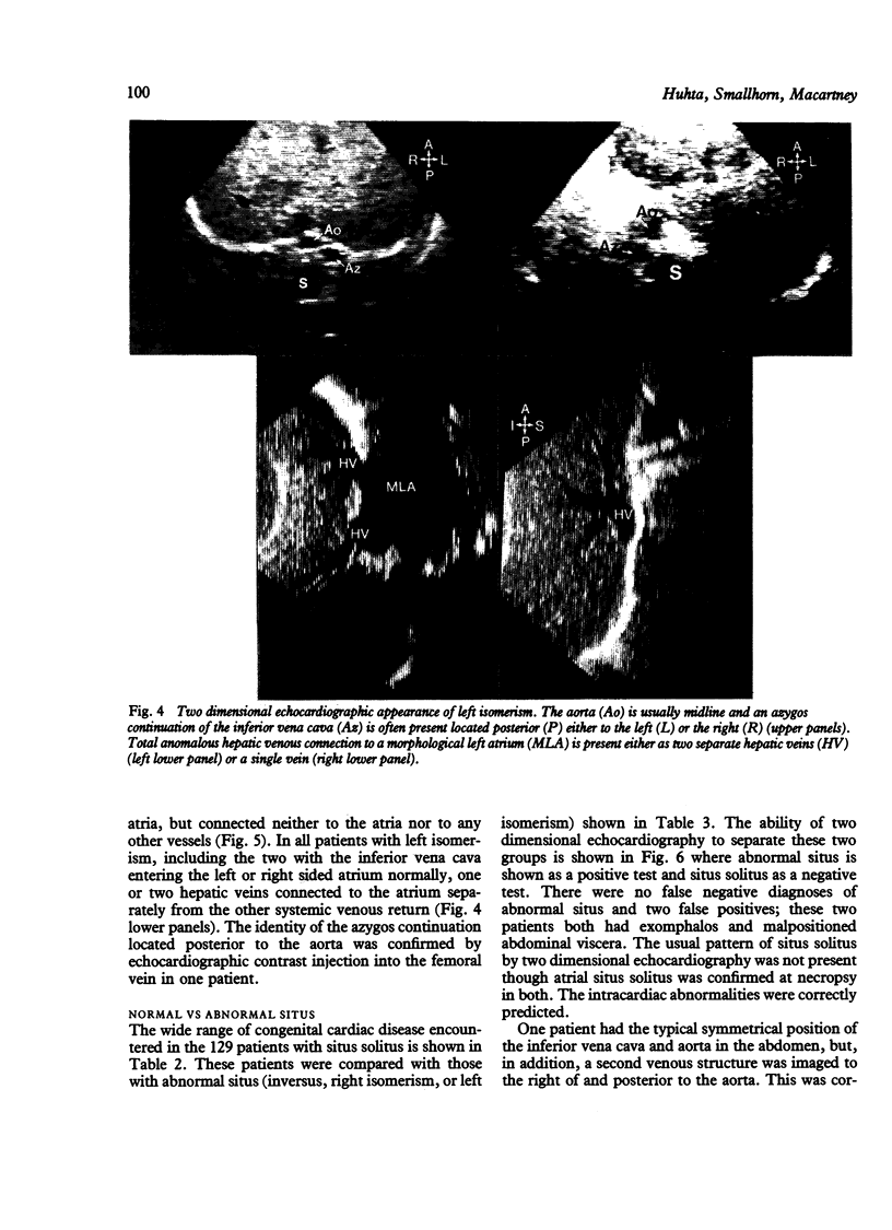
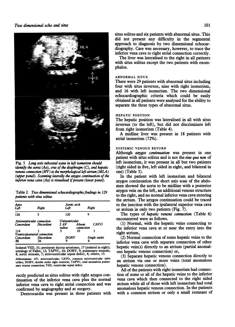
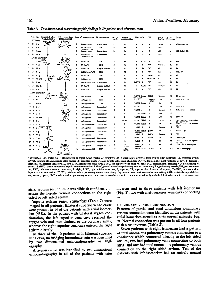
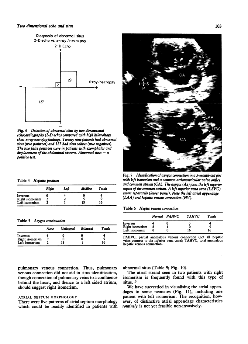
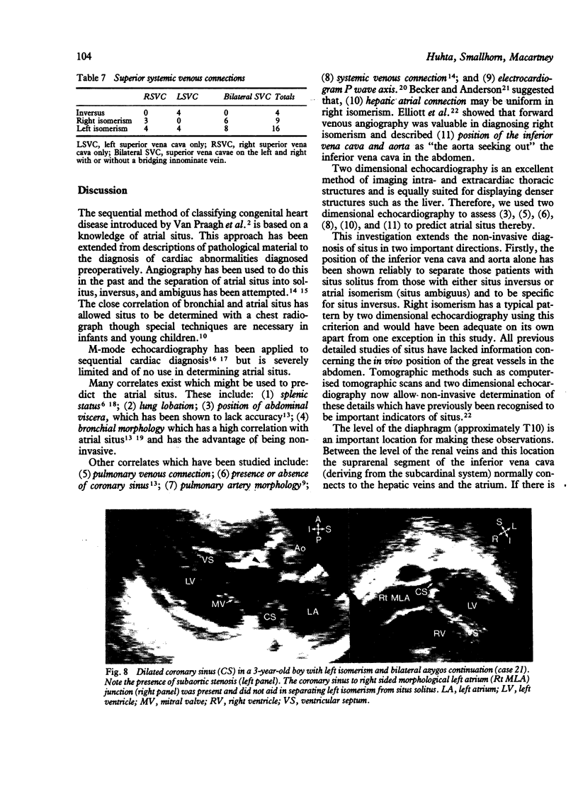
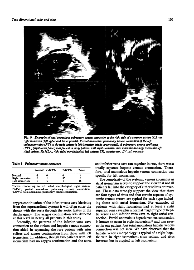
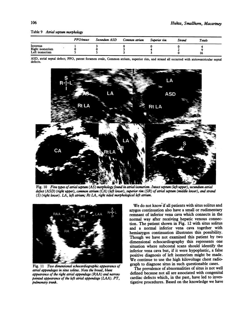
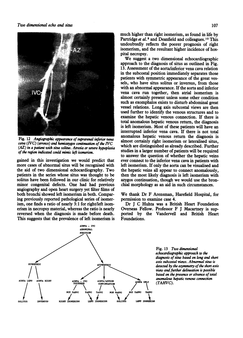
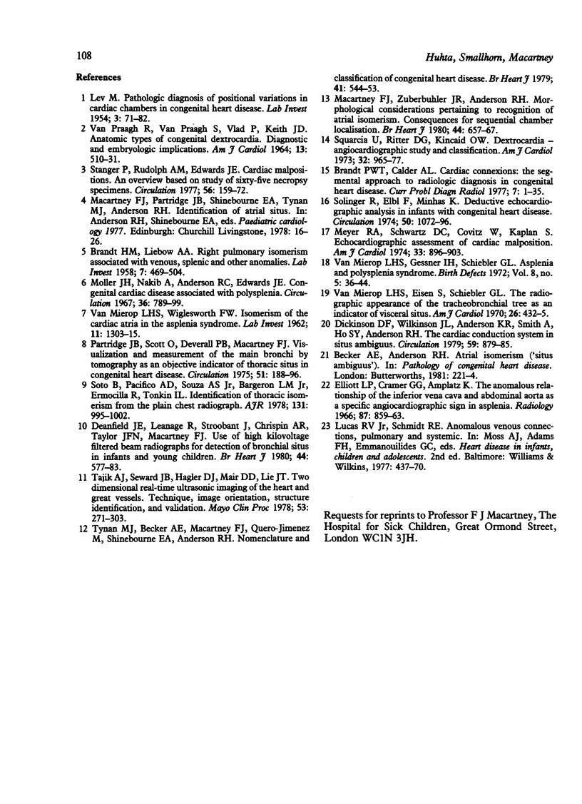
At present there is no reliable method of recognising atrial isomerism by two dimensional echocardiography. We therefore used two dimensional echocardiography to examine 158 patients including 25 with atrial isomerism and four with situs inversus. Particular attention was paid to the short and long axis subcostal scans of the abdomen. Using the position of the inferior vena cava and the aorta with respect to the spine it was possible to separate those with situs solitus from the others. Two false positives for abnormal situs had exomphalos. In situs solitus the aorta lay to the left of the spine and the inferior vena cava lay to the right. One patient with situs solitus and azygos continuation of the inferior vena cava also had inferior vena cava to right atrial connection. In the four patients with situs inversus the mirror image of the normal pattern was present. In nine patients with right isomerism the inferior vena cava and aorta ran together on one or other side of the spine. The inferior vena cava, anterior to the aorta at the level of the diaphragm, received at least the right hepatic veins (normal or partial anomalous hepatic venous connection). Of the 16 patients with left isomerism, 14 had azygos continuation of the inferior vena cava which was visualised posterior to the aorta in all but two. All patients with left isomerism had total anomalous hepatic venous connection to one or both atria via one or two separate veins. Two dimensional echocardiography therefore provides the means of detecting abnormal atrial situs and of diagnosing right or left isomerism in the great majority of patients, if not all.
Full text
PDF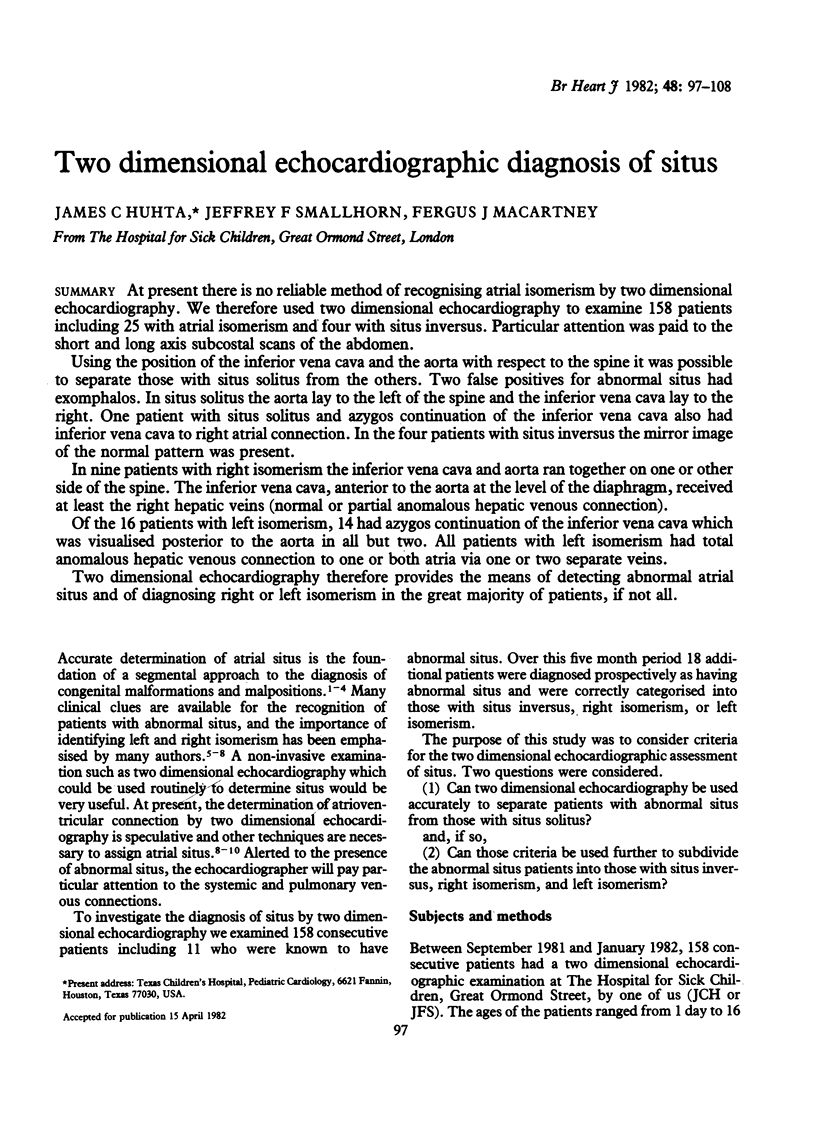
Images in this article
Selected References
These references are in PubMed. This may not be the complete list of references from this article.
- BRANDT H. M., LIEBOW A. A. Right pulmonary isomerism associated with venous, splenic, and other anomalies. Lab Invest. 1958 Sep-Oct;7(5):469–504. [PubMed] [Google Scholar]
- Brandt P. W., Calder A. L. Cardiac connections: the segmental approach to radiologic diagnosis in congenital heart disease. Curr Probl Diagn Radiol. 1977 May-Jun;7(3):1–35. doi: 10.1016/s0363-0188(77)80006-6. [DOI] [PubMed] [Google Scholar]
- Deanfield J. E., Leanage R., Stroobant J., Chrispin A. R., Taylor J. F., Macartney F. J. Use of high kilovoltage filtered beam radiographs for detection of bronchial situs in infants and young children. Br Heart J. 1980 Nov;44(5):577–583. doi: 10.1136/hrt.44.5.577. [DOI] [PMC free article] [PubMed] [Google Scholar]
- Dickinson D. F., Wilkinson J. L., Anderson K. R., Smith A., Ho S. Y., Anderson R. H. The cardiac conduction system in situs ambiguus. Circulation. 1979 May;59(5):879–885. doi: 10.1161/01.cir.59.5.879. [DOI] [PubMed] [Google Scholar]
- Elliott L. P., Cramer G. G., Amplatz K. The anomalous relationship of the interior vena cava and abdominal aorta as a specific angiocardiographic sign in asplenia. Radiology. 1966 Nov;87(5):859–863. doi: 10.1148/87.5.859. [DOI] [PubMed] [Google Scholar]
- LEV M. Pathologic diagnosis of positional variations in cardiac chambers in congenital heart disease. Lab Invest. 1954 Jan-Feb;3(1):71–82. [PubMed] [Google Scholar]
- Macartney F. J., Zuberbuhler J. R., Anderson R. H. Morphological considerations pertaining to recognition of atrial isomerism. Consequences for sequential chamber localisation. Br Heart J. 1980 Dec;44(6):657–667. doi: 10.1136/hrt.44.6.657. [DOI] [PMC free article] [PubMed] [Google Scholar]
- Meyer R. A., Schwartz D. C., Covitz W., Kaplan S. Echocardiographic assessment of cardiac malposition. Am J Cardiol. 1974 Jun;33(7):896–903. doi: 10.1016/0002-9149(74)90638-9. [DOI] [PubMed] [Google Scholar]
- Moller J. H., Nakib A., Anderson R. C., Edwards J. E. Congenital cardiac disease associated with polysplenia. A developmental complex of bilateral "left-sidedness". Circulation. 1967 Nov;36(5):789–799. doi: 10.1161/01.cir.36.5.789. [DOI] [PubMed] [Google Scholar]
- Partridge J. B., Scott O., Deverall P. B., Macartney F. J. Visualization and measurement of the main bronchi by tomography as an objective indicator of thoracic situs in congenital heart disease. Circulation. 1975 Jan;51(1):188–196. doi: 10.1161/01.cir.51.1.188. [DOI] [PubMed] [Google Scholar]
- Solinger R., Elbl F., Minhas K. Deductive echocardiographic analysis in infants with congenital heart disease. Circulation. 1974 Dec;50(6):1072–1096. doi: 10.1161/01.cir.50.6.1072. [DOI] [PubMed] [Google Scholar]
- Soto B., Pacifico A. D., Souza A. S., Jr, Bargeron L. M., Jr, Ermocilla R., Tonkin I. L. Identification of thoracic isomerism from the plain chest radiograph. AJR Am J Roentgenol. 1978 Dec;131(6):995–1002. doi: 10.2214/ajr.131.6.995. [DOI] [PubMed] [Google Scholar]
- Squarcia U., Ritter D. G., Kincaid O. W. Dextrocardia: angiocardiographic study and classificiation. Am J Cardiol. 1973 Dec;32(7):965–977. doi: 10.1016/s0002-9149(73)80166-3. [DOI] [PubMed] [Google Scholar]
- Stanger P., Rudolph A. M., Edwards J. E. Cardiac malpositions. An overview based on study of sixty-five necropsy specimens. Circulation. 1977 Aug;56(2):159–172. doi: 10.1161/01.cir.56.2.159. [DOI] [PubMed] [Google Scholar]
- Tajik A. J., Seward J. B., Hagler D. J., Mair D. D., Lie J. T. Two-dimensional real-time ultrasonic imaging of the heart and great vessels. Technique, image orientation, structure identification, and validation. Mayo Clin Proc. 1978 May;53(5):271–303. [PubMed] [Google Scholar]
- Tynan M. J., Becker A. E., Macartney F. J., Jiménez M. Q., Shinebourne E. A., Anderson R. H. Nomenclature and classification of congenital heart disease. Br Heart J. 1979 May;41(5):544–553. doi: 10.1136/hrt.41.5.544. [DOI] [PMC free article] [PubMed] [Google Scholar]
- VAN MIEROP L. H., WIGLESWORTH F. W. Isomerism of the cardiac atria in the asplenia syndrome. Lab Invest. 1962 Dec;11:1303–1315. [PubMed] [Google Scholar]
- VANPRAAGH R., VANPRAAGH S., VLAD P., KEITH J. D. ANATOMIC TYPES OF CONGENITAL DEXTROCARDIA: DIAGNOSTIC AND EMBRYOLOGIC IMPLICATIONS. Am J Cardiol. 1964 Apr;13:510–531. doi: 10.1016/0002-9149(64)90159-6. [DOI] [PubMed] [Google Scholar]
- Van Mierop L. H., Eisen S., Schiebler G. L. The radiographic appearance of the tracheobronchial tree as an indicator of visceral situs. Am J Cardiol. 1970 Oct;26(4):432–435. doi: 10.1016/0002-9149(70)90743-5. [DOI] [PubMed] [Google Scholar]