Molecular insights into Huntington’s disease
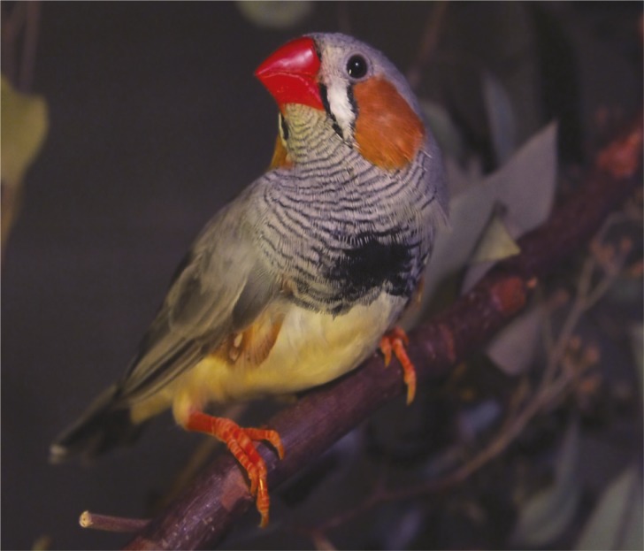
Zebra finch.
Huntington’s disease is marked by uncoordinated gestures and disorganized movements, but the precise links among the genetic mutation in the huntingtin protein, affected neural circuits, and movement deficits remain unclear. The neural circuitry underlying song production in zebra finches resembles circuits implicated in Huntington’s disease. Hence, Masashi Tanaka et al. (pp. E1720–E1727) engineered adult male zebra finches to selectively produce a mutant huntingtin fragment in the songbirds’ so-called area X neurons, which correspond to the human basal ganglia—brain structures implicated in the disease. Compared with normal finches, engineered birds vocalized a greater amount daily and sang songs with disorganized syllable sequences 1–2 months after intervention; the effect persisted for several months. Changes in song were accompanied by loss of area X medium spiny neurons, reduced number of inhibitory synapses on pallidal neurons, and altered activity of lateral magnocellular nucleus of the anterior nidopallium (LMAN) neurons, the human counterparts of all of which are tied to the disease. Reversible chemical blockade of LMAN neurons restored normal singing patterns in engineered birds. According to the authors, mutant huntingtin might trigger movement defects through its effects on temporal patterns of activity in the circuit involving the brain’s cortex and basal ganglia. In a related article, Guohao Wang et al. (pp. 3359–3364) explored the elusive function of huntingtin in adult tissues, in light of ongoing therapeutic attempts to hobble mutant huntingtin using genetic techniques in the adult mouse brain. The authors report that depleting huntingtin in adult mouse neurons did not trigger Huntington’s disease-related neurodegeneration or discernible symptoms. However, loss of huntingtin in 2-month-old mice proved lethal, likely due to acute pancreatitis, marked by the loss of pancreatic acinar cells, which undergo degeneration mediated by the digestive enzyme trypsin, normally kept in check by huntingtin. Engineering a truncated huntingtin protein into huntingtin-depleted mice ameliorated pancreatitis and prevented death. According to the authors, huntingtin might thus play age- and cell type-specific roles in humans, and the findings together point to potential time-sensitive therapeutic targets for the disease. — P.N.
Previously unidentified tuna spawning ground
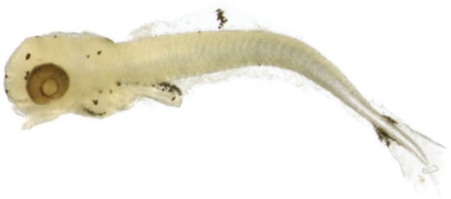
Small bluefin tuna larvae collected in 2013 from the Slope Sea.
Managing the Atlantic bluefin tuna fishery is challenging due to the Atlantic bluefin’s highly migratory lifestyle and the large number of nations that harvest the fish. Developing effective management strategies requires understanding the life history of the species. David Richardson et al. (pp. 3299–3304) collected 67 larval bluefin tuna from the Slope Sea, off the coast of the northeastern United States, between late June and early August 2013. A majority of the larvae had been spawned within 6 days of collection, indicating that Atlantic bluefin tuna spawn in the Slope Sea in addition to previously identified spawning grounds in the Mediterranean Sea and the Gulf of Mexico. Electronic tagging data indicate that bluefin migration patterns in the western Atlantic vary with fish size. Only the largest individuals migrate into the Gulf of Mexico, whereas small- to medium-sized individuals migrate between the North Sargasso Sea and the northeastern United States, suggesting that a significant fraction of western Atlantic bluefin spawning occurs outside of the Gulf of Mexico. The western Atlantic bluefin’s current life history model, which assumes that spawning occurs only in the Gulf of Mexico, might overestimate age at maturity, and western Atlantic bluefin might be less vulnerable to exploitation than previously thought, according to the authors. — B.D.
Mite–virus mutualism in honey bee colony loss
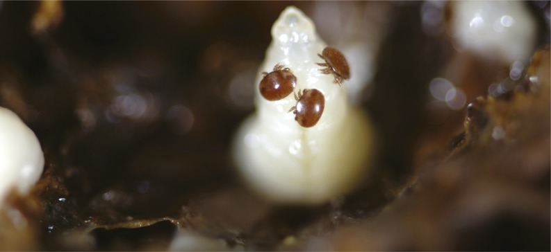
Honey bee pupa with Varroa mites. Image courtesy of Denis Anderson (Commonwealth Scientific and Industrial Research Organisation).
The parasitic mite Varroa destructor and the deformed wing virus, the mite’s viral passenger, have been implicated in the widespread losses of honey bee colonies that have taken a staggering toll on apiculture. Previous studies have found that the mite and its offspring feed on the haemolymph of honey bee pupae and help spread the virus, which suppresses honey bee immunity by tamping down signaling mediated by the NF-κB protein. Gennaro Di Prisco et al. (pp. 3203–3208) tested whether virus-induced immune suppression facilitates mite feeding, resulting in a mutualism between vector and pathogen. Because insects respond to intruding parasites by encasing them in a melanin-coated capsule that hampers continued feeding, the authors implanted a nylon thread in the bodies of virus-infected, fifth instar honey bee larvae and scored the degree of the thread’s melanization and encapsulation 1 day later. Melanization and encapsulation—as well as the expression levels of related genes—were negatively associated with virus levels in the larvae. Mirroring the feeding benefit, virus levels were positively correlated with mite reproduction on honey bee pupae, as revealed by numbers of viral genome copies and offspring mites, suggesting that viral infection enhances mite reproductive fitness. According to the authors, the mite–virus symbiosis, a relationship that boosts mite feeding and reproduction, holds a key to unlocking the mysterious etiology of honey bee colony loss. — P.N.
Movement of mitochondria between plant cells
Although many horizontal gene transfer events in plants involve mitochondrial sequences, evidence for cell-to-cell movement of mitochondria in plants is lacking. Csanad Gurdon et al. (pp. 3395–3400) investigated the cell-to-cell movement of mitochondria using two tobacco plants Nicotiana tabacum and Nicotiana sylvestris. The authors used an experimental system in which the cytoplasm of N. tabacum was replaced with the cytoplasm of Nicotiana undulata, which carries a sterility-causing mitochondrial genome that makes N. tabacum male flowers sterile. N. sylvestris flowers are fertile, and the authors could detect the rare transfer of mitochondrial DNA (mtDNA) from N. sylvestris to N. tabacum by monitoring flowering events in N. tabacum plants regenerated from graft junctions, as the transfer of mtDNA restored fertile flower anatomy. The authors identified branches with fertile flowers in regenerated plants, indicating cell-to-cell movement of mitochondria through the graft junction. When the authors analyzed the mitochondrial genomes, they found extensive recombination, and identified orf293 as the gene likely responsible for male sterility. The cell-to-cell movement of mitochondria during grafting may recapitulate the horizontal mtDNA transfer that occurred during evolution. According to the authors, such intercellular movement may be used to transfer mtDNA to other species. — S.R.
Regulation of cell surface composition
Cell surface components such as lipids and proteins must be spatially and temporally organized for the execution of several functions, including receptor signaling and vesicular trafficking. To investigate the role of active processes in cell surface organization, Darius Köster et al. (pp. E1645–E1654) used a reconstituted in vitro system consisting of a lipid bilayer, membrane-associated actin-binding components, actin filaments, and myosin motors. The authors found that the active mechanics of actomyosin complexes, which are made up of actin filaments and myosin motors, can lead to the clustering of membrane components and regulate the local composition of the cell surface. By systematically changing actin and myosin concentrations and actin filament length, the authors revealed distinct states of actomyosin organization at the membrane surface after ATP consumption. The ATP-dependent actin and myosin dynamics produced transient accumulations of actin-binding membrane molecules, similar to that seen in living cells. The results suggest that active actomyosin processes can affect the organization of membrane proteins and drive local membrane composition. According to the authors, the actomyosin-driven organization of cell membrane components may have implications for cell signaling. — S.R.


