Abstract
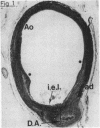
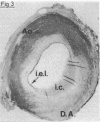
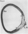
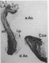
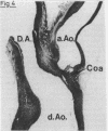
Localised coarctation of the aorta was studied histologically in 45 cases. The age range of the patients, from 2 weeks to 40 years, enabled us to make a comparison between early and late findings. The basic histological architecture of all coarctations was identical. The ridge consisted mainly of thickening plus some infolding of the aortic wall. Ductal tissue formed the inner part of the ridge for more than half of its total circumference. In older cases masses of secondary intimal proliferation were present, which narrowed the residual lumen of the coarctation. The coarctation was located preductally in most young patients and postductally in the majority of older patients. It is concluded that ductal tissue is invariably present in the ridge and that all coarctations develop in a preductal position. Secondary changes include a gradual shift of the ridge from a preductal to a postductal position, and progressive narrowing of the residual lumen by intimal proliferation.
Full text
PDF
Images in this article
Selected References
These references are in PubMed. This may not be the complete list of references from this article.
- Becker A. E., Becker M. J., Edwards J. E. Anomalies associated with coarctation of aorta: particular reference to infancy. Circulation. 1970 Jun;41(6):1067–1075. doi: 10.1161/01.cir.41.6.1067. [DOI] [PubMed] [Google Scholar]
- Bruins C. Twelfth Edgar Mannheimer lecture. Competition between aortic isthmus and ductus arteriosus; reciprocal influence of structure and flow. Eur J Cardiol. 1978 Aug;8(1):87–97. [PubMed] [Google Scholar]
- Burrington J. D. Response to experimental coarctation of the aorta and pulmonic stenosis in the fetal lamb. J Thorac Cardiovasc Surg. 1978 Jun;75(6):819–826. [PubMed] [Google Scholar]
- CLAGETT O. T., KIRKLIN J. W., EDWARDS J. E. Anatomic variations and pathologic changes in coarctation of the aorta; a study of 124 cases. Surg Gynecol Obstet. 1954 Jan;98(1):103–114. [PubMed] [Google Scholar]
- Freed M. D., Heymann M. A., Lewis A. B., Roehl S. L., Kensey R. C. Prostaglandin E1 infants with ductus arteriosus-dependent congenital heart disease. Circulation. 1981 Nov;64(5):899–905. doi: 10.1161/01.cir.64.5.899. [DOI] [PubMed] [Google Scholar]
- Gittenberger-de Groot A. C. Persistent ductus arteriosus: most probably a primary congenital malformation. Br Heart J. 1977 Jun;39(6):610–618. doi: 10.1136/hrt.39.6.610. [DOI] [PMC free article] [PubMed] [Google Scholar]
- Heymann M. A., Berman W., Jr, Rudolph A. M., Whitman V. Dilatation of the ductus arteriosus by prostaglandin E1 in aortic arch abnormalities. Circulation. 1979 Jan;59(1):169–173. doi: 10.1161/01.cir.59.1.169. [DOI] [PubMed] [Google Scholar]
- Hutchins G. M. Coarctation of the aorta explained as a branch-point of the ductus arteriosus. Am J Pathol. 1971 May;63(2):203–214. [PMC free article] [PubMed] [Google Scholar]
- Levin D. L., Mills L. J., Parkey M. Morphologic development of the pulmonary vascular bed in experimental coarctation of the aorta. Circulation. 1979 Aug;60(2):349–354. doi: 10.1161/01.cir.60.2.349. [DOI] [PubMed] [Google Scholar]
- Rudolph A. M., Heymann M. A., Spitznas U. Hemodynamic considerations in the development of narrowing of the aorta. Am J Cardiol. 1972 Oct;30(5):514–525. doi: 10.1016/0002-9149(72)90042-2. [DOI] [PubMed] [Google Scholar]
- Talner N. S., Berman M. A. Postnatal development of obstruction in coarctation of the aorta: role of the ductus arteriosus. Pediatrics. 1975 Oct;56(4):562–569. [PubMed] [Google Scholar]
- Tinkelenberg J. Graphic reconstruction and stereoscopy. J Audiov Media Med. 1980 Apr;3(2):68–71. doi: 10.3109/17453058009154269. [DOI] [PubMed] [Google Scholar]


