

Abstract
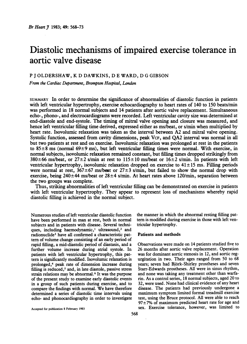
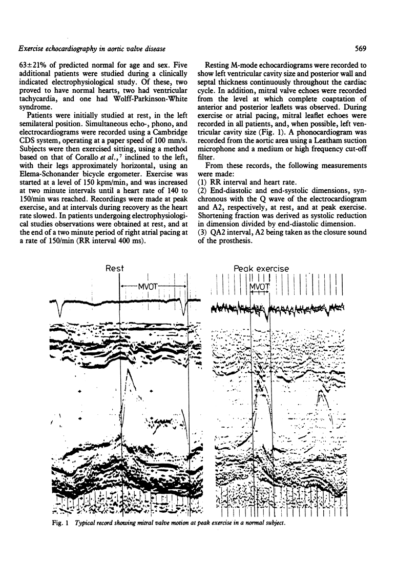
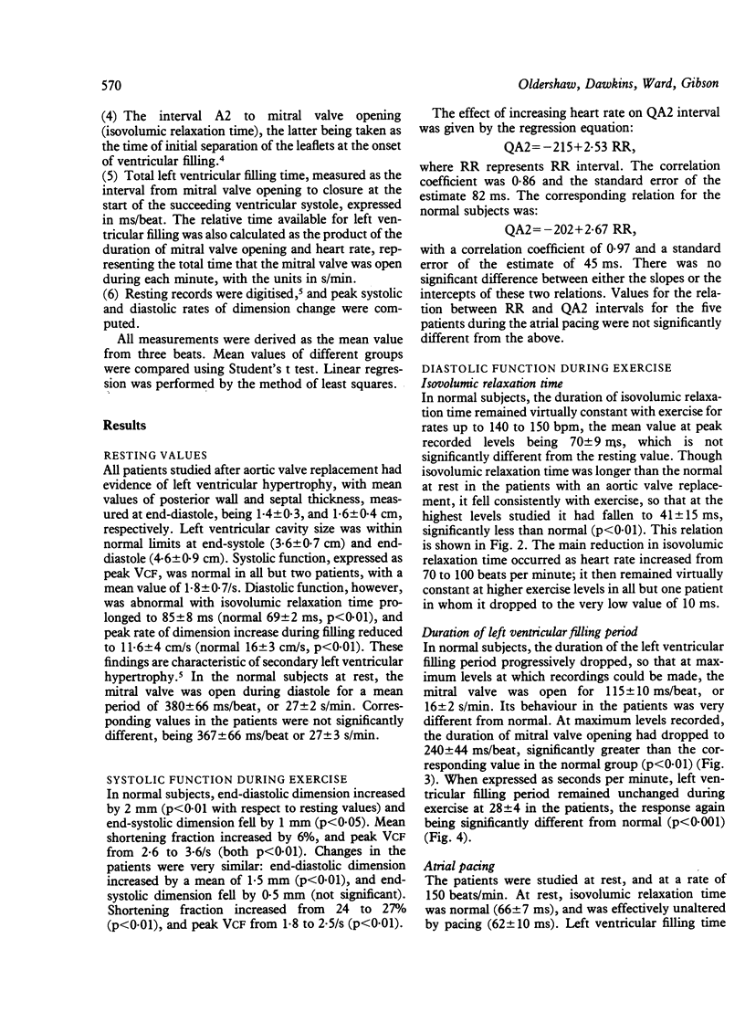
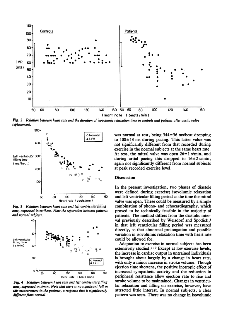
In order to determine the significance of abnormalities of diastolic function in patients with left ventricular hypertrophy, exercise echocardiography to heart rates of 140 to 150 beats/min was performed in 18 normal subjects and 14 patients after aortic valve replacement. Simultaneous echo-, phono-, and electrocardiograms were recorded. Left ventricular cavity size was determined at end-diastole and end-systole. The timing of mitral valve opening and closure was measured, and hence left ventricular filling time derived, expressed either as ms/beat, or s/min when multiplied by heart rate. Isovolumic relaxation was taken as the interval between A2 and mitral valve opening. Systolic function, assessed from cavity dimensions, peak VCF, and QA2 interval was normal in all but two patients at rest and on exercise. Isovolumic relaxation was prolonged at rest in the patients to 85 +/- 8 ms (normal 69 +/- 9 ms), but left ventricular filling times were normal. With exercise, in normal subjects, isovolumic relaxation remained constant, but filling times dropped strikingly from 380 +/- 66 ms/beat, or 27 +/- 2 s/min at rest to 115 +/- 10 ms/beat or 16 +/- 2 s/min. In patients with left ventricular hypertrophy, isovolumic relaxation dropped on exercise to 41 +/- 15 ms. Filling periods were normal at rest, 367 +/- 67 ms/beat or 27 +/- 3 s/min, but failed to show the normal drop with exercise, being 240 +/- 44 ms/beat or 28 +/- 4 s/min. At heart rates above 120/min, separation between the two groups was complete. Thus, striking abnormalities of left ventricular filling can be demonstrated on exercise in patients with left ventricular hypertrophy. They appear to represent loss of mechanisms whereby rapid diastolic filling is achieved in the normal subject.
Full text
PDF
Selected References
These references are in PubMed. This may not be the complete list of references from this article.
- ASTRAND P. O., CUDDY T. E., SALTIN B., STENBERG J. CARDIAC OUTPUT DURING SUBMAXIMAL AND MAXIMAL WORK. J Appl Physiol. 1964 Mar;19:268–274. doi: 10.1152/jappl.1964.19.2.268. [DOI] [PubMed] [Google Scholar]
- Bonow R. O., Bacharach S. L., Green M. V., Kent K. M., Rosing D. R., Lipson L. C., Leon M. B., Epstein S. E. Impaired left ventricular diastolic filling in patients with coronary artery disease: assessment with radionuclide angiography. Circulation. 1981 Aug;64(2):315–323. doi: 10.1161/01.cir.64.2.315. [DOI] [PubMed] [Google Scholar]
- Chen W., Gibson D. Relation of isovolumic relaxation to left ventricular wall movement in man. Br Heart J. 1979 Jul;42(1):51–56. doi: 10.1136/hrt.42.1.51. [DOI] [PMC free article] [PubMed] [Google Scholar]
- Gibson D. G., Traill T. A., Hall R. J., Brown D. J. Echocardiographic features of secondary left ventricular hypertrophy. Br Heart J. 1979 Jan;41(1):54–59. doi: 10.1136/hrt.41.1.54. [DOI] [PMC free article] [PubMed] [Google Scholar]
- Mattheos M., Shapiro E., Oldershaw P. J., Sacchetti R., Gibson D. G. Non-invasive assessment of changes in left ventricular relaxation by combined phono-, echo-, and mechanocardiography. Br Heart J. 1982 Mar;47(3):253–260. doi: 10.1136/hrt.47.3.253. [DOI] [PMC free article] [PubMed] [Google Scholar]
- Mirsky I. Assessment of passive elastic stiffness of cardiac muscle: mathematical concepts, physiologic and clinical considerations, directions of future research. Prog Cardiovasc Dis. 1976 Jan-Feb;18(4):277–308. doi: 10.1016/0033-0620(76)90023-2. [DOI] [PubMed] [Google Scholar]
- Sonnenblick E. H. The structural basis and importance of restoring forces and elastic recoil for the filling of the heart. Eur Heart J. 1980;Suppl A:107–110. doi: 10.1093/eurheartj/1.suppl_1.107. [DOI] [PubMed] [Google Scholar]
- Upton M. T., Gibson D. G. The study of left ventricular function from digitized echocardiograms. Prog Cardiovasc Dis. 1978 Mar-Apr;20(5):359–384. doi: 10.1016/0033-0620(78)90003-8. [DOI] [PubMed] [Google Scholar]
- Weisdorf D., Spodick D. H. Duration of diastole versus cycle length as correlates of left ventricular ejection time. Br Heart J. 1976 Mar;38(3):282–284. doi: 10.1136/hrt.38.3.282. [DOI] [PMC free article] [PubMed] [Google Scholar]