Abstract
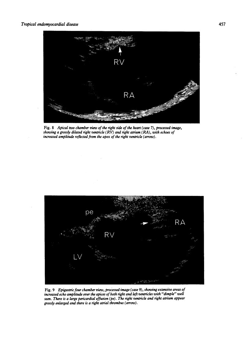
Fifteen patients with tropical endomyocardial disease which had been proved angiographically were studied using M-mode and cross-sectional echocardiography to determine the extent to which specific features of this disease could be recognised by these non-invasive methods. Tethering of the posterior mitral valve leaflet to the ventricular wall in combination with areas of echo-dense material in the posterior left ventricular wall and associated papillary muscle appeared to be a constant diagnostic feature of this disease. Colour coding of regional echo amplitude showed high intensity echoes in a distribution corresponding closely to that of the fibrosis known to occur in this condition. Though M-mode echocardiography did not contribute diagnostic information, it was useful in defining the functional consequences of myocardial or mitral valve disease. Digitisation of records allowed a restrictive pattern of left ventricular filling to be observed. It was concluded that cross-sectional echocardiography, particularly when supplemented by colour coded amplitude processing, can make a confident non-invasive diagnosis of tropical endomyocardial disease and so could be useful in assessing its progression or response to treatment.
Full text
PDF




Images in this article
Selected References
These references are in PubMed. This may not be the complete list of references from this article.
- Acquatella H. Two-dimensional echocardiography in endomyocardial disease. Postgrad Med J. 1983 Mar;59(689):157–159. doi: 10.1136/pgmj.59.689.157. [DOI] [PMC free article] [PubMed] [Google Scholar]
- Chew C. Y., Ziady G. M., Raphael M. J., Nellen M., Oakley C. M. Primary restrictive cardiomyopathy. Non-tropical endomyocardial fibrosis and hypereosinophilic heart disease. Br Heart J. 1977 Apr;39(4):399–413. doi: 10.1136/hrt.39.4.399. [DOI] [PMC free article] [PubMed] [Google Scholar]
- Chivers R. C. Tissue characterization. Ultrasound Med Biol. 1981;7(1):1–20. doi: 10.1016/0301-5629(81)90018-1. [DOI] [PubMed] [Google Scholar]
- Davies J., Gibson D. G., Foale R., Heer K., Spry C. J., Oakley C. M., Goodwin J. F. Echocardiographic features of eosinophilic endomyocardial disease. Br Heart J. 1982 Nov;48(5):434–440. doi: 10.1136/hrt.48.5.434. [DOI] [PMC free article] [PubMed] [Google Scholar]
- Dienot B., Ekra A., Bertrand E. L'échocardiographie dans 23 cas de fibroses endomyocardiques constrictives droites ou bilatérales. Arch Mal Coeur Vaiss. 1979 Oct;72(10):1101–1107. [PubMed] [Google Scholar]
- Gibson D. G., Brown D. Measurement of instantaneous left ventricular dimension and filling rate in man, using echocardiography. Br Heart J. 1973 Nov;35(11):1141–1149. doi: 10.1136/hrt.35.11.1141. [DOI] [PMC free article] [PubMed] [Google Scholar]
- Gottdiener J. S., Maron B. J., Schooley R. T., Harley J. B., Roberts W. C., Fauci A. S. Two-dimensional echocardiographic assessment of the idiopathic hypereosinophilic syndrome. Anatomic basis of mitral regurgitation and peripheral embolization. Circulation. 1983 Mar;67(3):572–578. doi: 10.1161/01.cir.67.3.572. [DOI] [PubMed] [Google Scholar]
- Haertel J. C., Castro I. Avaliaço ecocardiográfica da fibrose endomiocárdica. Arq Bras Cardiol. 1980 Dec;35(6):475–480. [PubMed] [Google Scholar]
- Hess O. M., Turina M., Senning A., Goebel N. H., Scholer Y., Krayenbuehl H. P. Pre- and postoperative findings in patients with endomyocardial fibrosis. Br Heart J. 1978 Apr;40(4):406–415. doi: 10.1136/hrt.40.4.406. [DOI] [PMC free article] [PubMed] [Google Scholar]
- Logan-Sinclair R., Wong C. M., Gibson D. G. Clinical application of amplitude processing of echocardiographic images. Br Heart J. 1981 Jun;45(6):621–627. doi: 10.1136/hrt.45.6.621. [DOI] [PMC free article] [PubMed] [Google Scholar]
- Olsen E. G. Cardiomyopathies. Cardiovasc Clin. 1972;4(2):239–261. [PubMed] [Google Scholar]
- Parrillo J. E., Borer J. S., Henry W. L., Wolff S. M., Fauci A. S. The cardiovascular manifestations of the hypereosinophilic syndrome. Prospective study of 26 patients, with review of the literature. Am J Med. 1979 Oct;67(4):572–582. doi: 10.1016/0002-9343(79)90227-4. [DOI] [PubMed] [Google Scholar]
- Pernod J., Gerbaux A., Vervin P., Terdjman M., Lelguen C., Droniou J. Apport de l'échocardiographie dans le diagnostic des fibroses endomyocardiques. Arch Mal Coeur Vaiss. 1980 Feb;73(2):139–146. [PubMed] [Google Scholar]
- Tajik A. J., Seward J. B., Hagler D. J., Mair D. D., Lie J. T. Two-dimensional real-time ultrasonic imaging of the heart and great vessels. Technique, image orientation, structure identification, and validation. Mayo Clin Proc. 1978 May;53(5):271–303. [PubMed] [Google Scholar]
- Upton M. T., Gibson D. G. The study of left ventricular function from digitized echocardiograms. Prog Cardiovasc Dis. 1978 Mar-Apr;20(5):359–384. doi: 10.1016/0033-0620(78)90003-8. [DOI] [PubMed] [Google Scholar]


