Abstract
Aim:
The aim of this study was to compare the efficacy of hand file, nickel titanium rotary instrument, and two reciprocating instruments for removing gutta-percha and sealer from the root canals.
Materials and Methods:
Eighty-eight mandibular premolar teeth were used. The root canals were shaped and filled with gutta-percha and a resin-based sealer. The specimens were divided into four groups according to the technique by which the root filling material was removed: Group 1 — Wave One; Group 2 — Reciproc; Group 3 — ProTaper; and Group 4 — Gates-Glidden burs and stainless steel hand file. Then teeth were split longitudinally and photographed. The images were transferred to a computer. The ratio of remaining filling material to the root canal periphery was calculated with the aid of ImageJ software. Statistical analysis was performed using Kruskal–Wallis and Mann–Whitney tests.
Results:
A significant difference was found among all groups (P < 0.001). The WaveOne group demonstrated significantly less remaining filling material. The greatest amount of filling material was found in specimens where gutta-percha was removed with Gates-Glidden burs and stainless steel hand file.
Conclusion:
The reciprocating files were found to be significantly more effective in removing the filling material from the canal walls compared to the rotational file and hand file.
Keywords: Reciprocating motion, retreatment, rotary systems
INTRODUCTION
Complete removal of the root canal content and access to the apical foramen in a retreatment are mandatory for the proper cleaning and reobturation.[1] Gutta-percha and endodontic sealer are widely used as filling materials, and their effective removal in endodontic retreatment is considered essential for success.[2] Gutta-percha is usually removed with Hedstrom files alone or in combination with Gates Glidden drills with or without solvents.[3] However, this conventional method can be a tedious and time-consuming process, especially when the root filling material is well condensed.[4]
Rotary nickel–titanium (NiTi) instruments have also been used for the removal of filling materials from root canals and various studies have reported their efficacy and safety.[5,6] The ProTaper Universal Retreatment system (Dentsply Maillefer, Ballaigues, Switzerland) was specifically developed for the retreatment that included three instruments as follows: D1-30/.09-16 mm, D2-25/.08-18 mm, and D3-20/.07-22 mm, which are used at 500 rpm.[7] These retreatment files have been found to be effective in removing root filling material,[8] when compared to manual procedures. However, ProTaper retreatment files were found to be unable to render the canals free of root filling material. Indeed, retreatment files as well as the Hedström files left substantial amounts of material on the canal walls.[9]
A new concept has been introduced for shaping the root canal from start to finish with one single file.[10,11] The reciprocating files are made of M-Wire NiTi alloy that offers greater flexibility and greater resistance to cyclic fatigue compared to traditional NiTi alloys.[12] Recently, two brands of reciprocating files have become available in the market; Reciproc and WaveOne. Reciproc is comprised of three file sizes (R25, R40 and R50) used with an electric motor while WaveOne is comprised of a small (21.06), a primary (25.08) and a large file (40.08).[13,14] The same technique is also indicated for retreatment purposes, in which the instruments are used with a brushing motion against the lateral walls of the canal to remove any residual filling material.[11,14]
The aim of this study was to compare the efficacy of hand file, nickel titanium rotary instrument, and two reciprocating instruments for removing filling material from extracted human teeth. The null hypothesis was that there was no significant difference among the three techniques tested in the efficacy of removing filling material.
MATERIALS AND METHODS
Tooth selection
Eighty-eight freshly extracted single-rooted mandibular premolar teeth were selected. Preoperative mesiodistal and buccolingual radiographs were taken to verify the presence of a single straight canal in each tooth. The teeth were verified radiographically as having patent canals with curvatures <10° according to the Schneider method.[15] All teeth were analyzed radiographically to confirm the presence of the mature root and the absence of root filling, resorption, or calcifications. Soft tissue and calculus were removed from the root surfaces and the teeth were stored in a 0.9% physiological saline solution until use.
The coronal access cavity was prepared by using a high-speed diamond bur with copious water spray. After the removal of the pulp tissue, patency was assured with a size 10 K-file (VDW Antaeos, Munich, Germany), and the working length was established 1 mm short of the apical foramen. To standardize the samples, the crowns were abraded to obtain root canals with a working length of 16 mm.
Root canal preparation and filling
The coronal portion of the canal was flared with sizes 2-3 Gates-Glidden burs (Dentsply Maillefer, Ballaigues, Switzerland). The tooth was further prepared with a step-back technique to apical size 40 using 0.02 taper K-files (VDW Antaeos, Munich, Germany). After instrumentation, 2 mL of 2.5% NaOCl was used and 17% ethylenediaminetetraacetic acid (EDTA) was applied for 3 min to remove the smear layer. The root canals were dried and obturation was done using cold lateral compaction with gutta-percha (DiaDent, DiaDent Group, Burnaby, Canada) and root canal sealer (AH 26, Dentsply DeTrey, Konstanz, Germany). Following the root canal obturation, a heated plugger was used to remove the excess gutta-percha to a level 2 mm short of the canal orifice followed by vertical compaction with a cold plugger. Teeth were radiographed in the buccolingual and mesiodistal directions to confirm the radiographic adequacy of root filling, using the following criteria: Reaching working length, uniform radiopacity, and no voids. The coronal orifice of each canal was sealed with a temporary filling material (Cavit G, 3M-ESPE, Seefeld, Germany), and the teeth were stored at 37°C in 100% humidity for 1 month to allow complete setting of the sealer.
Retreatment procedures
The temporary fillings were removed. All specimens were coded and randomly assigned to 4 groups of 22 specimens in each group. In each group, the bulk of the gutta-percha in the coronal third of the root canals were removed with sizes 2-3 Gates-Glidden burs.
Group 1 (Reciproc group)
A size 40, 0.06 taper Reciproc single file (VDW, Munich, Germany) was used in a slow, reciprocating, in-and-out pecking motion with an electric motor (VDW Silver Reciproc, VDW GmbH, Munich, Germany), according to the manufacturer's instructions. The flutes of the instrument were cleaned with 1% sodium hypochlorite after each set of three pecks.
Group 2 (WaveOne group)
A size 40, 0.08 taper WaveOne single file (Dentsply Maillefer, Ballaigues, Switzerland) was used with a WaveOne motor (Dentsply Maillefer, Ballaigues, Switzerland) and operated with a 6:1 reducing handpiece. The preprogrammed motor was set for the angle of reciprocation and the speed for WaveOne instruments, according to the manufacturer's instructions. The files were used with a progressive up-and-down movement no more than four times with minimal apical pressure. The files were then removed and wiped clean.
Group 3 (ProTaper universal retreatment group)
The files (Dentsply Maillefer, Ballaigues, Switzerland) were operated with a speed and torque-controlled electric motor (X-Smart; Dentsply Maillefer, Ballaigues, Switzerland) at a constant speed of 400 rpm and a torque of 3 Ncm for the D2 and D3 instruments. The D1 instrument was used for cervical third, the D2 instrument was used for the middle third, and the D3 was used for the apical third. The instruments were used in a crown-down manner to reach working length, using a brushing motion, according to the manufacturers’ instructions. Preparation of root canal was completed when the D3 instrument reached working length, and no additional root canal filling material could be recovered on the instrument (Bramante and Betti 2000).
Group 4 (Hand instrument group)
Hand instrumentation was carried out using Hedström files (VDW Antaeos, Munich, Germany) in a circumferential motion. A step-back procedure with Hedström files was then completed coronally in 1-mm increments to file size 40.
During retreatment procedures, a total of 2.5 mL 2.5% NaOCl solution was used per tooth. Irrigation with 5 mL 17% EDTA was performed for 3 min to remove the smear layer from each tooth followed by final irrigation with 5 mL 2.5% NaOCl for each specimen.
Evaluation of residual material
The amount of residual material was evaluated using a method described by Zuolo et al.[16] Removal of filling materials was considered complete when the working length was reached, and no more gutta-percha could be seen on the last instrument used. To evaluate the residual filling material, the teeth were grooved buccolingually using a double-sided diamond disc and sectioned longitudinally using a chisel. Both halves of the root canal were photographed (Olympus DP20, Olympus, Tokyo, Japan) under a stereomicroscope (Olympus SZ61, Olympus, Melville, NY, US) at ×8 magnification. The photographs of the samples obtained were captured as JPEG images. The remaining gutta-percha and sealer on the split root halves were measured using ImageJ 1.33 software (National Institutes of Health, Bethesda, MD). The evaluation of the specimens was performed by two operators blinded to the techniques and the devices used for retreatment. In case of disagreement, the specimen was reevaluated by both operators. For each specimen, the arithmetical means of the area of the canal and of remaining gutta-percha and sealer (in square millimeters), obtained by the two operators, were used to determine the percentage of remaining filling materials for all specimens.
Statistical analysis for the area of residual filling material was performed using the Kruskal–Wallis and Mann–Whitney tests with a significance level of <0.05. Statistical tests were performed using the software, Statistical Packages for Social Sciences (SPSS) (version 19.0) for Windows (SPSS Inc., Chicago, IL).
RESULTS
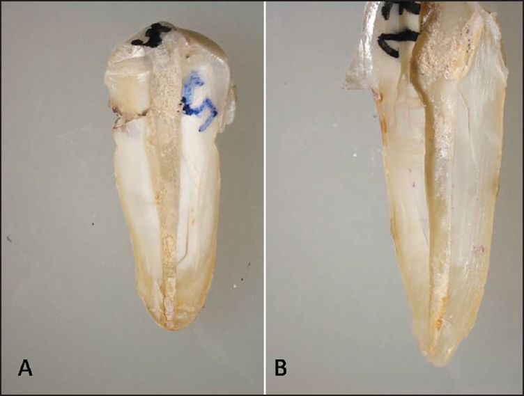
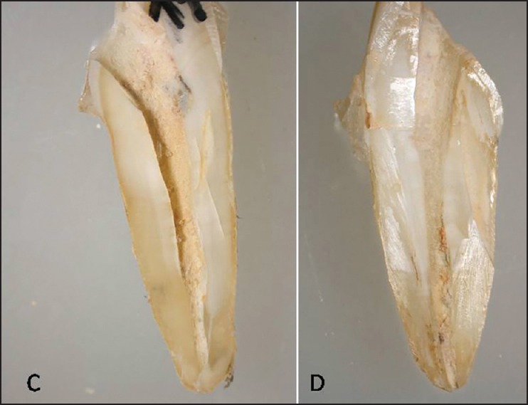
None of the files completely removed the filling material and remnants of filling material were observed in all samples regardless of the groups examined. Means and standard deviations of the area of residual filling material after instrumentation were presented in Table 1. A significant difference was found among all groups (P < 0.001). The specimens prepared with WaveOne demonstrated significantly less remaining filling material [Figure 1a]. The Reciproc group [Figure 1b] demonstrated significantly less remaining material than the ProTaper Universal Retreatment file group [Figure 2a]. The highest amount of filling material was found among specimens in which gutta-percha was removed using Gates-Glidden burs and stainless steel hand file [Figure 2b].
Table 1.
The measurement of the mean and standard deviation of the area of residual filling material by canal region after instrumentation. Different superscript letters represents statistically significant differences (P < 0,001)

Figure 1.

A specimen representative of WaveOne reciprocating file group (a) and Reciproc reciprocating file group (b)
Figure 2.

A specimen representative of ProTaper Universal Retreatment group (a) and hand instrument group (b)
No procedural errors including perforations, blockages, ledges, and deformations or fractures of any instrument were noted.
DISCUSSION
In the present study, three different instrumentation techniques were compared to evaluate whether the instrumentation techniques using with reciprocation could remove the filling material from root canals more effectively than other methods. In this study, mandibular premolar teeth with single-rooted straight canals were used. Similarly, the majority of experimental studies comparing the efficacy of retreatment techniques have been performed using straight root canals to simplify the standardization of the specimens.[16,17]
Various studies have compared the efficacy of rotary NiTi instruments to that of stainless steel hand files for removing gutta-percha and sealer from root canals.[5,8] Studies in which the efficacy of rotary NiTi instrumentation and manual instrumentation were compared in terms of the amount of remaining filling material demonstrated conflicting results. Some studies have revealed similar amounts of residual root filling material and sealer after rotary NiTi and manual instrumentation in root canals.[18,19] In contrast, some studies have reported the superiority of rotary NiTi instrumentation over manual instrumentation in terms of the amount of remaining filling material,[6,20] while other studies have reported the superiority of manual instrumentation over rotary NiTi instrumentation.[21,22] Bueno et al.[23] reported that NiTi rotary instruments were less effective in removing filling material from root canal walls than manual instruments in straight root canals. Our results contradicted this conclusion and showed that rotary NiTi instrumentation with a retreatment file was superior to the manual instrumentation technique. The difference between the results could be related to the NiTi files used in these two studies. In the present study, a NiTi rotary file specifically manufactured for retreatment procedures was used. On the other hand, Bueno et al.[21] and Aydin et al.[22] used K3 and Hero 642 files, respectively, which were manufactured as root canal preparation files but not as retreatment files. Similar to our results, according to Giuliani et al.,[24] the ProTaper Universal System for retreatment files left cleaner root canal walls root canal walls compared to both hand and rotary instruments.
The current literature contains limited data about the efficacy of the reciprocating systems in retreatment procedures. These files are made of M-Wire NiTi alloy that offers greater flexibility and greater resistance to cyclic fatigue compared to the traditional NiTi alloy.[15] The main advantage is that the working time is four times less than the traditional NiTi systems, eliminating cross-contamination between patients, once the instrument is discarded after use.[12,23,25] Recently, two brands of NiTi instruments advocating the reciprocation concept were introduced in the market: Reciproc and WaveOne. Although both instruments work according to the same principles, there are some differences in their preparation motions. The manufacturers claim that Reciproc works in the reciprocal motion with rotation of 150° counterclockwise, then 30° clockwise rotation, whereas WaveOne works with rotation of 170° counterclockwise, then 50° clockwise rotation. Additionally, these files have different tapers and different reciprocating rotation rates of 300 rpm and 350 rpm for Reciproc and WaveOne, respectively.[12] As a methodological limitation, two reciprocating instruments with different tapers were compared in the present study. Although using instruments with different tapers may seem like a lack of standardization, the most appropriate apical file size compatible with the prepared root canal size before obturation was selected.
Another limitation of this study was the technique that was used to determine the amount of remaining filling material. Taking digital images after splitting teeth longitudinally provided direct visualization of the filling material but the splitting procedure may have caused loss of gutta-percha. However, several studies have reported that the use of vertical split roots is an adequate technique, and is more accurate than radiographic examinations, which produce only two-dimensional images of the samples.[16,26]
In the present study, the WaveOne group demonstrated less residual filling materials when compared to the Reciproc group, albeit significantly. We considered that this result could be related to clockwise–counterclockwise motion, the smaller taper (.06) and less rotation rate (300 rpm) of the Reciproc system. Both reciprocating files left significantly less filling materials on the canal walls than the full-rotation NiTi file. This result is in accordance with a recent study by Zuolo et al.,[17] which found that reciprocating instruments removed more filling material from the canal walls than retreatment files (Mtwo R).
CONCLUSION
Samples in every experimental group demonstrated remnants of filling material. WaveOne was significantly more effective than Reciproc in removing the root canal filling. The reciprocating technique was the most efficient method for removing gutta-percha and sealer, followed by the rotary technique and the hand file technique.
Financial support and sponsorship
Nil.
Conflicts of interest
No potential conflict of interest relevant to this article was reported.
REFERENCES
- 1.Salebrabi R, Roststein I. Epidemiologic evaluation of the outcomes of orthograde endodontic retreatment. J Endod. 2010;36:790–2. doi: 10.1016/j.joen.2010.02.009. [DOI] [PubMed] [Google Scholar]
- 2.Duncan HF, Chong BF. Removal of root filling materials. Endod Topics. 2011;19:33–57. [Google Scholar]
- 3.Dalton BC, Orstavik D, Phillips C, Pettiette M, Trope M. Bacterial reduction with nickel-titanium rotary instrumentation. J Endod. 1998;24:763–7. doi: 10.1016/S0099-2399(98)80170-2. [DOI] [PubMed] [Google Scholar]
- 4.de Oliveira DP, Barbizam JV, Trope M, Teixeira FB. Comparison between gutta-percha and resilon removal using two different techniques in endodontic retreatment. J Endod. 2006;32:362–4. doi: 10.1016/j.joen.2005.12.006. [DOI] [PubMed] [Google Scholar]
- 5.Imura N, Kato AS, Hata GI, Uemura M, Toda T, Weine F. A comparison of the relative efficacies of four hand and rotary instrumentation techniques during endodontic retreatment. Int Endod J. 2000;33:361–6. doi: 10.1046/j.1365-2591.2000.00320.x. [DOI] [PubMed] [Google Scholar]
- 6.Saad AY, Al-Hadlaq SM, Al-Katheeri NH. Efficacy of two rotary NiTi instruments in the removal of Gutta-Percha during root canal retreatment. J Endod. 2007;33:38–41. doi: 10.1016/j.joen.2006.08.012. [DOI] [PubMed] [Google Scholar]
- 7.Bramante CM, Fidelis NS, Assumpção TS, Bernardineli N, Garcia RB, Bramante AS, et al. Heat release, time required, and cleaning ability of MTwo R and ProTaper universal retreatment systems in the removal of filling material. J Endod. 2010;36:1870–3. doi: 10.1016/j.joen.2010.08.013. [DOI] [PubMed] [Google Scholar]
- 8.Gu LS, Ling JQ, Wei X, Huang XY. Efficacy of ProTaper Universal rotary retreatment system for gutta-percha removal from root canals. Int Endod J. 2008;41:288–95. doi: 10.1111/j.1365-2591.2007.01350.x. [DOI] [PubMed] [Google Scholar]
- 9.Taşdemir T, Er K, Yildirim T, Celik D. Efficacy of three rotary NiTi instruments in removing gutta-percha from root canals. Int Endod J. 2008;41:191–6. doi: 10.1111/j.1365-2591.2007.01335.x. [DOI] [PubMed] [Google Scholar]
- 10.Prichard J. Rotation or reciprocation: A contemporary look at NiTi instruments? Br Dent J. 2012;212:345–6. doi: 10.1038/sj.bdj.2012.268. [DOI] [PubMed] [Google Scholar]
- 11.Yared G. Canal preparation using only one Ni-Ti rotary instrument: Preliminary observations. Int Endod J. 2008;41:339–44. doi: 10.1111/j.1365-2591.2007.01351.x. [DOI] [PubMed] [Google Scholar]
- 12.Kim HC, Kwak SW, Cheung GS, Ko DH, Chung SM, Lee W. Cyclic fatigue and torsional resistance of two new nickel-titanium instruments used in reciprocation motion: Reciproc versus WaveOne. J Endod. 2012;38:541–4. doi: 10.1016/j.joen.2011.11.014. [DOI] [PubMed] [Google Scholar]
- 13.Alapati SB, Brantley WA, Iijma M, Clark WA, Kovarik L, Buie C, et al. Metallurgical characterization of a new nickel-titanium wire for rotary endodontic instruments. J Endod. 2009;35:1589–93. doi: 10.1016/j.joen.2009.08.004. [DOI] [PubMed] [Google Scholar]
- 14.Gutmann JL, Gao Y. Alteration in the inherent metallic and surface properties of nickel-titanium root canal instruments to enhance performance, durability and safety: A focussed review. Int Endod J. 2012;45:113–28. doi: 10.1111/j.1365-2591.2011.01957.x. [DOI] [PubMed] [Google Scholar]
- 15.Schneider SW. A comparison of canal preparations in straight and curved root canals. Oral Surg Oral Med Oral Pathol. 1971;32:271–5. doi: 10.1016/0030-4220(71)90230-1. [DOI] [PubMed] [Google Scholar]
- 16.Zuolo AS, Mello JE, Jr, Cunha RS, Zuolo ML, Bueno CE. Efficacy of reciprocating and rotary techniques for removing filling material during root canal retreatment. Int Endod J. 2013;46:947–53. doi: 10.1111/iej.12085. [DOI] [PubMed] [Google Scholar]
- 17.Rödig T, Hausdörfer T, Konietschke F, Dullin C, Hahn W, Hülsmann M. Efficacy of D-RaCe and ProTaper Universal Retreatment NiTi instruments and hand files in removing gutta-percha from curved root canals-a micro-computed tomography study. Int Endod J. 2012;45:580–9. doi: 10.1111/j.1365-2591.2012.02014.x. [DOI] [PubMed] [Google Scholar]
- 18.Akpınar KE, Altunbaş D, Kuştarcı A. The efficacy of two rotary NiTi instruments and H-files to remove gutta-percha from root canals. Med Oral Patol Oral Cir Bucal. 2012;17:e506–11. doi: 10.4317/medoral.17582. [DOI] [PMC free article] [PubMed] [Google Scholar]
- 19.Somma F, Cammarota G, Plotino G, Grande NM, Pameijer CH. The effectiveness of manual and mechanical instrumentation for the retreatment of three different root canal filling materials. J Endod. 2008;34:466–9. doi: 10.1016/j.joen.2008.02.008. [DOI] [PubMed] [Google Scholar]
- 20.Hülsmann M, Bluhm V. Efficacy, cleaning ability and safety of different rotary NiTi instruments in root canal retreatment. Int Endod J. 2004;37:468–76. doi: 10.1111/j.1365-2591.2004.00823.x. [DOI] [PubMed] [Google Scholar]
- 21.Bueno CE, Delboni MG, de Araújo RA, Carrara HJ, Cunha RS. Effectiveness of rotary and hand files in gutta-percha and sealer removal using chloroform or chlorhexidine gel. Braz Dent J. 2006;17:139–43. doi: 10.1590/s0103-64402006000200011. [DOI] [PubMed] [Google Scholar]
- 22.Aydin B, Köse T, Calişkan MK. Effectiveness of HERO 642 versus Hedström files for removing gutta-percha fillings in curved root canals: An ex vivo study. Int Endod J. 2009;42:1050–6. doi: 10.1111/j.1365-2591.2009.01624.x. [DOI] [PubMed] [Google Scholar]
- 23.Gavini G, Caldeira CL, Akisue E, Candeiro GT, Kawakami DA. Resistance to flexural fatigue of Reciproc R25 files under continuous rotation and reciprocating movement. J Endod. 2012;38:684–7. doi: 10.1016/j.joen.2011.12.033. [DOI] [PubMed] [Google Scholar]
- 24.Giuliani V, Cocchetti R, Pagavino G. Efficacy of ProTaper universal retreatment files in removing filling materials during root canal retreatment. J Endod. 2008;34:1381–4. doi: 10.1016/j.joen.2008.08.002. [DOI] [PubMed] [Google Scholar]
- 25.Bürklein S, Hinschitza K, Dammaschke T, Schäfer E. Shaping ability and cleaning effectiveness of two single-file systems in severely curved root canals of extracted teeth: Reciproc and WaveOne versus Mtwo and ProTaper. Int Endod J. 2012;45:449–61. doi: 10.1111/j.1365-2591.2011.01996.x. [DOI] [PubMed] [Google Scholar]
- 26.Rios Mde A, Villela AM, Cunha RS, Velasco RC, De Martin AS, Kato AS, et al. Efficacy of 2 reciprocating systems compared with a rotary retreatment system for gutta-percha removal. J Endod. 2014;40:543–6. doi: 10.1016/j.joen.2013.11.013. [DOI] [PubMed] [Google Scholar]


