

Abstract
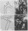
The echocardiographic appearances in a case of coarctation of the aorta, first suspected at 21 weeks of intrauterine life, changed progressively during this time. Early right ventricular hypertrophy and increasing aortic arch hypoplasia were evident and suggest the possible sequence of evolution of the coarctation in this case.
Full text
PDF
Images in this article
Selected References
These references are in PubMed. This may not be the complete list of references from this article.
- Rudolph A. M., Heymann M. A., Spitznas U. Hemodynamic considerations in the development of narrowing of the aorta. Am J Cardiol. 1972 Oct;30(5):514–525. doi: 10.1016/0002-9149(72)90042-2. [DOI] [PubMed] [Google Scholar]
- Shinebourne E. A., Alseed A. M. Developmental explanation for absence of coarctation in conditions with reduced pulmonary flow from birth. Br Heart J. 1973 Aug;35(8):866–866. [PubMed] [Google Scholar]
- Smallhorn J. F., Anderson R. H., Macartney F. J. Morphological characterisation of ventricular septal defects associated with coarctation of aorta by cross-sectional echocardiography. Br Heart J. 1983 May;49(5):485–494. doi: 10.1136/hrt.49.5.485. [DOI] [PMC free article] [PubMed] [Google Scholar]
- Wielenga G., Dankmeijer J. Coarctation of the aorta. J Pathol Bacteriol. 1968 Jan;95(1):265–274. doi: 10.1002/path.1700950131. [DOI] [PubMed] [Google Scholar]