Abstract
Purpose
This study assessed the accuracy of age estimates produced by a regression equation derived from lower third molar development in a Thai population.
Materials and Methods
The first part of this study relied on measurements taken from panoramic radiographs of 614 Thai patients aged from 9 to 20. The stage of lower left and right third molar development was observed in each radiograph and a modified Gat score was assigned. Linear regression on this data produced the following equation: Y=9.309+1.673 mG+0.303S (Y=age; mG=modified Gat score; S=sex). In the second part of this study, the predictive accuracy of this equation was evaluated using data from a second set of panoramic radiographs (539 Thai subjects, 9 to 24 years old). Each subject's age was estimated using the above equation and compared against age calculated from a provided date of birth. Estimated and known age data were analyzed using the Pearson correlation coefficient and descriptive statistics.
Results
Ages estimated from lower left and lower right third molar development stage were significantly correlated with the known ages (r=0.818, 0.808, respectively, P≤0.01). 50% of age estimates in the second part of the study fell within a range of error of ±1 year, while 75% fell within a range of error of ±2 years. The study found that the equation tends to estimate age accurately when individuals are 9 to 20 years of age.
Conclusion
The equation can be used for age estimation for Thai populations when the individuals are 9 to 20 years of age.
Keywords: Age Determination by Teeth; Molar, Third; Radiography, Panoramic; Forensic Dentistry
Introduction
Age estimation has many applications. Important applications include estimation of age for those who do not know their birth date and the use of age estimation in identification of bodies. Estimation methods include using radiographs to examine epiphysial fusion of long bones,1 to examine hands and wrists2 and to examine cervical vertebrae,3 observing changes in the pubic symphysis, 4 and evaluating fusion of the skull's sutures.5
Teeth are another structure in the body that can be used for age estimation. Age estimation from teeth can be accomplished using an understanding of dental eruption and tooth formation.6 Few age estimation studies of Thai subjects, which use these methods, exist.7,8,9,10 Raungpaka8 found a high correlation between dental age and chronological age when she evaluated the development of 14 permanent teeth (seven types of permanent teeth in the right maxilla and mandible, not including third molars) in panoramic radiographs using the dental development score proposed by Gat et al.11 Krailassiri et al.10 reported a relationship between dental calcification stage and skeletal maturity stage using Demirjian scores12 for dental calcification. However, they found that calcification stage in third molars had the lowest correlation when used to predict skeletal maturity. In general, age estimation from erupted and erupting teeth proves useful only up until age 14. When all teeth, except third molars, have erupted and are completely formed, estimation is difficult. Therefore, between the ages of 14 and 21, only third molar development provides information on which age estimates can be based.13,14,15,16,17,18,19 Consequently, third molar development has been the basis of many age estimation studies.
While such studies have been performed in various countries, the literature includes reference to only one such study in Thailand.9 We performed our study to estimate the relationship between chronological age and third molar development stage in a Northern Thai population and to assess the accuracy of estimates based on the resulting equation.
Materials and Methods
This retrospective study made use of the patient archive at our Radiology clinic. Two stages comprised this study. The first estimated the relationship between chronological age and stage of third molar development and was based upon panoramic radiographs of Thai patients at the Faculty of Dentistry, Chiang Mai University. In the first stage of the study, a total of 614 panoramic radiographs of 341 female and 273 male patients aged 9 to 20 years who came to the Radiology Clinic of the Faculty of Dentistry, Chiang Mai University during January to December 2005 were evaluated. The panoramic radiographs were made using a Planmeca Proline machine (Planmeca Proline 2002, Planmeca Oy, Helsinki, Finland). To be included in the study, patients had to be healthy and free of systemic disease, fall within the study's age range and show no developmental disturbances of the teeth. Patients' medical records confirmed that the subjects met these inclusion criteria.
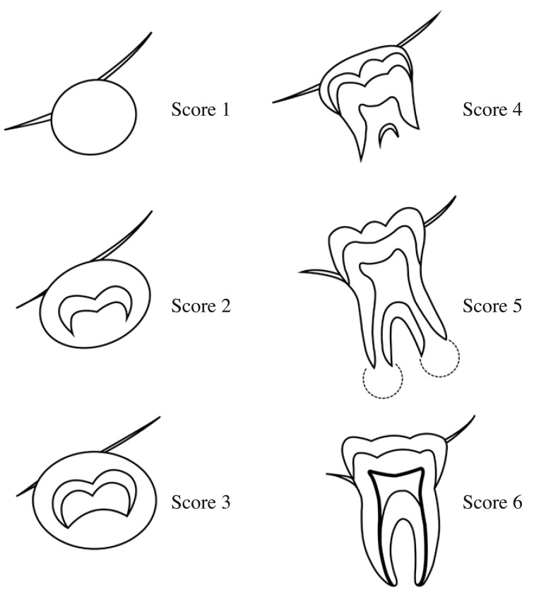
The panoramic radiographs were evaluated to determine the development stage of each subject's third molars. Our study modified Gat's scoring system with the development of third molars catagorized as follows: score 0: no development of the third molar follicle, score 1: radiolucent area of the third molar seen, but without calcification, score 2: calcification of the third molar, up to half of the crown, score 3: calcification of the third molar, from more than half of the crown to complete crown formation, but without root formation, score 4: root formation begun, but less than half of the root length formed, score 5: root formation, more than half and up to full root length formed, but the apex remains open, score 6: completion of root apex closure. Figure 1 presents the drawings representing the stage of development corresponding to each score.
Fig. 1. The drawings represent the stage of development of the third molar which is the modified Gat's scoring system.
The first stage of the study was carried out by three observers who scored the stage of third molar development. The observers used a viewing box in a room with subdued light. A mask was placed around the area of interest on each radiograph. Observers viewed the radiographs twice with a week between each session. They were trained and their scoring calibrated by means of practice scoring sessions so that there was no significant difference among the observers. If a difference arose in scoring a particular radiograph, a discussion followed to produce a consensus score.
The chronological age of each patient was calculated by subtracting the patient's date of birth from the date of radiographic examination, followed by rounding. If a patient was six months older than his or her nominal age, the nominal age was rounded up to the next whole number. Otherwise, the patient's nominal age was recorded. For example, the recorded age for a patient aged 10 years and 4 months would have been 10 years. The recorded age of another patient, aged 10 years and 7 months would have been 11 years. This rounding was introduced because, in legal matters, only whole-numbered ages are considered in Thailand.
The Mann-Whitney U test was employed to detect differences in the relationship between tooth development stage and age for males and females and a step-wise regression analysis was used to formulate the equation.
The second stage of the study then tested the accuracy of the regression equation in predicting chronological age. The second stage sample consisted of 539 subjects, aged 9 to 24 years, who visited the Faculty of Dentistry, Chiang Mai University between June 2006 and June 2008. Panoramic radiographs of the patients were made using the Planmeca Proline machine (Planmeca Proline 2002, Planmeca Oy). As in the first stage, patients included in the study had to be healthy and free of systemic disease, fall within the study's age range and show no developmental disturbances of the teeth. Patients' medical records confirmed that the subjects met these inclusion criteria.
Stage-of-development scores for the mandibular third molars were determined by two oral and maxillofacial radiologists. They evaluated the radiographs and then assigned each molar a modified Gat score.
A calibration session for the two observers was conducted before subject radiographs were scored. In the calibration session, a random sample of 100 panoramic radiographs was selected for evaluation using a viewing box in a room with subdued light and a mask around the area of interest on each radiograph. Each observer evaluated the panoramic radiographs twice with a two-week interval between evaluations. Scores from both observers were compared using Kappa analysis. In cases where the observers assigned different scores, both observers reviewed the radiograph together and agreed upon a final score for tooth development in that case.
After the calibration session, observation sessions were conducted in the same fashion with no more than 100 radiographs reviewed per session to prevent visual fatigue.
Finally, analysis was performed using data from the second part of the study. Application of the first part's regression equation to scores assigned in the second part produced an estimated age for each patient. Correlation between estimated ages and known chronological ages of subjects in the study's second part was analyzed by means of the Pearson correlation coefficient. Accuracy of the equation's age estimates was assessed allowing a ±1 year range of error and allowing a ±2 years range of error, both centered on each subject's known chronological age. In addition, the accuracy of age estimates based on mandibular left and right third molars was compared.
Results
The results from the first stage of the study show a strong relationship between the stage of development of the mandibular left third molar and the chronological age (r=0.818). The results also show a difference between the sexes in the rate of third molar development. Males exhibited faster development than their female counterparts in all four third molars. Stepwise regression analysis supported inclusion of the independent variables modified Gat score of the mandibular third molar and sex in the regression equation. Estimation of parameters using the first stage data set produced the following equation: Y=9.309+1.673 mG+0.303S (Y=estimated age, mG=modified Gat score, S=sex (male=0, female=1)).
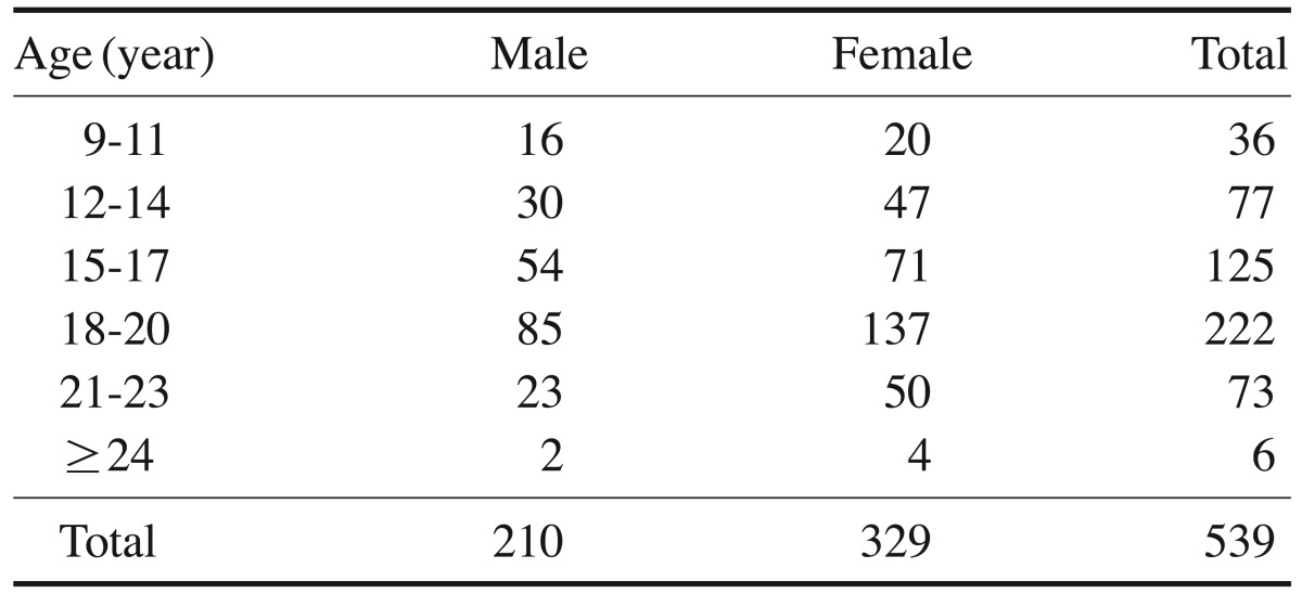
In the second stage, 539 subjects with panoramic radiographs in the archive met the inclusion criteria and were included in the study. There were 210 male and 329 female patients in this set of subjects. The distribution of age and sex in the set is shown in Table 1.
Table 1. Sex and age distribution of the study group.
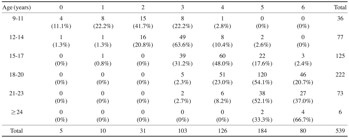
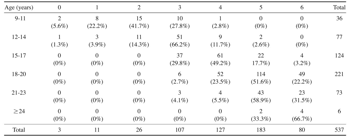
Observers scored the development of 539 mandibular left and 537 mandibular right third molars. The panoramic radiographs of two subjects exhibited severe distortion of the right third molar image, preventing them from being scored. Tables 2 and 3, respectively, present the distribution by age of mandibular left and right third molar development in the set.
Table 2. Frequency of modified Gat score for the left mandibular third molar, by subject age group.
Table 3. Frequency of modified Gat score for the right mandibular third molar, by subject age group.
The degree of intra-observer agreement ranged from good to very good, with Kappa scores ranging from 0.786 to 0.853. The degree of inter-observer agreement was very good, with a Kappa score of 0.817.
Our analysis found a significant correlation between ages estimated by the regression equation introduced in this article and the subjects' chronological ages. Age estimation by means of the mandibular left third molar resulted in r=0.818 and by means of the mandibular right third molar resulted in r=0.808, with p≤0.01 in both cases.
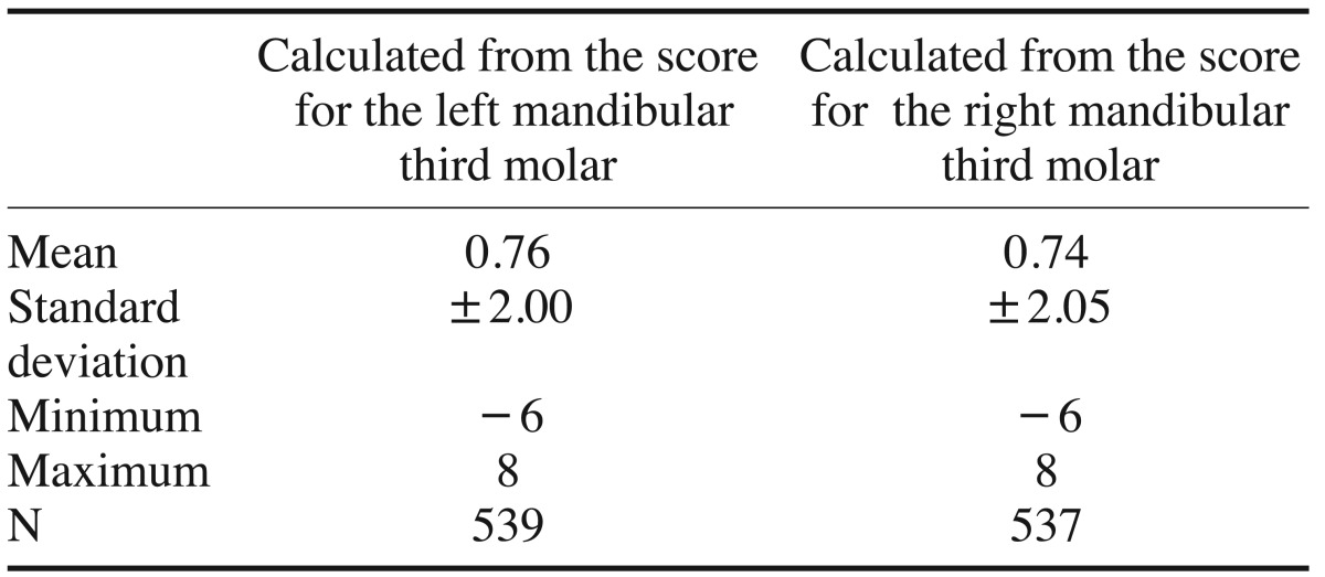
On average, the difference between estimated and actual ages in the second part of the study was 0.76 years using the mandibular left third molar and 0.74 years using the mandibular right third molar. The standard deviations of the differences were 2.00 years and 2.05 years, respectively (Table 4). Allowing for a range of error of ±1 year, 51% and 49% of ages estimated by means of the mandibular left and right third molars, respectively. were deemed accurate. Allowing for a range of error of ±2 years, 77% and 76% of ages estimated by means of the mandibular left and right third molars, respectively, were deemed accurate (Table 5).
Table 4. Descriptive statistics for the differences between real age and age estimated by the equation.
Table 5. Accuracy of the age estimates, left/right third molars, ranges of error ±1 year/±2 years.
Using actual age ±2 years as our measure of accuracy, this study found that between 61.1% and 96.0% of mandibular left third molar age estimates were accurate across the four three-year age ranges used to group subjects aged 9 to 20 years. For estimates based upon mandibular right third molar development, accuracy ranged from 55.6% to 96.8% (Table 6). Accuracy varied little between estimates based on left and right mandibular third molar development. However, accuracy dropped dramatically for patients older than 20. None of the estimates for subjects 24 and older were deemed accurate.
Table 6. Accuracy of the age calculated from the score allowing a range of error of ±2 years, by subject age range.
When estimates were grouped by modified Gat score, with estimates falling within two years of actual age considered accurate, we found consistently high accuracy (66.7%-87.1% left side, 66.7%-86.3% right side) across the range of scores. The modified Gat score of 4 presented the lowest level of accuracy for estimates based on mandibular left third molar development and the second lowest level of accuracy for estimates based on mandibular right third molar development (Table 7).
Table 7. Accuracy of the age calculated from the score allowing a range of error of ±2 years, by modified Gat score.
Discussion
In the first part of our study, we developed a regression equation that relates age to stage of third molar development and sex. Third molar development was classified using a scoring system modified from the work of Gat. Gat's study compared third molar development scores in boys and girls aged 5 to 13 years, but did not estimate an equation relating age to score. Gat's tooth development scores classified stages of crown calcification and root formation and ranged from 0 to 5. For mathematical reasons, we modified Gat's scoring, identifying 7 stages of development, scored on a scale from 0 to 6.
Mesotten et al.20 presented equations for age estimation in their study of third molar crown and root development in the Belgian population. These equations based their estimates on the devlopmental stages of all four third molars with tooth development divided into 10 stages. The separate equations for age estimation in males and females follow: age estimate for males=10.2000+0.5122UL+0.5273LL, Age estimate for females=13.6206+0.1933UR+0.5080LR, where UR=the tooth development score for the maxillary right third molar, UL =the tooth dvelopment score for the maxillary left third molar, LR=the tooth development score for the mandibular right third molar, LL=the tooth development score for the mandibular left third molar. They found that these equations worked well only when estimating ages for subjects aged 16 to 18 years. When compared to the equations in Mesotten et al., our equation requires examination of only one mandibular third molar, making it simpler to use. For cases in which a subject's mandibular left third molar is missing, our equation can be applied to a modified Gat score based on mandibular right third molar development with an expectation that the application will produce a similar rate of accuracy. The correlation between age estimates and actual age differed little when mandibular left third molar (r=0.818) and mandibular right third molar (r=0.808) were the basis of scores. Moreover, we found our equation accurate within ±2 years when estimating age across a wider range of ages (9 to 20 years old), with accuracy >80% when the subjects' ages fell within the range 12 to 20 years old (Table 6).
The standard deviation of age estimation errors was 2.00 when scores were based on mandibular left third molar development and 2.05 when scores were based upon mandibular right third molar development. These results were similar to results in other studies. Thevissen et al.9 resported standard deviations ranging from 1.74 to 1.90 for age estimation based on scoring the development of third molars in the Thai population. In the study by Mesotten et al., cited earlier, they found standard deviations of 1.52 and 1.56, respectively, for male and female age estimates produced by their equations. Willerhausen et al.21 reported a margin of error of ±2-4 years in their study using third molars to estimate age in a group of Europeans aged 15 to 24 years. Darji et al.22 reported that they found a strong positive correlation between third molar developmental stage and chronological age in an Indian population with a standard deviation of 2 years at each stage. Bagherpour et al.23 studied young Iranian adults and presented formulas for male and female age estimation. They reported a standard deviation of 1.4 years for males and of 1.56 years for females.
Although our scoring system, modified from Gat's study, was simple to use because it divided third molar development into only seven stages, its specification might have contributed to the high standard deviation found in our study. In particular, the seven stages used might not provide the correct degree of refinement to ideally distinguish age difference. Table 7 shows that age estimates associated with a score of 4 proved least, or next to least, accurate. We speculate that a score of 4, which is associated with root formation up to no more than half of the root length, cannot be consistently assigned. In practice, this length can only be measured when root formtaion is complete. Before root formation is complete, final root length cannot be known, perhaps leading to incorrect assignment of development stage.
Our equation produced its lowest rate of accuracy for subjects aged 9 to 11 years. Even when we allowed acceptance of estimates within two years of actual age, only 61.1% of left side and 55.6% of right side estimates fell within the range of error. We speculate that this lower level of accuracy relates to the wide range of scores (from 0 to 4) observed in subjects falling within this age group. This finding is consistent with results in studies by Uzamis et al.16 and Balonos et al.17 They reported wide ranges of third molar development rates in the early stages of development. Moreover, the number of subjects falling within this age range in our study was small, accounting for only 8.9% of the number studied. Further investigation with a larger sample drawn from this age group is recommended.
We note that the methods of our study produced poor age estimates for those older than 20 years of age. For subjects older than 20, root formation was complete, which meant that these subjects were all assigned a Gat score of 6. Consequently, age estimates were all identical.
The subjects in this study were all drawn from a Northern Thai population. Differences in environment, culture and the ethnic composition of different parts of the country might affect the distribution of tooth development rates. Therefore, the accuracy of our equation should be evaluated for groups living elsewhere in Thailand and for populations elsewhere in the ASEAN community.
In summary, this study's equation is simple to apply, requiring only two pieces of information: sex and a score indicating stage of lower third molar development. We found this equation suitable for estimating the age of people whose actual age ranged from 9 to 20 years, with approximately 50% of age estimates falling within one year of actual age and approximately 75% of age estimates in our validation study falling within two years of actual age.
We recommend further testing of this study's estimation equation and adjustments to improve its accuracy. Factors that might improve accuracy include patients' height and the eruption height of impacted third molars.24
Acknowledgements
The authors wish to thank Dr. M. Kevin O Carroll, Professor Emeritus, School of Dentistry, University of Mississippi, and Faculty Consultant, Faculty of Dentistry, Chiang Mai University, for his assistance in editing this manuscript.
Footnotes
This study was supported by a research funding from the Faculty of Dentistry, Chiang Mai University.
References
- 1.Crowder C, Austin D. Age ranges of epiphyseal fusion in the distal tibia and fibula of contemporary males and females. J Forensic Sci. 2005;50:1001–1007. [PubMed] [Google Scholar]
- 2.Cameriere R, De Luca S, Biagi R, Cingolani M, Farronato G, Ferrante L. Accuracy of three age estimation methods in children by measurements of developing teeth and carpals and epiphyses of the ulna and radius. J Forensic Sci. 2012;57:1263–1270. doi: 10.1111/j.1556-4029.2012.02120.x. [DOI] [PubMed] [Google Scholar]
- 3.Varshosaz M, Ehsani S, Nouri M, Tavakoli MA. Bone age estimation by cervical vertebral dimensions in lateral cephalometry. Prog Orthod. 2012;13:126–131. doi: 10.1016/j.pio.2011.09.003. [DOI] [PubMed] [Google Scholar]
- 4.Lottering N, Macgregor DM, Meredith M, Alston CL, Gregory LS. Evaluation of the Suchey-Brooks method of age estimation in an Australian subpopulation using computed tomography of the pubic symphyseal surface. Am J Phys Anthropol. 2013;150:386–399. doi: 10.1002/ajpa.22213. [DOI] [PubMed] [Google Scholar]
- 5.Wolff K, Vas Z, Sótonyi P, Magyar LG. Skeletal age estimation in Hungarian population of known age and sex. Forensic Sci Int. 2012;223:374.e1–374.e8. doi: 10.1016/j.forsciint.2012.08.033. [DOI] [PubMed] [Google Scholar]
- 6.Halcrow SE, Tayles N, Buckley HR. Age estimation of children from prehistoric Southeast Asia: are the dental formation methods used appropriate? J Archaeol Sci. 2007;34:1158–1168. [Google Scholar]
- 7.Kamalanathan GS, Hauck HM. Dental development of children in a Siamese village, Bang Chan, 1953. J Dent Res. 1960;39:455–461. doi: 10.1177/00220345600390030601. [DOI] [PubMed] [Google Scholar]
- 8.Raungpaka S. The study of tooth-development age of Thai children in Bangkok. J Dent Assoc Thai. 1988;38:72–81. [PubMed] [Google Scholar]
- 9.Thevissen PW, Pittayapat P, Fieuws S, Willems G. Estimating age of majority on third molars developmental stages in young adults from Thailand using a modified scoring technique. J Forensic Sci. 2009;54:428–432. doi: 10.1111/j.1556-4029.2008.00961.x. [DOI] [PubMed] [Google Scholar]
- 10.Krailassiri S, Anuwongnukroh N, Dechkunakorn S. Relationships between dental calcification stages and skeletal maturity indicators in Thai individuals. Angle Orthod. 2002;72:155–166. doi: 10.1043/0003-3219(2002)072<0155:RBDCSA>2.0.CO;2. [DOI] [PubMed] [Google Scholar]
- 11.Gat H, Sarnat H, Bjorvath K, Dayan D. Dental age evaluation. A new six-developmental-stage method. Clin Prev Dent. 1984;6:18–22. [PubMed] [Google Scholar]
- 12.Demirjian A, Goldstein H, Tanner JM. A new system of dental age assessment. Hum Biol. 1973;45:211–227. [PubMed] [Google Scholar]
- 13.Mincer HH, Harris EF, Berryman HE. The A.B.F.O. study of third molar development and its use as an estimator of chronological age. J Forensic Sci. 1993;38:379–390. [PubMed] [Google Scholar]
- 14.Kullman L. Accuracy of two dental and one skeletal age estimation method in Swedish adolescents. Forensic Sci Int. 1995;75:225–236. doi: 10.1016/0379-0738(95)01792-5. [DOI] [PubMed] [Google Scholar]
- 15.Nykanen R, Espeland L, Kvaal SI, Krogstad O. Validity of the Demirjian method for dental age estimation when applied to Norwegian children. Acta Odontol Scand. 1998;56:238–244. doi: 10.1080/00016359850142862. [DOI] [PubMed] [Google Scholar]
- 16.Uzamis M, Kansu O, Taner TU, Alpar R. Radiographic evaluation of third-molar development in a group of Turkish children. ASDC J Dent Child. 2000;67:136–141. [PubMed] [Google Scholar]
- 17.Bolanos MV, Moussa H, Manrique MC, Bolanos MJ. Radiographic evaluation of third molar development in Spanish children and young people. Forensic Sci Int. 2003;133:212–219. doi: 10.1016/s0379-0738(03)00037-9. [DOI] [PubMed] [Google Scholar]
- 18.Gunst K, Mesotten K, Carbonez A, Willems G. Third molar root development in relation to chronological age: a large sample sized retrospective study. Forensic Sci Int. 2003;136:52–57. doi: 10.1016/s0379-0738(03)00263-9. [DOI] [PubMed] [Google Scholar]
- 19.Olze A, Taniguchi M, Schmeling A, Zhu BL, Yamada Y, Maeda H, et al. Comparative study on the chronology of third molar mineralization in a Japanese and a German population. Leg Med (Tokyo) 2003;5(Suppl 1):S256–S260. doi: 10.1016/s1344-6223(02)00143-8. [DOI] [PubMed] [Google Scholar]
- 20.Mesotten K, Gunst K, Carbonez A, Willems G. Dental age estimation and third molars: a preliminary study. Forensic Sci Int. 2002;129:110–115. doi: 10.1016/s0379-0738(02)00245-1. [DOI] [PubMed] [Google Scholar]
- 21.Willershausen B, Löffler N, Schulze R. Analysis of 1202 orthopantograms to evaluate the potential of forensic age determination based on third molar developmental stages. Eur J Med Res. 2001;6:377–384. [PubMed] [Google Scholar]
- 22.Darji JA, Govekar G, Kalele SD, Hariyani H. Age estimation from third molar development: A radiological study. J Indian Acad Forensic Med. 2011;33:130–134. [Google Scholar]
- 23.Bagherpour A, Anbiaee N, Partovi P, Golestani S, Afzalinasab S. Dental age assessment of young Iranian adults using third molars: a multivariate regression study. J Forensic Leg Med. 2012;19:407–412. doi: 10.1016/j.jflm.2012.04.009. [DOI] [PubMed] [Google Scholar]
- 24.Mahasantipiya PM, Pramojanee S, Thaiupathump T. Image analysis of the eruptive positions of third molars and adjacent second molars as indicators of age evaluation in Thai patients. Imaging Sci Dent. 2013;43:289–293. doi: 10.5624/isd.2013.43.4.289. [DOI] [PMC free article] [PubMed] [Google Scholar]


