

Abstract
Objective: To assess the early complications in the orthopedic treatment of metastatic bone lesions and the factors associated with these complications. Method: There were assessed, retrospectively, 64 patients that underwent surgical treatment for bone metastases, analyzing the complications that occurred in the pre-operative and early post- operative period and associating them with the tumor origin, type of procedure done, the need of blood reposition before the surgery, the need of new surgical procedures and the mortality due to the complications. Results: Early complications in the treatment were observed in 17 (26.6%) patients, of which six (35.2%) ended up dying due to these complications. Regarding the type, 15 (23.8%) cases were due to surgical complications, four (6.3%) clinical and three (4.7%) patients showed clinical and surgical complications. There was no significant difference in the frequency of complications or mortality when assessed the type of reconstruction or affected region. The tumors with a renal origin needed more blood reposition and showed a bigger frequency of complications. Conclusion: The complications occurred in 26.6%. The complications are not related to the kind of treatment performed or to the region affected. The renal origin tumors showed a higher risk of hemorrhage.
Keywords: Bone neoplasms; Oncology; Fractures, spontaneous; Postoperative complications; orthopedic surgery
INTRODUCTION
Metastatic bone disease is the most common malignant neoplasia of the skeleton, the bone being the third most common site of the spread of adenocarcinomas, preceded only by the lung and the liver(1). Bone's predisposition to metastases is explained by the high blood flow under low pressure in the areas of red marrow and because bone matrix is a fertile ground for the implantation of tumor cells(2). Advances in adjuvant and neoadjuvant therapy, particularly hormone therapy and chemotherapy, have improved the survival of cancer patients and increased the prevalence of patients living with bone metastases (BM). This makes control of BM a key factor in improving the quality of life, pain control, and in maintaining patient independence1, 3.
Although most cases of BM are treated clinically, orthopedic follow-up is able to identify lesions early that compromise the mechanical stability of the skeleton, preventing or treating fractures, controlling pain, and reducing morbidity(3). Besides prevention, orthopedic treatment can restore the ability to walk in 94% of patients with pathological fracture(2).
The complications of this treatment are common and influenced by the patient's clinical conditions, stage of disease, and the surgical procedure. They can occur perioperatively, in the immediate postoperative period, or in the late postoperative period and include clinical and surgical complications1, 2, 3.
The aim of this study is to evaluate the early complications of patients undergoing orthopedic treatment of metastatic bone lesions and the factors associated with these complications.
METHODS
Patients
We reviewed the medical records of patients registered at the Department of Orthopedic Oncology, Hospital das Clínicas, Universidade Federal de Minas Gerais (UFMG) and Santa Casa de Belo Horizonte between January 2002 and March 2007 who underwent orthopedic surgical treatment of bone injuries secondary to extraskeletal malignant neoplasia. The study was approved by the ethics committee of the respective hospitals. The study included 64 from a total of 90 patients. Twenty-six patients were excluded due to insufficient data in the medical record (n = 12) and exclusion of tumors of medullary origin: multiple myeloma (n = 9), leukemia (n = 3), and lymphomas (n = 2).
The average age was 56.63 ± 14.26 years (29 to 85 years), and 23 (35.9%) of the patients were males and 41 were females (64.1%). Twelve (18.8%) patients were melanoderm, 24 (37.5%) were Mulatto and 28 (43.8%) were Caucasian. The mean survival was 9.20 ± 11.96 months (median 6.6 months).
Radiotherapy was previously performed in 27 (42.1%) patients and chemotherapy was previously performed in 56 (87.5%) patients. The reason for referral to orthopedic treatment was fracture in 42 (65.6%) patients, pain in 20 (31.2%), and changes in tests in asymptomatic patients in two (3.2%) cases.
Tumors
All BM located in the limbs, pelvic and scapular girdle, and requiring surgical treatment were included in the study. Primary bone tumors and those of medullary origin were excluded in the study. Regarding the organ of origin, the most common was the breast tumor in 23 (35.9%) patients (Table 1). Regarding the histological type, the BM were adenocarcinomas in 34 (53.1%) cases, carcinoma in 23 (35.9%) patients, sarcomas in five (7.8%), and in two (3.1%) the cell type was undetermined.
Table 1.
Frequency of metastases according to the organ of origin.
| ORIGIN | PATIENTS (n) | ABSOLUTE FREQUENCY (%) | ACCUMULATED FREQUENCY (%) |
|---|---|---|---|
| BREAST | 23 | 35.9 | 35.9 |
| RENAL | 8 | 12.5 | 48.4 |
| PROSTATE | 8 | 12.5 | 60.9 |
| LUNG | 4 | 6.3 | 67.2 |
| COLORECTAL | 4 | 6.3 | 73.4 |
| THYROID | 3 | 4.7 | 78.3 |
| OTHER | 12 | 18.8 | 96.9 |
| UNKNOWN | 2 | 3.1 | 100 |
| TOTAL | 64 | 100 | 100 |
Source: SAME, Hospital das Clínicas, UFMG, Santa Casa, BH.
The most common site was the lower limbs in 52 (81.3%) patients, seven cases were located in the pelvis (10.9%), and five (7.9%) in the upper limbs. The lesions were solitary in 22 (34.3%) patients and in multiple locations in the skeleton in 42 (65.7%) (Table 2).
Table 2.
Anatomical distribution of lesions submitted to surgical treatment of bone metastases.
| LOCALIZATION | PATIENTS (n) | FREQUENCY (%) | ACCUMULATED FREQUENCY (%) |
|---|---|---|---|
| PELVIS | 7 | 10.9 | 10.9 |
| FEMORAL NECK | 7 | 10.9 | 21.9 |
| TROCHANTER | 25 | 39.1 | 60.9 |
| SUBTROCHANTER | 13 | 20.3 | 81.3 |
| FEMORAL DIAPHYSIS | 4 | 6.3 | 87.5 |
| DISTAL FEMUR | 2 | 3.1 | 90.6 |
| TIBIA | 1 | 1.6 | 92.2 |
| PROXIMAL HUMERUS | 3 | 4.7 | 96.9 |
| HUMERAL DIAPHYSIS | 1 | 1.6 | 98.4 |
| DISTAL HUMERUS | 1 | 1.6 | 100.0 |
| TOTAL | 64 | 100.0 | 100.0 |
Source: SAME, Hospital das Clínicas, UFMG, Santa Casa, BH.
Orthopedic treatment
The indications for orthopedic surgery were pathological fractures in 43 (67.2%) patients, impending fractures in 17 (26.6%), and simple tumor resection with curative goal in four (5.3%).
The goal of surgery was the rigid stabilization of established or impending fracture, allowing for the immediate ambulation of the patient when possible, or wide resection of the lesion with a curative goal in isolated lesions without other organs affected and with a good oncological prognosis. Arthroplasties (conventional, hemiarthroplasty, and endoprostheses) were performed in 24 (37.5%) patients, osteosynthesis (plates and intramedullary nails) was performed in 36 (56.3%) and four (6.3%) did not undergo reconstruction (Table 3).
Table 3.
Methods of fixation and reconstruction used in the treatment of bone metastases.
| TREATMENT | PATIENTS (n) | FREQUENCY (%) | ACCUMULATED FREQUENCY (%) |
|---|---|---|---|
| PLATE + PMMA | 23 | 35.9 | 35.9 |
| IMN + PMMA | 11 | 17.2 | 53.1 |
| TOTAL PROSTHESIS | 9 | 14.1 | 67.2 |
| ENDOPROSTHESIS | 13 | 20.3 | 87.5 |
| HEMIARTHROPLASTY | 2 | 3.1 | 90.6 |
| ISOLATED PLATE | 1 | 1.6 | 92.1 |
| ISOLATED IMN | 1 | 1.6 | 93.7 |
| RESECTION | 4 | 6.3 | 100.0 |
| TOTAL | 64 | 100.0 | 100.0 |
Source: SAME, Hospital das Clínicas, UFMG, Santa Casa, BH.
The mean preoperative blood hemoglobin concentration was 10.2 ± 1.9 mg/dL and the average perioperative replacement concentration of red blood cells was 1.48 ± 1.2 units per patient (range from zero to six units); only 17 (26.6%) patients did not require replacement.
Complications
All complications that occurred perioperatively and in the first month after surgery were evaluated, separated into clinical and surgical. Clinical complications included the systemic consequences of treatment and surgical complications were due to local effects during the operative procedure and the first 30 postoperative days. We analyzed the frequency of complications, mortality, the need for blood replacement measured by the number of units of packed red blood cells used in the perioperative and the need for additional surgical procedures.
These data were correlated with the following parameters: the origin of the tumor, the type of procedure performed (arthroplasty X osteosynthesis), the location of the lesion (lower limbs X upper limbs) and the specific surgical site.
Statistical analysis
Data were analyzed using SPSS® software 12.0 (Chicago, USA). Univariate analysis was performed using chi-square (x2) for qualitative variables or Fisher's exact test when one of the frequencies was less than five. For quantitative variables we used analysis of variance (ANOVA), considering p values < 0.05 as significant.
RESULTS
At the time of the study, 33 (51.6%) patients had progressed to death. Of the 64 patients who underwent surgical treatment for bone metastases, 17 (26.6%) had some early complication in the treatment, and six (35.2%) died as a result of this complication. Surgical complications were observed in ten (15.6%) patients, clinical complications in four (6.3%), and three cases (4.7%) had both clinical and surgical complications. Of the patients who developed complications, four (23.5%) required a new surgical procedure.
The clinical complications observed were deep vein thrombosis (n = 3), pulmonary thromboembolism (n = 1), acute myocardial infarction (n = 1), and pneumonia (n = 2). As for surgical complications, the most frequent was bleeding (n = 8), superficial infections (n = 2), deep infection (n = 1), dislocation of the prosthesis (n = 1), and breach of osteosynthesis (n = 1) (Figure 1).
Figure 1.
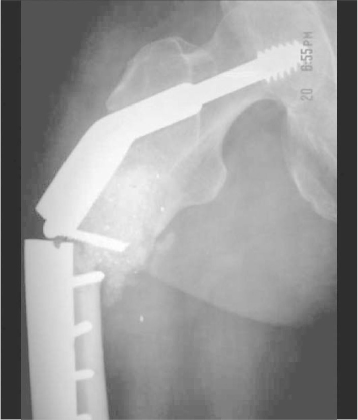
Postoperative complication characterized by breach of osteosynthesis and refracture after three weeks of surgery.
When comparing the type of surgical procedure, five (20.8%) patients undergoing arthroplasty and 11 (30.5%) undergoing osteosynthesis presented complications, a difference that is not significant (p = 0.404). Regarding mortality, two (8.3%) patients undergoing arthroplasty and three (8.3%) undergoing osteosynthesis died, a difference that is not significant (p = 1.000).
The average number of units of red blood cell replacement was 1.54 unit in the group undergoing arthroplasty and 1.44 unit in the osteosynthesis group, a difference that is not significant (p = 0.759). When the primary tumor was evaluated, tumors of a renal origin showed an increased risk of perioperative red blood cell replacement in comparison with other tumors (p = 0.0001).
As for the location of the lesion in the skeleton, there were no significant differences; surgical procedures performed on the lower limbs resulted in 16 (27.1 %) complications and were observed in one (20%) patient with upper limb injuries (p = 0.890). When evaluating each specific operative site, despite the trend toward greater frequency of complications in the subtrochanteric region, there was no significant difference (p = 0.079).
DISCUSSION
Orthopedic treatment aims at controlling pain, stabilizing impending or pathologic fractures, maintaining mobility, gait, and preventing the complications of prolonged recumbency1, 3, 4, 5.
Surgical treatment options include conventional prostheses, endoprostheses, simple or associated osteosynthesis with use of polymethylmethacrylate (bone cement) and, more rarely, amputation. The choice of procedure depends on the condition of the patient, the extent of disease, and the site involved1, 4, 5.
Postoperative complications are more frequent in these patients, who are usually debilitated, malnourished, and who have metabolic and hematological disorders1, 6.
Harrington et al.(2) analyzed 375 pathological fractures by BM and observed as complications: 13 infections, 11 thromboembolic complications, two cases of disseminated intravascular coagulation and one case of metastatic spread on the surgical site.
Park et al.(7) observed that in 58 patients undergoing surgical treatment of BM with arthroplasty infection occurred in 8.6% of cases and dislocation in 12.9% in the first two postoperative months.
Thai et al.(8), evaluating 96 BM surgeries of the humerus, found radial nerve palsy to be the most common complication, in 6% of cases, and as a clinical complication, pulmonary thromboembolism in two cases.
In our study we observed a relatively high incidence of complications (26.6%) compatible with most of the series studied3, 4, 5, 6, with surgical complications being the most common, especially hemorrhage. The occurrence of increased bleeding is due to hypervascularity, systemic effects of the tumor and of the chemotherapy and radiotherapy1, 5. The tumors of renal origin presented significant risk of hemorrhage and required more blood replacement than other tumors, which suggests the need for routine preoperative embolization.
Infections occurred in 4.8% of cases, which is consistent with other studies in the literature4, 5, 6, 7, 8, 9, 10. Jacofsky et al.(11), evaluating 42 cases of hip arthroplasty for the treatment of primary and metastatic malignant tumors, found deep infection to be the most common complication, with an incidence of almost 10% of the series.
Mechanical failure of the reconstruction was observed in one case of osteosynthesis of a subtrochanteric fracture. Because the proximal femur is a region of high mechanical stress, several authors have suggested that the reconstruction should be done with unconventional prostheses, avoiding the complications of osteosynthesis indications7, 10, 11, as seen in Figure 1.
From a clinical standpoint, the most common complication was thromboembolism, which occurred in 7.7% of cases, with one case of massive pulmonary embolism resulting in death in the immediate postoperative period. These findings are similar to those observed by Marco et al.(9), which reported deep vein thrombosis and superficial infections as being responsible for 67% of the early complications of surgical treatment of acetabular BM. Nathan et al.(12) suggest that pharmacological prophylaxis in these patients should be complemented by the use of mechanical methods due to the high risk of venous thrombosis.
In our series, complications were frequent and resulted in 35.2% deaths and 23.5% new surgical interventions, which suggests that they are usually more severe and with increased morbidity in these patients.
Most studies do not compare the early complications between the different orthopedic procedures8, 9, 10, and the evaluations are made from the point of view of the function and the late failure of the fixation method. Our study focused only early complications, in order to compare the magnitude of the surgery and its consequences in patients with metastatic disease. Unlike some studies that suggest a lower rate of complications with the use of arthroplasty when compared with osteosynthesis8, 9, 10, our study showed no difference in the incidence of complications, mortality, or the need for blood replacement, suggesting that both procedures are compatible in terms of perioperative complications.
CONCLUSION
The incidence of complications associated with the orthopedic treatment of BM was 26.6%, with 35.2% mortality; bleeding, infection, and thromboembolic events were the most frequent complications. The complications do not depend on the type of procedure or the area affected. The tumors of renal origin had a higher risk of bleeding and hemorrhage.
Footnotes
Study conducted at the Hospital das Clínicas, Universidade Federal de Minas Gerais (UFMG) and the Santa Casa de Belo Horizonte, Minas Gerais.
REFERENCES
- 1.Finn HA. General considerations. In: Simon MA, Springfield D, editors. Surgery for bone and soft tissue tumors. Lippincott-Raven; Philadelphia: 1998. pp. 609–613. [Google Scholar]
- 2.Harrington KD, Sim FH, Enis JE, Johnston JO, Diok HM, Gristina AG. Methylmethacrylate as an adjuvanct in internal fixation of pathological fractures: experience with three hundred and seventy-five cases. J Bone and Joint Surg Am. 1976;58(8):1047–1055. [PubMed] [Google Scholar]
- 3.British Orthopaedic Association and the British Orthopaedic Oncology Society. Metastatic bone disease: a guide to good practice. 2001 [Google Scholar]
- 4.Healey JH, Brown HK. Complications of bone metastases: surgical management. Cancer. 2000;88(12 Suppl):2940–2951. doi: 10.1002/1097-0142(20000615)88:12+<2940::aid-cncr10>3.0.co;2-w. [DOI] [PubMed] [Google Scholar]
- 5.Capanna R, Campanacci D. The treatment of metastases in the appendicular skeleton. J Bone Joint Surg Br. 2001;83(4):471–481. doi: 10.1302/0301-620x.83b4.12202. [DOI] [PubMed] [Google Scholar]
- 6.Bibbo C, Patel DV, Benevenia J. Perioperative considerations in patients with metastatic bone disease. Orthop Clin North Am. 2000;31(4):577–593. doi: 10.1016/s0030-5898(05)70177-2. [DOI] [PubMed] [Google Scholar]
- 7.Park DH, Jaiswal PK, Al-Hakin W, Aston WJS, Pollock RC, Skinner JA. The use of massive endoprosthesis for the treatment of bone metastases. Sarcoma. 2007;2007:62151. doi: 10.1155/2007/62151. [DOI] [PMC free article] [PubMed] [Google Scholar]
- 8.Thai DM, Kitagawa Y, Choong PFM. Outcome of surgical management of bony metastases to the humerus and shoulder girdle: a retrospective analysis of 93 patients. Intern Semin Surg Oncol. 2006;3:5. doi: 10.1186/1477-7800-3-5. http://www.isoonline.com/content/3/1/5 doi:10.1186/1477-7800-3-5. [DOI] [PMC free article] [PubMed] [Google Scholar]
- 9.Marco RA, Sheth DS, Boland PJ, Wunder JS, Siegel JA, Healey JH. Functional and oncological outcome of acetabular reconstruction for the treatment of metastatic disease. J Bone Joint Surg Am. 2000;82(5):642–651. doi: 10.2106/00004623-200005000-00005. [DOI] [PubMed] [Google Scholar]
- 10.Josin P, Dutka J. Clinical and radiological evaluation of mechanical sufficiency of the operative treatment of pathological fractures in bone metastases. Orthop Traumatol Rehabil. 2003;5(3):290–296. [PubMed] [Google Scholar]
- 11.Jacofsky DJ, Haidukewych GJ, Zhang H, Sim F. Complications and results of arthroplasty for salvage of failed treatment of malignant pathologic fractures of the hip. Clin Orthop Relat Res. 2004;(427):52–56. doi: 10.1097/01.blo.0000143572.96021.93. [DOI] [PubMed] [Google Scholar]
- 12.Nathan SS, Simmons KA, Lin PP, Hann LE, Morris CD, Athanasian EA. Proximal deep vein venous thrombosis after hip replacement for oncologic indications. J Bone Joint Surg Am. 2006;88(5):1066–1070. doi: 10.2106/JBJS.D.02926. [DOI] [PubMed] [Google Scholar]