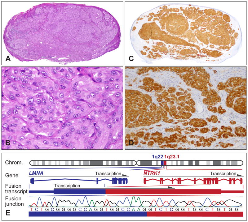
Fig. 6.
Spitz tumours with a NTRK1 kinase fusion. (A) Oval-shaped, compound melanocytic tumour with moderate epidermal hyperplasia from the upper arm of a 9-year-old girl. (B) Large epithelioid melanocytes with vesicular nuclei and prominent nucleoli, and moderate nuclear pleomorphism. (C,D) The neoplastic melanocytes are positive for NTRK1 immunohistochemistry and show cytoplasmic staining. (E) The LMNA–NTRK1 kinase fusion is caused by a 743 kb intrachromosomal deletion on chromosome 1q, joining the first two exons of LMNA with exon 11–17 of NTRK1. Sanger sequencing confirms the in-frame junction of the fusion transcript.

