Abstract
Cutaneous sporotrichosis, also known as “Rose Gardener's disease,” caused by dimorphic fungus Sporothrix schenkii, is usually characterized by indolent nodular or nodulo-ulcerative lesions arranged in a linear pattern. We report bizarre nonlinear presentation of Sporotrichosis, in an immunocompetent adult occurring after a visit to Amazon rain forest, speculating infection with more virulent species of Sporothrix. The diagnosis was reached with the help of periodic acid-Schiff positive yeast cells and cigar shaped bodies seen in skin biopsy along with the therapeutic response to potassium iodide.
Keywords: Herpetiform, potassium iodide, sporotrichosis
What was known?
Sporotrichosis is a subacute or chronic infection caused by Sporothrix schenckii.
It is characterized by indolent nodular or nodulo-ulcerative lesions arranged in a linear pattern. The de-novo presentation of ulcers in a bizarre nonlinear pattern with no prior nodules posed diagnostic difficulty in our patient.
Introduction
Sporotrichosis is a subacute or chronic infection caused by the saprophytic dimorphic fungus Sporothrix schenckii. Although only one species of Sporothrix was classically described, phenotypic and genetic studies have identified additional Sporothrix species. It is usually characterized by indolent nodular or nodulo-ulcerative lesions arranged in a linear pattern.
Case Report
A 54-year-old healthy male, presented with multiple, variably sized, nonhealing ulcers over his right lower leg since 3 months. It started as a single coin-sized ulcer over the shin, which rapidly increased in size with the simultaneous appearance of multiple new similar lesions and swelling over the right leg. The patient had received multiple courses of oral and intravenous antibiotics despite of which his disease was progressing. There was no history of apparent trauma or evidence of an immunocompromised state. History of excursion to Amazon rain forest 1-month prior to the onset of ulceration was present.
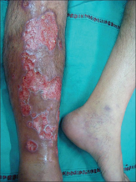
Cutaneous examination revealed irregularly shaped ulcers ranging in size from pinpoint to 9 cm × 5 cm, covered with seropurulent slough [Figure 1] distributed nonlinearly over the extensor aspect of the right leg. Few of them were herpetiform in morphology. The involved limb presented a cellulitic picture with diffuse erythema and induration. There was no evidence of any preceding nodulation. A single thickened lymphatic cord was palpable on the medial aspect of the thigh in the later course of his disease.
Figure 1.

Multiple irregular shaped ulcers distributed randomly over the leg
All the hematological and biochemical investigations were normal except for a raised erythrocyte sedimentation rate (45 mm/h). Magnetic resonance imaging lower leg showed findings consistent with cellulitis with no deep extension, abscess, osteomyelitis, or muscular involvement.
Histopathology and tissue culture samples were sent with a differential diagnosis of pyoderma gangrenosum, mycobacterial infection, cutaneous leishmaniasis, and deep fungal infection. Repeated cultures for the same were negative. Histopathology revealed multiple, poorly formed, epithelioid cell granulomas along with diffuse infiltrates of lympho-histiocytes, plasma cells, and neutrophils in the deeper dermis. Special stain (periodic acid-Schiff [PAS] and Gomori Methenamine silver stain) showed small budding and occasional elongated yeast cells in the dermis [Figure 2]. PAS stain from tissue imprints revealed cigar-shaped bodies suggesting Sprorothrix infection.
Figure 2.

Tissue section showing periodic acid-Schiff positive yeast forms with budding at places (high power view)
Patient was started on itraconazole capsules 200 mg twice daily monotherapy, with only marginal response. He developed multiple subcutaneous nodules and an ulcer over the right thigh even after 6 weeks of treatment. Thus, saturated solution of potassium iodide (SSKI) was added with complete clearance of the lesions in 10 weeks.
Discussion
A classical lymphocutaneous form of sporotrichosis presents with linearly arranged nodulo-ulcerative lesions, along a lymphatic tract.[1] This case presented unusually with multiple, irregular shaped, randomly distributed progressive ulceration, some with herpetiform morphology, not preceded by nodules. They had an inflamed, cellulitic background, unlike typical sporotrichosis. Various atypical forms of Sporotrichosis such as rosacea, acneiform, cellulitis, erysipeloids, and pyoderma gangrenosum have been documented from different countries.[2,3,4] Most of these reports were in immunocompromised patients, or in those wrongly treated with immunosuppressives. To the best of our knowledge, bizarre atypical presentation of sporotrichosis, in an immunocompetent is a rare presentation. The dissemination of infection in our patient, could best be explained by multiple traumatic implantations as also described by de Lima Barros et al.[5] It could also be attributed to more virulent species like Sporothrix brasiliensis which is known to cause atypical chronic disseminated cutaneous presentations. S. brasiliensis is the most virulent species in terms of mortality, tissue burden, and tissue damage.[6,7] Moreover, S. brasiliensis is the most common species in hyperendemic areas like Brazil, and our patient had a history of visit to the Amazon forest bordering Brazil 1-month before the onset of the ulcers. Hence, we can only postulate that S. brasiliensis could be the probable species for the atypical presentation in our patient. However, species identification could not be done due to culture negativity and lack of other resources.
Histopathology is usually nonspecific and variable. Demonstration of cigar shaped, oval to round or single budding forms of the yeast is diagnostic,[8] and isolation of the organism on cultures is confirmatory.[9] In the present case, the cultures were repeatedly negative. Diagnosis was reached with the help of histopathology, demonstration of cigar shaped bodies in tissue imprints and the clinical response to potassium iodide. A favorable response to SSKI is considered highly supportive of the disease especially in the absence of mycological support.[2,10]
Itraconazole was preferred over potassium iodide because of its lesser side effects and a much better outcome. As our patient was not showing any marked improvement with itraconazole alone even after 4–6 weeks, we added potassium iodide. This case highlights the efficacy of combining, itraconazole, and potassium iodide, in the management of widespread lesions of sporotrichosis.
Such unusual cases pose a diagnostic challenge and often lead to wrong management. This emphasizes that a high degree of clinical suspicion should always be kept for an unusual presentation of a not so unusual fungal disease, especially if a history of travel to the endemic area is present.
What is new?
Sporotrichosis can present as atypical nonlinear circumferentially arranged, herpetiform and bizarre shaped ulcers without prior nodular stage in an immunocompetent person
This could be attributed to virulent species of endemic areas like Sporothrix brasiliensis
Saturated solution of potassium iodide-less commonly used these days, still holds an important position in the treatment
High degree of clinical suspicion should always be kept for an unusual presentation of a not so unusual fungal disease, especially if a history of travel to the endemic area is present.
Footnotes
Source of Support: Nil
Conflict of Interest: Nil.
References
- 1.Vaishampayan S, Borde P. An unusual presentation of sporotrichosis. Indian J Dermatol. 2013;58:409. doi: 10.4103/0019-5154.117350. [DOI] [PMC free article] [PubMed] [Google Scholar]
- 2.Mahajan VK, Sharma NL, Sharma RC, Gupta ML, Garg G, Kanga AK. Cutaneous sporotrichosis in Himachal Pradesh, India. Mycoses. 2005;48:25–31. doi: 10.1111/j.1439-0507.2004.01058.x. [DOI] [PubMed] [Google Scholar]
- 3.Sharma NL, Mehta KI, Mahajan VK, Kanga AK, Sharma VC, Tegta GR. Cutaneous sporotrichosis of face: Polymorphism and reactivation after intralesional triamcinolone. Indian J Dermatol Venereol Leprol. 2007;73:188–90. doi: 10.4103/0378-6323.32745. [DOI] [PubMed] [Google Scholar]
- 4.Ravikumar BC, Kumar B. Fixed cutaneous sporotrichosis masquerading as lupus vulgaris. Indian J Dermatol. 1998;43:24–25. [Google Scholar]
- 5.de Lima Barros MB, de Oliveira Schubach A, Galhardo MC, Schubach TM, dos Reis RS, Conceição MJ, et al. Sporotrichosis with widespread cutaneous lesions: Report of 24 cases related to transmission by domestic cats in Rio de Janeiro, Brazil. Int J Dermatol. 2003;42:677–81. doi: 10.1046/j.1365-4362.2003.01813.x. [DOI] [PubMed] [Google Scholar]
- 6.Almeida-Paes R, de Oliveira MM, Freitas DF, do Valle AC, Zancopé-Oliveira RM, Gutierrez-Galhardo MC. Sporotrichosis in Rio de Janeiro, Brazil: Sporothrix brasiliensis is associated with atypical clinical presentations. PLoS Negl Trop Dis. 2014;8:e3094. doi: 10.1371/journal.pntd.0003094. [DOI] [PMC free article] [PubMed] [Google Scholar]
- 7.Freitas DF, Santos SS, Almeida-Paes R, de Oliveira MM, do Valle AC, Gutierrez-Galhardo MC, et al. Increase in virulence of Sporothrix brasiliensis over five years in a patient with chronic disseminated sporotrichosis. Virulence. 2015;6:112–20. doi: 10.1080/21505594.2015.1014274. [DOI] [PMC free article] [PubMed] [Google Scholar]
- 8.Kwon-Caung KJ, Bennett JE. Pennsylvania, USA: Lea and Febiger; 1992. Sporotrichosis. Medical Mycology; pp. 707–729. [Google Scholar]
- 9.Mahajan VK, Sharma NL, Shanker V, Gupta P, Mardi K. Cutaneous sporotrichosis: Unusual clinical presentations. Indian J Dermatol Venereol Leprol. 2010;76:276–80. doi: 10.4103/0378-6323.62974. [DOI] [PubMed] [Google Scholar]
- 10.Das S, Banerjee G, Biswas I. Fixed variant of cutaneous sporotrichosis: A rare entity in non-endemic belt. Indian J Dermatol. 2006;51:209–10. [Google Scholar]


