Abstract
The checkpoint inhibitor nivolumab is active in metastatic melanoma patients who have failed ipilimumab. In this phase I/II study, we assessed nivolumab's safety in 92 ipilimumab refractory patients with unresectable stage III or IV melanoma, including those who experienced grade 3-4 drug related toxicity to ipilimumab. We report long-term survival, response duration, and biomarkers in these patients after nivolumab treatment (3 mg/kg) every 2 weeks for 24 weeks, then every 12 weeks for up to 2 years, with or without a multipeptide vaccine. Response rate for ipilimumab-refractory patients was 30% (95%CI: 21% - 41%). Median duration of response was 14.6 months, median progression-free survival was 5.3 months, and median overall survival was 20.6 months, when followed up a median of 16 months. One and two year survivals were 68.4% and 31.2%, respectively. Ipilimumab-naïve and -refractory patients showed no significant difference in survival. The 21 patients with prior grade 3–4 toxicity to ipilimumab that was managed with steroids, tolerated nivolumab well, with 62% (95%CI: 38% - 82%) having complete or partial remissions or stabilized disease at 24 weeks. High numbers of myeloid-derived suppressor cells (MDSCs) were associated with poor survival. Thus, survival and long-term safety were excellent in ipilimumab-refractory patients treated with nivolumab. Prior grade 3-4 immune-related adverse effects from ipilimumab were not indicative of nivolumab toxicities, and patients had a high overall rate of remission or stability at 24 weeks. Prospectively evaluating MDSC numbers before treatment could help assess the expected benefit of nivolumab.
Keywords: antibody, suppressor cell, immunity, checkpoint, toxicity
Introduction
Nivolumab, an IgG4 human antibody that blocks the programmed death-1 (PD-1) receptor on T and B cells, has significant clinical activity in previously treated melanoma patients that are ipilimumab (anti–CTLA-4) naïve (1,2). In a phase III trial, anti–PD-1 had a higher objective response rate (ORR) and superior toxicity profile when compared with investigator-chosen chemotherapy, in patients that had progressed after treatment with ipilimumab alone or with a BRAF inhibitor (3). Those data supported the FDA approval of nivolumab in ipilimumab refractory melanoma in late 2014, but follow-up was short, and overall survival data were not mature at the time of publication.
A trial in treatment-naïve melanoma patients showed that nivolumab had superior survival compared to dacarbazine as front-line therapy, albeit with a median duration of follow-up of less than a year (4). Long-term follow-up studies in a phase I cohort of nivolumab-naïve, treatment refractory melanoma patients who had not been exposed to ipilimumab showed that 3 year overall survival (OS) was 41%, with a median survival of 22 months (5, 6). These data established the utility of nivolumab in ipilimumab-naïve and -refractory melanoma patients, but long-term toxicity, response duration, and overall survival have not been described in ipilimumab-refractory patients treated with nivolumab. It is still not clear how patients who had severe or dose-limiting (grades 3-4) immune-related adverse events (irAEs) from ipilimumab, would respond to nivolumab, because those patients have generally been excluded from trials of the PD-1 antibodies, such as nivolumab and pembrolizumab (7,8). Predictive markers for the utility of nivolumab and other PD-1/PD-L1 blocking antibodies have not been well defined, although in multiple studies in melanoma and other tumor histologies, programmed-death ligand-1 (PD-L1) staining of the tumor, stroma, or combinations of those two tissues appear to be associated with improved overall survival after treatment with PD-1 antibodies (9-12).
In this report we expand upon a previous report on the treatment of 90 patients with ipilimumab-naïve or -refractory melanoma with nivolumab with or without a peptide vaccine (13), and provide long-term duration of response and survival data from a cohort of 92 patients treated with nivolumab that had progressed after treatment with ipilimumab. We demonstrate that patients who finished a two and a half year regimen of nivolumab with a complete response (CR), partial response (PR), or stable disease do not progress after stopping therapy, and show that patients who had grades 3 or 4 immune related adverse events from ipilimumab and did not receive infliximab did not recapitulate the same toxicity when treated with nivolumab. The regimen of the current cohort included treatment with nivolumab at 3 mg/kg every other week for 24 weeks, but patients then received drug every 12 weeks for the next 96 weeks, a schedule that differs from other nivolumab phase II/III trials in which the drug was given every other week for at least 96 weeks, or until progression (3-6). Analysis of the pre-treatment peripheral blood in the current trial showed that higher levels of myeloid-derived suppressor cells (MDSCs) were associated with lower response rate, higher rates of progression of disease and shorter survival.
Materials and Methods
Patients
One hundred and twenty six patients were enrolled at Moffitt Cancer Center onto a trial approved by the University of South Florida Institutional Review Board (ClinicalTrials.gov identifier: NCT01176461), of which 92 had progressed after ipilimumab without response or stable disease and were deemed ipilimumab refractory; they are the principal subject of this report. Inclusion criteria included written informed consent; age 16 years or older; histologic diagnosis of unresectable stage III or IV melanoma with measurable disease by modified World Health Organization (mWHO) criteria; progressive disease after at least one previous systemic treatment; positive tumor staining in at least 10% of tumor cells for gp100, NY-ESO-1, and/or MART-1; Eastern Cooperative Oncology Group performance status of 0 or 1; and adequate hepatic, renal, and hematologic function. Patients were prescreened for HLA- A*0201 by allele-specific polymerase chain reaction for cohorts 1 to 5, whose patients also received a multi-peptide vaccine (supplemental table 1). Patients with treated brain metastases were allowed if they were radiolographically stable 8 weeks after treatment; patients with untreated brain metastases were allowed in cohort 6. Patients in cohorts 4-6 were required to start nivolumab 8 or more weeks after prior ipilimumab. Any number of prior therapies was allowed; treatment with prior anti–PD-1 or anti–PD-L1 was not. Patients in cohort 5 had grades 3 or 4 irAEs with ipilimumab but could not have had grade 4 colitis, nor could they have received infliximab. The 92 patients in cohorts 4-6 (ipilimumab refractory) all had progressive disease without responding to prior ipilimumab. Cohorts 4 and 5 were consecutively accrued and received nivolumab with peptide vaccine, and cohort 6 received nivolumab alone and accrued concurrently with cohorts 4 to 5; in cohorts 4 to 6, one patient withdrew consent but was included in the analysis for safety and efficacy, and was replaced per protocol. No patients were ineligible or lost to follow-up. The assignment of patients by cohort is shown in supplemental table 1
Study Design and Treatment
Nivolumab was provided by Bristol-Myers Squibb (Princeton, NJ). The gp100209-217 (210M; National Service Center [NSC] No. 683472) and MART- 126-35 (27L; NSC No. 709401) peptides were provided by the Cancer Therapy Evaluation Program of the National Cancer Institute. The good manufacturing practice grade gp100280-288 (288V; NSC No. 683473) and NY-ESO-1157- 165 (165V; NSC No. 717388) peptides were produced by Clinalfa (Zurich, Switzerland). All peptides were emulsified in Montanide ISA 51 VG (Seppic, Paris, France) and were included to assess the effects of PD-1 blockade on antigen-specific T-cell reactivity. The protocol was conducted under Investigational New Drug number BB 13704 with the Food and Drug Administration(13). Primary end points were toxicity and tolerability, and secondary endpoints were objective response rate, duration of response, progression-free and overall survival, as well as correlative immune assays.
Assessment of Response and Adverse Effects
Tumor assessments included chest, abdomen, and pelvis computed tomography (CT) and brain magnetic resonance imaging with contrast every 12 weeks. Objective response (CR and PR) was evaluated using mWHO and immune-related response criteria (14); immune-related response criteria were only used to determine whether patients should remain on treatment in case of a mixed response. Patients were assessed with history and physical examinations every 2 weeks for up to 24 weeks and then every 12 weeks thereafter. Leukapheresis was performed before treatment, at week 12, and at week 24 in cohorts 4 and 5, and 80 mL of peripheral blood was collected at the same time points from patients in cohort 6. Patients were discontinued from treatment for progression, dose-limiting nivolumab- or vaccine-related adverse events as defined in the Supplementary Materials and Methods, or upon withdrawal of consent.
Flow Cytometry Analysis for MDSC
Peripheral blood mononuclear cells were collected by leukapheresis as previously described and purified using Lymphoprep (Stemcell Technologies, Vancouver, BC) gradient, then frozen in liquid nitrogen prior to thawing and analysis (13). Phenotypic markers of MDSCs were evaluated by flow cytometry with a lineage-marker negative population gated to exclude CD3-, CD19-, or CD56-expressing cells using antibodies to CD11b, CD14, HLA-DR, and CD33 from BD Biosciences (San Jose, CA), except where indicated. The lineage-marker positive cells (CD3+, CD19+, and CD56+) highly expressed HLA-DR, which was used as a reference to set the HLA-DRlow gate which included the cells below the bottom edge of the clearly positive expression of that molecule from the lineage positive cells. Peripheral blood mononuclear cells were stained with Live/Dead violet dye (Invitrogen, Carlsbad, CA) to gate on live cells. Data were acquired on an LSR II flow cytometer (BD Biosciences) and analyzed with Flowjo software (TreeStar, Ashland, OR). Data were analyzed by the principal investigator, J.S.W., and biostatisticians X.Z. and Y.A.C.
Statistical Analysis
The primary objective of this study for cohorts 4-6 was to assess the safety and tolerability of nivolumab with or without a peptide vaccine in ipilimumab-refractory patients. The secondary objectives were to evaluate the ORR, PFS, OS, and changes in immunity. Toxicity rate was calculated by using the number of patients with grade 3 or greater toxicity divided by all patients. The ORR was estimated using the number of CRs and PRs at 24 weeks divided by the total number of patients treated. Patients were required to be observed for at least 24 weeks to be declared a confirmed PR, CR, or stable. The stable disease rate was estimated using the number of patients with stable disease (SD) for at least 24 weeks divided by the total number of patients. Progression-free survival rate was calculated as the sum of the ORR and stable disease rate. Duration of response is defined from first response to progression or last follow up for continuous responders. To visualize the response overall time, we plotted the response from on-treatment time in swimmer's plots. A Wilcoxon rank sum test was performed to determine whether the number of MDSCs before treatment (baseline) differed between those with a response and stable disease (responder + stable, R + S), and those who did not respond (nonresponder, NR), with all groups evaluated at 24 weeks. The Kaplan-Meier product-limit analysis and log-rank test were performed to investigate the association between overall survival and amounts of pretreatment monocytic–myeloid-derived suppressor cells (M-MDSCs). Proportions of M-MDSCs were dichotomized using the median as the cut point. Tumor change was calculated as sum of maximum tumor shrinkage and minimum tumor increase in size from baseline for each target lesion. Waterfall plots were used to visualize the maximum tumor reduction or minimum increase in size. An alpha level of .05 was used to declare statistical significance. The binomial CIs were calculated using the exact Clopper-Pearson method. We used SAS 9.2 (SAS Institute, Cary, NC) and Matlab 2015 (MathWorks, Natick, MA) for the statistical analyses.
Results
Baseline Patient Characteristics
Between August 2010 and December 2013, 152 patients were screened for all 6 cohorts, and 126 patients were enrolled. 92 patients were enrolled in cohorts 4-5-6 and all had progressed without response after receiving ipilimumab. All 92 patients were evaluated for toxicity and for response. Thirty-four patients in cohorts 1-2-3 have been previously described (13). In cohorts 4 and 5, 15 patients received nivolumab (3 mg/kg) with peptide vaccine; an additional 16 patients in cohort 5, and 61 patients in cohort 6 received nivolumab (3 mg/kg) alone. Three patients dropped out of cohort 6 for early progression of disease, and were replaced, but were evaluable for survival and toxicity. Median age was 60 years. Sixty-five percent of patients were male, and eighty-six percent (80/92) had American Joint Commission on Cancer M1c disease. Sixty patients (60/92 = 65%) received two or more prior therapies for metastatic disease. Eighty-five patients had primary cutaneous melanoma. Three patients had ocular melanoma, and four patients had an unknown primary. BRAF mutational status was known for 69 tumors, and 20 tumors (20/69 = 28%) were BRAF mutated. Four patients had experienced progression after a BRAF-targeted therapy before enrollment. Ten patients had radiated brain metastases, and six additional patients in cohort 6 had untreated brain metastases. Patient characteristics at trial entry are listed in Table 1.
Table 1.
Demographics: ipilimumab refractory cohorts 4-5-6
| Number | Percentage | Median | |
|---|---|---|---|
| Gender | |||
| Male | 60 | 65 | |
| Female | 32 | 35 | |
| Age | 60 | ||
| Prior regimens | 2 | ||
| Chemotherapy | 47 | 51 | |
| Immunotherapy (not ipilimumab) | 65 | 70 | |
| Interleukin-2 | 26 | 28 | |
| Targeted | 25 | 27 | |
| BRAF status | |||
| + | 20 | ||
| - | 49 | ||
| Unknown | 23 | ||
| Subtype | |||
| Cutaneous | 85 | ||
| Ocular | 3 | ||
| Unknown | 4 | ||
| Stage | |||
| IIIC | 4 | ||
| IVa | 4 | ||
| IVb | 4 | ||
| IVc | 80 | ||
| Brain metastases | 15 | 16 | - |
Safety
Treatment-related adverse events are listed in Table 2 by cohort. The most common adverse events were rash and pruritis, fatigue, arthralgias, and diarrhea across all cohorts. Most events were mild to moderate in severity and easily managed by supportive treatment. Dose-limiting grade 3-4 colitis was not seen in this trial. In the 92 patients in ipilimumab-refractory cohorts 4 to 6, one dose limiting toxicity (grade 3 rash) was observed in cohort 5 in a patient that had previously had grade 3 colitis with ipilimumab, that resolved completely with a 6-week prednisone taper from 60 mg. One episode of grade 3 pneumonitis was observed in a cohort 5 patient that had prior grade 4 transaminase elevation with ipilimumab, requiring prednisone tapers from 120 mg lasting 3 to 4 months for complete resolution, but which occurred after the DLT period of 12 weeks (at week 14). Both patients fully recovered to baseline without sequelae. One patient in cohort 5 had late onset grade 3 arthralgias that caused him to stop treatment at week 96, and one additional patient had late grade 3 rash after week 48 that did not require treatment discontinuation. An additional patient that had grade 4 necrotizing fasciitis after ipilimumab that required amputation had grade 2 rash with nivolumab. No other treatment–related grade 3 immune-related adverse events were seen in cohort 5. More grade 1 or 2 infusion reactions were observed in cohorts 4 to 6 (13 of 92 patients, 14%) than in cohorts 1 to 3 (one of 34 patients, 3%), although this was not statistically significant (P = 0.08). The overall toxicity profile for all 92 ipilimumab refractory patients in cohorts 4-5-6 by grades 1-2 and 3-4 for all three cohorts is shown in table 2. No patient discontinued nivolumab as a result of an infusion reaction in 92 ipiliumumab-refractory patients, and no treatment-related deaths were observed.
Table 2.
Drug-related toxicities for ipilimumab-refractory cohorts 4–5–6 (includes any irAEs or >5% of total)
| Cohort4 (n=10) | Cohort5 (n=21) | Cohort6 (n=61) | Cohort4+5+6 (n=92) | |||||
|---|---|---|---|---|---|---|---|---|
| Grades 1-2 | Grades 3-4 | Grades 1-2 | Grades 3-4 | Grades 1-2 | Grades 3-4 | Grades 1-2 | Grades 3-4 | |
| Adrenal insufficiency | 1 (5%) | 2 (3%) | 2 (3%) | 3 (3%) | 2 (2%) | |||
| Alanine aminotransferase increased | 2 (10%) | 4 (7%) | 6 (7%) | |||||
| Alkaline phosphatase increased | 4 (19%) | 3 (5%) | 7 (8%) | |||||
| Allergic reaction | 1 (5%) | 1 (2%) | 2 (2%) | |||||
| Anemia | 7 (70%) | 1 (10%) | 7 (11%) | 1 (2%) | 14 (15%) | 2 (2%) | ||
| Anorexia | 1 (10%) | 1 (5%) | 8 (13%) | 1 (2%) | 10 (11%) | 1 (1%) | ||
| Arthralgia | 8 (80%) | 9 (43%) | 1 (5%) | 11 (18%) | 28 (30%) | 1 (1%) | ||
| Aspartate aminotransferase increased | 2 (10%) | 3 (5%) | 5 (5%) | |||||
| Chills | 1 (10%) | 5 (24%) | 5 (8%) | 11 (12%) | ||||
| Colitis | 1 (10%) | 1 (1%) | ||||||
| Confusion | 1 (2%) | 1 (1%) | ||||||
| Constipation | 1 (10%) | 2 (10%) | 4 (7%) | 7 (8%) | ||||
| Cough | 3 (14%) | 1 (2%) | 4 (4%) | |||||
| Creatinine increased | 3 (5%) | 3 (3%) | ||||||
| Dehydration | 1 (10%) | 1 (5%) | 1 (2%) | 1 (2%) | 2 (2%) | 2 (2%) | ||
| Diarrhea | 5 (50%) | 14 (67%) | 20 (33%) | 39 (42%) | ||||
| Dry eye | 3 (5%) | 3 (3%) | ||||||
| Dry mouth | 1 (10%) | 2 (10%) | 4 (7%) | 7 (8%) | ||||
| Dry skin | 1 (5%) | 4 (7%) | 5 (5%) | |||||
| Dyspnea | 1 (10%) | 3 (14%) | 4 (7%) | 8 (9%) | ||||
| Endocrine disorders — Other, specify | 1 (10%) | 6 (29%) | 8 (13%) | 15 (16%) | ||||
| Erythema multiforme | 2 (3%) | 2 (2%) | ||||||
| Fatigue | 5 (50%) | 16 (76%) | 32 (52%) | 1 (2%) | 53 (58%) | 1 (1%) | ||
| Fever | 5 (24%) | 1 (5%) | 10 (16%) | 15 (16%) | 1 (1%) | |||
| Flu-like symptoms | 4 (19%) | 4 (4%) | ||||||
| Gastrointestinal disorders — Other, specify | 1 (5%) | 1 (2%) | 2 (2%) | |||||
| Generalized muscle weakness | 2 (10%) | 3 (5%) | 5 (5%) | |||||
| Hyperglycemia | 1 (5%) | 1 (5%) | 1 (1%) | 1 (1%) | ||||
| Headache | 4 (40%) | 3 (14%) | 8 (13%) | 15 (16%) | ||||
| Hyperhidrosis | 1 (10%) | 1 (5%) | 1 (2%) | 3 (3%) | ||||
| Hyperthyroidism | 3 (14%) | 3 (5%) | 6 (7%) | |||||
| Hyponatremia | 1 (5%) | 8 (13%) | 1 (2%) | 8 (9%) | 2 (2%) | |||
| Hypothyroidism | 2 (20%) | 4 (19%) | 5 (8%) | 11 (12%) | ||||
| Immune system disorders — Other, specify | 1 (10%) | 5 (24%) | 10 (16%) | 16 (17%) | ||||
| Infusion related reaction | 3 (30%) | 5 (24%) | 5 (8%) | 13 (14%) | ||||
| Injection-site reaction | 7 (70%) | 8 (38%) | 15 (16%) | |||||
| Lipase elevated | 1 (5%) | 1 (5%) | 1 (1%) | 1 (1%) | ||||
| Localized edema | 2 (10%) | 2 (3%) | 4 (4%) | |||||
| Lymphocyte count decreased | 7 (70%) | 1 (10%) | 1 (2%) | 1 (2%) | 8 (9%) | 2 (2%) | ||
| Mucositis oral | 1 (10%) | 2 (10%) | 1 (2%) | 4 (4%) | ||||
| Myalgia | 3 (14%) | 1 (2%) | 4 (4%) | |||||
| Nausea | 1 (10%) | 1 (10%) | 6 (29%) | 10 (16%) | 17 (18%) | 1 (1%) | ||
| Neutrophil count decreased | 8 (80%) | 2 (20%) | 2 (3%) | 10 (11%) | 2 (2%) | |||
| Pain | 1 (10%) | 2 (10%) | 1 (2%) | 4 (4%) | ||||
| Pain in extremity | 2 (10%) | 1 (2%) | 3 (3%) | |||||
| Pancreatitis | 1 (5%) | 1 (2%) | 2 (2%) | |||||
| Pneumonitis | 2 (10%) | 1 (5%) | 2 (2%) | 1 (1%) | ||||
| Platelet count decreased | 3 (30%) | 2 (20%) | 1 (2%) | 4 (4%) | 2 (2%) | |||
| Pruritus | 7 (70%) | 7 (33%) | 34 (56%) | 1 (2%) | 48 (52%) | 1 (1%) | ||
| Rash acneiform | 1 (2%) | 1 (1%) | ||||||
| Rash maculo-papular | 5(50%) | 14(67%) | 1 (5%) | 44 (72%) | 4 (7%) | 75 (82%) | 6 (7%) | |
| Skin & subcutaneous tissue disorders — Other, specify | 1 (10%) | 1 (10%) | 7 (33%) | 4 (7%) | 12 (13%) | 1 (1%) | ||
| Stomach pain | 3 (5%) | 3 (3%) | ||||||
| Vomiting | 1 (10%) | 2 (10%) | 5 (8%) | 7 (8%) | 1 (1%) | |||
| Weight loss | 2 (10%) | 3 (5%) | 5 (5%) | |||||
| White blood cell decreased | 1(10%) | 1 (10%) | 2 (10%) | 4 (7%) | 19 (21%) | 1 (1%) | ||
Clinical Activity
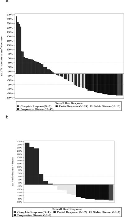
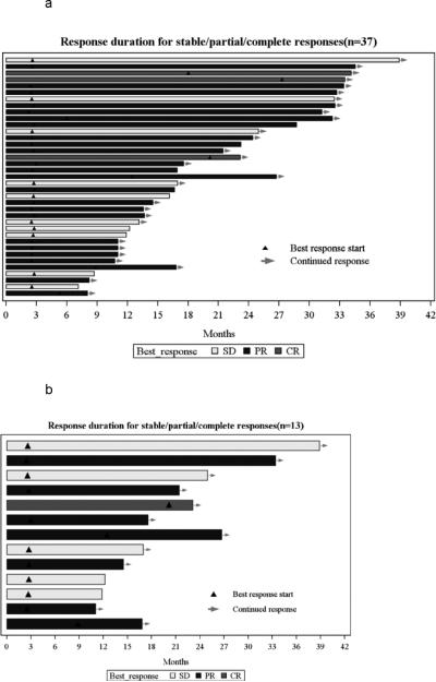
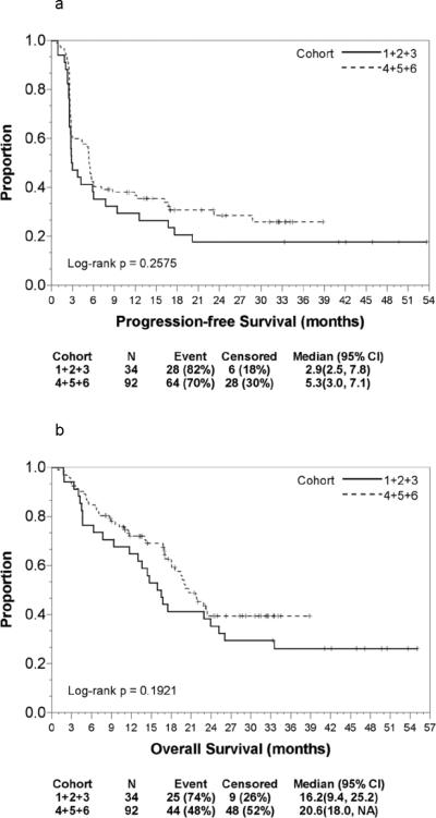
The confirmed objective response rate by mWHO criteria in 92 ipilimumab refractory patients that received nivolumab was 29%, with 3% CR and 26% PR. An additional 11% of patients had confirmed stable disease at week 24. The waterfall plot in Fig. 1A shows a transition point at 63%. For cohort 5 in Fig. 1B, the transition point was at 68%. Out of 92 patients, 10 patients progressed before week 24, and no post-treatment scans were obtained. Median of the maximum tumor change for all 82 patients (with pre- and post-scans) was shrinkage of 36%. The swimmer's plot in Fig. 2A shows that with a median follow-up of 16 months, 37/92 or 40% of patients had a complete or partial response or stable disease, of which 28/37 were ongoing as of the database lock on May 1, 2015, indicated by the arrow at the end of each line in ongoing patients. The median duration of response or stability (PR+CR+SD) in cohorts 4-5-6 was 14.3 months while the median duration of response (CR+PR) was 14.6 months (95% CI 2.8, 31.9). The median duration of SD was 12.0 months. Forty nine patients in the entire group of 126 treated ipilimumab-naïve or –refractory patients had stable disease, a complete or partial response confirmed at week 24, and 15 of those (15/49=30.6%) either discontinued nivolumab due to toxicity or continued on treatment and reached their final treatment date at 120 weeks; of those 15, none have progressed to date. Nine of those were in the ipilimumab-refractory cohorts 4-5-6. In the cohort of 21 patients in cohort 5 that had grade 3 or 4 toxicity to prior ipilimumab, there were 8 confirmed responders (1 complete and 7 partial) and 5 with stable disease, all confirmed at 24 weeks for a disease control rate of 62%. Only two of the 13 patients with disease control in cohort 5 have progressed. In addition to the 13 patients, three additional progressors are also alive. For the 16 patients who are still alive, the minimum, median, and maximum follow up times are 11 months, 20 months, and 38.9 months, respectively. The waterfall plot of those cohort 5 patients is shown in Fig. 1B, and the swimmer's plot is shown in Fig. 2B. Six patients with at least one previously untreated brain metastasis were treated on this trial in cohort 6, and there was one confirmed PR, one confirmed patient with stable disease, and four who progressed. For all 92 ipilimumab refractory patients in cohorts 4-6, with a median follow up of 16 months, median PFS was 5.3 months, and the median overall survival was 20.6 months, both shown in the Kaplan-Meyer plot for PFS in Fig. 3A, and for OS in Fig. 3B, respectively. One and two year survivals were 68.4% and 31.2%, respectively.
Figure 1.
Waterfall plots in 82 ipilimumab-refractory patients receiving nivolumab (3 mg/kg) with (14) or without (82) a peptide vaccine. Ten of the 92 patients in cohorts 4-5-6, and 2 of those are in cohort 5, progressed before week 12,which precluded collection of their post-treatment data.. A, Cohorts 4-5-6 patients that were refractory to ipilimumab and received nivolumab The transition point is noted by the arrow at 63%. B, Cohort 5's 19 patients who were refractory to ipilimumab, and who experienced grades 3-4 immune-related adverse events after treatment with ipilimumab
Figure 2.
Swimmer's plots for ipilimumab-refractory patients receiving nivolumab. Bar length indicates duration of stability or response. Triangles show time point when response or stable disease was achieved. Arrowheads indicate patients whose stable disease or response was ongoing at the time of data analysis. A, Patients (n = 37) in cohorts 4-5-6 that were stable, or had a partial or complete response at week 24. B, Patients (n = 13) in cohorts 5 that were stable, or had a partial or complete response; 11 were sustained at week 24.
Figure 3.
Kaplan-Meyer plot comparison of cohorts 1-2-3 to cohorts 4-5-6 of the 92 ipilimumab-refractory patients receiving nivolumab. A, Months of progression-free survival and. B, length of overall survival. P values determined by log-rank test. Data for each cohort displayed beneath each plot
Immune Biomarkers
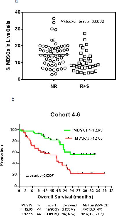
Myeloid-derived suppressor cells have been described as immature, myeloid derived cells that have immunoregulatory properties (15). In the cancer-bearing host MDSCs are diverted from normal differentiation pathways to become potent suppressors of innate and adaptive immunity. They are broadly grouped into granulocytic and monocytic categories. Monocytic MDSC were measured in frozen PBMCs that were thawed and then rested briefly, and subjected to flow cytometry analysis for a lineage negative CD11b+/CD14+/HLA DRlow population. MDSCs were measured as a proportion of total live cells within the total l blood mononuclear cells (PBMC). The gating strategy for MDSCs is shown in Supplemental Fig. S1. Figure 4A shows the association between ipilimumab refractory patients in cohorts 4-6 that received nivolumab, and had a complete or partial and response, or stable disease, at week 24 (responder + stable, R + S), and the proportion of MDSCs in peripheral blood, compared to those who did not respond (nonresponder, NR). The results indicate a significant association between response and stable disease (R + S) and fewer pretreatment MDSCs (P = 0.003). The association between survival and the proportion of MDSCs measured in the peripheral blood before treatment (Fig. 4B) shows that for 88 patients with available PBMCs in cohorts 4-6, the proportion of pretreatment M-MDSCs and overall survival were significantly inversely associated (P = 0.0007) with the proportion of MDSCs separated at the median value of 12.6%. There was also a significant association between the proportion of MDSCs in peripheral blood before treatment and median and progression-free survival (P = 0.002, data not shown). T-cell function could be suppressed by M-MDSCs (Supplemental Fig. S2).
Figure 4.

Myeloid-derived suppressor cells in ipilimumab-refractory and -naïve patients receiving nivolumab. A, Proportion of CD14+/CD11b+/HLA DRlow MDSC cells present before nivolumab treatment, as a percent of total live cells, in patients grouped as NR (nonresponders) and responders + stable patients (R+S). B, Kaplan-Meyer plot ofrelation of overall survival to proportion of CD14+/CD11b+/HLA DRlow MDSC cells before treatment. Cut-point was at the median (12.65 months). Red curve is survival for patients below the median, Green shows survival for those patients above the median. Data for groups displayed beneath plot.
Discussion
These data provide toxicity and survival information with the longest follow-up in nivolumab-treated patients that have progressed after prior ipilimumab. The results of this trial make a number of points important for patients with metastatic melanoma receiving immunotherapy. The PD-1 antibody nivolumab was well tolerated in ipilimumab naïve or refractory patients, and was also well tolerated in those who had prior dose-limiting toxicity to ipilimumab and did not require secondary immune suppression with infliximab. The duration of response for patients with confirmed partial or complete regression and stable disease in this trial was highly clinically meaningful, with a median duration of 14.6 months. Progression-free survival was 5.4 months in this group of 92 patients in cohorts 4-6, with median overall survival of 20.6 months. These data suggest that even after failing prior immunotherapy with another checkpoint protein inhibitor, responses of long duration may be seen with nivolumab, as has been observed in treatment-naïve melanoma and in other histologies. Fifteen patients in the overall trial cohort of 126 ipilimumab-naïve or -refractory patients completed two-and-a-half years of therapy, or stopped treatment due to toxicity, and had stable disease, a partial, or a complete response. None of those 15 patients has progressed to this date, including 9 in the ipilimumab-refractory group in cohorts 4-6. Minimum, median, and maximum follow-up time is 31.2, 33.5, and 53.7 months, respectively. All of the patients are still alive without progression.
Long-term toxicity data from this trial suggest that grade 2 toxicities like fatigue and arthralgias may linger in patients treated with nivolumab for over 2 years, and that some patients may develop persistent, cumulative, dose-limiting toxicity that is not of grade 3 or higher but may be debilitating and result in discontinuing therapy. Three patients in the current study stopped therapy due to unacceptable grade 2 fatigue or arthralgias. Prospective studies of the quality of life in future trials of nivolumab given alone or in combination will shed more light on this issue.
The encouraging short and long-term toxicity results from a cohort of 21 patients treated with nivolumab that had prior dose-limiting grades 3 and 4 irAEs, other than grade 4 colitis, with ipilimumab confirm that toxicities seen with one drug are not recapitulated with the other, and that the overall side effect profile with nivolumab is not worsened in such patients. Although no patients that received infliximab were treated on this trial, we are currently treating patients who had been administered prior infliximab for severe colitis in an additional expansion cohort. The response rate of 38% (8/21) in cohort 5 with 3 additional patients that were stable at week 24 provides intriguing preliminary data on the potential for increased benefit with nivolumab in those who have had irAEs with prior ipilimumab; we hope to further test this by treating additional patients in that cohort. Those data are consistent with results of toxicity analyses in nivolumab-treated patients, which suggested that clinical benefit from nivolumab may be associated with incidence of irAEs (16).
The regimen used in the current trial utilized every two-week nivolumab dosing for only 24 weeks, at which time the drug was then administered every 12 weeks for an additional 2 years, then discontinued. In contrast, many other trials of nivolumab or pembrolizumab used continuous dosing every 2 or 3 weeks until progression of disease, which are the recommended schedules in the package inserts for those two drugs (16-20). Nonetheless, the median survival in our cohort of 92 ipilimumab-refractory patients was equivalent to the data from a recent trial of second or later line nivolumab in similar populations treated until 96 weeks or unacceptable toxicity (6). Our encouraging data also demonstrate that no patient has progressed after stopping treatment, and/or completing the two and a half year regimen if stable, or in partial or complete remission. This raises the issue of how long to treat patients with PD-1 blocking antibodies once stability or response is achieved, and whether one may be able to shift to a maintenance regimen of drug every 12 weeks after an intensive induction regimen. These issues can only be resolved by the conduct of a randomized trial.
Limitations of this work include the fact that PD-1 inhibitors are increasingly being utilized as first-line treatment for metastatic melanoma due to a more favorable response rate and side effect profile compared to ipilimumab, so the future of ipilimumab as front line treatment may be limited.
The biomarker data from peripheral blood samples in this trial show that plentiful M-MDSCs (CD14+, CD11b+, and HLA DRlow) before treatment were associated with a lower likelihood of partial or complete response or stable disease, and poorer progression-free and overall survival, particularly for the 92 patients that were ipilimumab-refractory in cohorts 4-6. Monocytic–myeloid-derived suppressor cells have a variety of mechanisms by which they can alter T-cell responses in cancer, which may limit the clinical utility of PD-1 blockade (21). They can deplete nutrients, generate reactive oxygen species, interfere with lymphocyte trafficking and viability, and promote the function of regulatory T cells. The presence of MDSCs is associated with worse survival in melanoma (22-24). They may be prognostic in melanoma, or a potential predictive marker for treatment with ipilimumab (25-28). Data from this work indicate that M-MDSCs are associated with a poorer outcome with nivolumab. The number of M-MDSCs before treatment are also inversely associated with objective response to nivolumab (Fig. 4A) and express S100A9, phosphorylated STAT3, and arginase (22, 29). Treatment with nivolumab does not impact MDSC suppressive function at week 12 (data not shown), but the ability to modulate MDSC function might be of use in patients treated with nivolumab.
Taken together, these data suggest that in previously treated, ipilimumab-refractory patients, nivolumab demonstrated an excellent safety profile, a high response rate with excellent duration of response, and median survival similar to that seen in previously treated ipilimumab-naive patients. These data raise a number of questions, including whether checkpoint protein inhibitors that block PD-1 and CTLA-4 can be given sequentially to achieve a high rate of durable responses, or whether it is necessary to administer them concurrently, which has been shown in several trials to result in high response rates and excellent 1 and 2-year survival, albeit with high rates of toxicity and immune related adverse events (30-33). The results of this trial also call into question whether continuous treatment with nivolumab, given every other week until progression, is necessary to achieve long-lasting clinical benefit and raise the issue whether regimens with shorter or more intermittent exposure of drug are worthy of being tested in a prospective fashion.
Supplementary Material
Acknowledgements
We are grateful to Joyce Lampasona, Rasa Hamilton, and Amy Giordano who provided exemplary administrative support, to Kate Shapland and the Moffitt Flow Cytometry Core for their tireless dedication, and to Drs. James Mulé, David Feltquate, Ian Waxman, Mary Ruisi, and Arvin Yang who read and commented on the final article. We also wish to acknowledge the Biostatistics Core and the Flow Cytometry Core Facilities supported by Cancer Center Support Grant P30 CA076292-14 to the H Lee Moffitt Comprehensive Cancer Center and Research Institute and the Donald A Adam Comprehensive Melanoma Research Center.
This work was supported by grants to J.S. Weber and Y.A. Chen from the National Cancer Institute RO1 CA 129594-01A2 and from the Donald A. Adam Comprehensive Melanoma Research Center
Footnotes
Conflict of Interest: Jeffrey Weber and Geoffrey Gibney have accepted honoraria from BMS
References
- 1.Brahmer JR, Drake CG, Wollner I, Powderly J, Picus J, Sharfman WH, et al. Phase I study of single-agent anti-programmed death-1 (MDX-1106) in refractory solid tumors: Safety, clinical activity, pharmacodynamics, and immunologic correlates. J Clin Oncol. 2010;28:3167–3175. doi: 10.1200/JCO.2009.26.7609. [DOI] [PMC free article] [PubMed] [Google Scholar]
- 2.Topalian SL, Hodi FS, Brahmer JR, Gettinger SN, Smith DC, McDermott DF, et al. Safety, activity, and immune correlates of anti-PD-1 antibody in cancer. N Engl J Med. 2012;366:2443–2453. doi: 10.1056/NEJMoa1200690. [DOI] [PMC free article] [PubMed] [Google Scholar]
- 3.Weber JS, D'Angelo SP, Minor D, Hodi FS, Gutzmer R, Neyns B, et al. Nivolumab versus chemotherapy in patients with advanced melanoma who progressed after anti-CTLA-4 treatment (CheckMate 037): a randomised, controlled, open-label, phase 3 trial. The Lancet Oncol. 2015;16(4):375–84. doi: 10.1016/S1470-2045(15)70076-8. [DOI] [PubMed] [Google Scholar]
- 4.Robert C, Long GV, Brady B, Dutriaux C, Maio M, Mortier L, et al. Nivolumab in previously untreated melanoma without BRAF mutation. N Engl J Med. 2015;372(4):320–30. doi: 10.1056/NEJMoa1412082. [DOI] [PubMed] [Google Scholar]
- 5.Sznol M, Kluger HM, Hodi FS, et al. Survival and long-term follow-up of safety and response in patients (pts) with advanced melanoma (MEL) in a phase I trial of nivolumab (anti-PD-1; BMS-936558; ONO-4538). J Clin Oncol. 2013;31(suppl):549s. abstr CRA9006. [Google Scholar]
- 6.Topalian SL, Sznol M, McDermott DF, Kluger HM, Carvajal RD, Sharfman WH, et al. Survival, durable tumor remission, and long-term safety in patients with advanced melanoma receiving nivolumab. J Clin Oncol. 2014;32(10):1020–30. doi: 10.1200/JCO.2013.53.0105. [DOI] [PMC free article] [PubMed] [Google Scholar]
- 7.Hamid O, Robert C, Daud A, Hodi FS, Hwu WJ, Kefford R, et al. Safety and tumor responses with lambrolizumab (anti-PD-1) in melanoma. N Engl J Med. 2013;369:134–144. doi: 10.1056/NEJMoa1305133. [DOI] [PMC free article] [PubMed] [Google Scholar]
- 8.Patnaik A, Kang SP, Rasco D, Papadopoulos KP, Elassaiss-Schaap J, Beeram M, et al. Phase I Study of Pembrolizumab (MK-3475; Anti-PD-1 Monoclonal Antibody) in Patients With Advanced Solid Tumors. Clin Cancer Res. 2015 doi: 10.1158/1078-0432.CCR-14-2607. pii: clincanres.2607.2014. [Epub ahead of print] [DOI] [PubMed] [Google Scholar]
- 9.Taube JM, Anders RA, Young GD, Xu H, Sharma R, McMiller TL, et al. Co-localization of inflammatory response with B7-H1 expression in human melanocytic lesions supports an adaptive resistance mechanism of immune escape. Sci Transl Med. 2012;4:127ra37. doi: 10.1126/scitranslmed.3003689. [DOI] [PMC free article] [PubMed] [Google Scholar]
- 10.Taube JM, Klein A, Brahmer JR, Xu H, Pan X, Kim JH, et al. Association of PD-1, PD-1 ligands, and other features of the tumor immune microenvironment with response to anti-PD-1 therapy. Clin Cancer Res. Oct 1. 2014;20(19):5064–74. doi: 10.1158/1078-0432.CCR-13-3271. [DOI] [PMC free article] [PubMed] [Google Scholar]
- 11.Tumeh PC, Harview CL, Yearley JH, Shintaku IP, Taylor EJ, Robert L, et al. PD-1 blockade induces responses by inhibiting adaptive immune resistance. Nature. 2014;515(7528):568–71. doi: 10.1038/nature13954. [DOI] [PMC free article] [PubMed] [Google Scholar]
- 12.Taube JM, Young GD, McMiller TL, Chen S, Salas JT, Pritchard TS, et al. Differential expression of immune-regulatory genes associated with PD-L1 display in melanoma: implications for PD-1 pathway blockade. Clin Cancer Res. 2015 doi: 10.1158/1078-0432.CCR-15-0244. pii: clincanres.0244.2015. [Epub ahead of print. [DOI] [PMC free article] [PubMed] [Google Scholar]
- 13.Weber JS, Kudchadkar RR, Yu B, Gallenstein D, Horak CE, Inzunza HD, et al. Safety, efficacy, and biomarkers of nivolumab with vaccine in ipilimumab-refractory or - naive melanoma. J Clin Oncol. 2013;31(34):4311–4318. doi: 10.1200/JCO.2013.51.4802. [DOI] [PMC free article] [PubMed] [Google Scholar]
- 14.Wolchok JD, Hoos A, O'Day S, Weber JS, Hamid O, Lebbé C, et al. Guidelines for the evaluation of immune therapy activity in solid tumors: immune-related response criteria. Clin Cancer Res. 2009;15(23):7412–20. doi: 10.1158/1078-0432.CCR-09-1624. [DOI] [PubMed] [Google Scholar]
- 15.Gabrilovich DI, Ostrand-Rosenberg S, Bronte V. Coordinated Regulation of Myeloid cells by Tumours. Nat Reviews Immunology. 2012;12(4):253–258. doi: 10.1038/nri3175. [DOI] [PMC free article] [PubMed] [Google Scholar]
- 16.Freeman-Keller M, Weber JS. Nivolumab in resected and unresectable melanoma: Immune-related adverse events and association with survival outcomes. J Clin Oncol. 2015;33(suppl) doi: 10.1158/1078-0432.CCR-15-1136. abstr 9028. [DOI] [PMC free article] [PubMed] [Google Scholar]
- 17.Ansell SM, Lesokhin AM, Borrello I, Halwani A, Scott EC, Gutierrez M, et al. PD-1 blockade with nivolumab in relapsed or refractory Hodgkin's lymphoma. N Engl J Med. 2015;372(4):311–9. doi: 10.1056/NEJMoa1411087. [DOI] [PMC free article] [PubMed] [Google Scholar]
- 18.Gettinger SN, Horn L, Gandhi L, Spigel DR, Antonia SJ, Rizvi NA, et al. Overall Survival and Long-Term Safety of Nivolumab (Anti-Programmed Death 1 Antibody, BMS-936558, ONO-4538) in Patients With Previously Treated Advanced Non-Small- Cell Lung Cancer. J Clin Oncol. 2015 doi: 10.1200/JCO.2014.58.3708. pii: JCO.2014.58.3708. [DOI] [PMC free article] [PubMed] [Google Scholar]
- 19.McDermott DF, Drake CG, Sznol M, Choueiri TK, Powderly JD, Smith DC, et al. Survival, Durable Response, and Long-Term Safety in Patients With Previously Treated Advanced Renal Cell Carcinoma Receiving Nivolumab. J Clin Oncol. 2015 doi: 10.1200/JCO.2014.58.1041. pii: JCO.2014.58.1041 Epub ahead of print. [DOI] [PMC free article] [PubMed] [Google Scholar]
- 20.Garon EB, Rizvi NA, Hui R, Leighl N, Balmanoukian AS, Eder JP, et al. KEYNOTE-001 Investigators. Pembrolizumab for the Treatment of Non-Small-Cell Lung Cancer. N Engl J Med. 2015 Apr 19; doi: 10.1056/NEJMoa1501824. Epub ahead of print. [DOI] [PubMed] [Google Scholar]
- 21.Rizvi NA, Mazières J, Planchard D, Stinchcombe TE, Dy GK, Antonia SJ, et al. Activity and safety of nivolumab, an anti-PD-1 immune checkpoint inhibitor, for patients with advanced, refractory squamous non-small-cell lung cancer (CheckMate 063): a phase 2, single-arm trial. The Lancet Oncol. 2015;16(3):257–65. doi: 10.1016/S1470-2045(15)70054-9. [DOI] [PMC free article] [PubMed] [Google Scholar]
- 22.Poschke I, Mougiakakos D, Hansson J, Masucci GV, Kiessling R. Immature immunosuppressive CD14+HLA-DR-/low cells in melanoma patients are Stat3hi and overexpress CD80, CD83, and DC-sign. Cancer Res. 2010;70(11):4335–4520. doi: 10.1158/0008-5472.CAN-09-3767. [DOI] [PubMed] [Google Scholar]
- 23.Gros A, Turcotte S, Wunderlich JR, Ahmadzadeh M, Dudley ME, Rosenberg SA. Myeloid cells obtained from the blood but not from the tumor can suppress T-cell proliferation in patients with melanoma. Clin Cancer Res. 2012;18(19):5212–23. doi: 10.1158/1078-0432.CCR-12-1108. [DOI] [PMC free article] [PubMed] [Google Scholar]
- 24.Kitano S, Postow MA, Ziegler CG, Kuk D, Panageas KS, Cortez C, et al. Computational algorithm-driven evaluation of monocytic myeloid-derived suppressor cell frequency for prediction of clinical outcomes. Cancer Immunol Res. 2014;2(8):812–21. doi: 10.1158/2326-6066.CIR-14-0013. [DOI] [PMC free article] [PubMed] [Google Scholar]
- 25.Pico de Coaña Y, Poschke I, Gentilcore G, Mao Y, Nyström M, Hansson J, et al. Ipilimumab treatment results in an early decrease in the frequency of circulating granulocytic myeloid-derived suppressor cells as well as their Arginase1 production. Cancer Immunol Res. 2013;1(3):158–62. doi: 10.1158/2326-6066.CIR-13-0016. [DOI] [PubMed] [Google Scholar]
- 26.Tarhini AA, Edington H, Butterfield LH, Lin Y, Shuai Y, Tawbi H, et al. Immune monitoring of the circulation and the tumor microenvironment in patients with regionally advanced melanoma receiving neoadjuvant ipilimumab. PLoS One. 2014;9(2):e87705. doi: 10.1371/journal.pone.0087705. [DOI] [PMC free article] [PubMed] [Google Scholar]
- 27.Meyer C, Cagnon L, Costa-Nunes CM, Baumgaertner P, Montandon N, Leyvraz L, et al. Frequencies of circulating MDSC correlate with clinical outcome of melanoma patients treated with ipilimumab. Cancer Immunol Immunother. 2014;63(3):247–57. doi: 10.1007/s00262-013-1508-5. [DOI] [PMC free article] [PubMed] [Google Scholar]
- 28.Weide B, Martens A, Zelba H, Stutz C, Derhovanessian E, Di Giacomo AM, et al. Myeloid-derived suppressor cells predict survival of patients with advanced melanoma: comparison with regulatory T cells and NY-ESO-1- or melan-A-specific T cells. Clin Cancer Res. 2014;20(6):1601–9. doi: 10.1158/1078-0432.CCR-13-2508. [DOI] [PubMed] [Google Scholar]
- 29.Zhao F, Hoechst B, Dufy A, Gamrekelashvili J, Fioravanti S, Manns MP, et al. S100A9: a new marker for monocytic human myeloid derived suppressor cells Immunology. 2012;136(2):176–183. doi: 10.1111/j.1365-2567.2012.03566.x. [DOI] [PMC free article] [PubMed] [Google Scholar]
- 30.Wolchok JD, Kluger H, Callahan MK, Postow MA, Rizvi NA, Lesokhin AM, et al. Nivolumab plus ipilimumab in advanced melanoma. N Engl J Med. 2013;369:122–133. doi: 10.1056/NEJMoa1302369. [DOI] [PMC free article] [PubMed] [Google Scholar]
- 31.Postow MA, Chesney J, Pavlick AC, Robert C, Grossmann K, McDermott D, et al. Nivolumab and Ipilimumab versus Ipilimumab in Untreated Melanoma. N Engl J Med. 2015 Apr 20; doi: 10.1056/NEJMoa1414428. Epub ahead of print. [DOI] [PMC free article] [PubMed] [Google Scholar]
- 32.Larkin J, Chiarion-Sileni V, Gonzalez R, Grob J-J, Cowey L, Lao CD, et al. Combined Nivolumab and Ipilimumab or Monotherapy in Previously Untreated Melanoma. N Engl J Med. 2015 doi: 10.1056/NEJMoa1504030. [DOI] [PMC free article] [PubMed] [Google Scholar]
- 33.Lipson EJ, Sharfman WH, Drake CG, Wollner I, Taube JM, Anders RA, et al. Durable cancer regression off-treatment and effective reinduction therapy with an anti-PD-1 antibody. Clin Cancer Res. 2013;19(2):462–8. doi: 10.1158/1078-0432.CCR-12-2625. [DOI] [PMC free article] [PubMed] [Google Scholar]
Associated Data
This section collects any data citations, data availability statements, or supplementary materials included in this article.


