Figure.

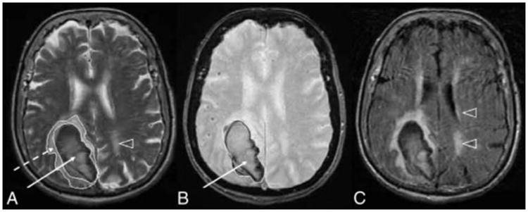
Examples of acute ICH and deep WMHs on MRI T2-weighted (A), GRE (B), and FLAIR (C) images in a patient with lobar ICH. Arrowheads point to WMHs; dashed arrow, to PHE; and solid arrow, to the hematoma.

Examples of acute ICH and deep WMHs on MRI T2-weighted (A), GRE (B), and FLAIR (C) images in a patient with lobar ICH. Arrowheads point to WMHs; dashed arrow, to PHE; and solid arrow, to the hematoma.