Abstract
Twist2 is a member of the basic helix-loop-helix (bHLH) family and plays a critical role in tumorigenesis. Growing evidence has proven that Twist2 is involved in tumor progression; however, the role of Twist2 in human kidney cancer and its underlying mechanisms remain unclear. Real-time polymerase chain reaction and Western blot analysis were used to detect the expression of Twist2 in kidney cancer cells and tissues. Cell proliferation, cell cycle, apoptosis, migration, and invasion assay were analyzed using the Cell Count Kit-8, flow cytometry, wound healing, and Transwell analysis, respectively. In this study, we showed that Twist2 was upregulated in human kidney cancer tissues compared with normal kidney tissues. Twist2 promoted cell proliferation, inhibited cell apoptosis, and augmented cell migration and invasion in human kidney-cancer-derived cells in vitro. Twist2 also promoted tumor growth in vivo. Moreover, we found that the knockdown of Twist2 decreased the levels of ITGA6 and CD44 expression. This result indicates that Twist2 may promote migration and invasion of kidney cancer cells by regulating ITGA6 and CD44 expression. Therefore, our data demonstrated that Twist2 is involved in kidney cancer progression. The identification of the role of Twist2 in the migration and invasion of kidney cancer provides a potential appropriate treatment for human kidney cancer.
Keywords: kidney cancer, Twist2, ITGA6, CD44
Introduction
Kidney cancer is one of the ten most common lethal cancers in adults, and approximately 27,000 patients are diagnosed with kidney cancer every year in the US.1–3 According to the morphology and immunohistochemistry of cancer cells, kidney cancer is divided into four types, which include clear-cell renal carcinoma (RCC), papillary renal carcinoma, chromophobe renal carcinoma, and renal oncocytoma.4 RCC is the most common type of adult kidney malignancy and constitutes about 80% of all primary renal neoplasms. Because of its resistance to conventional chemotherapy and radiation therapy, radical nephrectomy has been regarded as the only curative and standard treatment for RCC patients.5 However, a high risk of local or distant recurrence makes prognosis poor for RCC patients after surgical resection. Therefore, finding out the molecular mechanisms of cancer cell metastases from primary lesions is crucial for the improvement of treatment and prediction of prognosis.6
Twist2 belongs to the basic helix–loop–helix (bHLH) family that comprises a series of transcription factors controlling cell differentiation in different tissues during embryogenesis.7 The bHLH family members regulate gene expression by binding the gene’s promoters.8–10 The bHLH family members are divided into three classes: class A bHLH factors comprise E2-2, HEB, and E proteins; the tissue-restricted class B bHLH factors; and the inhibitory HLH proteins, constituted by the Id proteins, which lack DNA binding domain. Twist1, Twist2, Paraxis, Scleraxis, Hand1, and Hand2 form a subfamily of class B bHLH factors. Twist1 and Twist2 are highly homogeneous, suggesting that their functions might be redundant.8,11–13
Current evidence shows that some signaling pathways which are critical to early development also exhibit key regulatory effects on tumor biology, such as the Wnt, Notch, and Twist pathways.14,15 Identification of these signaling molecules, which play a physiological role in early developmental processes, provides an opportunity to define the biological cause for various tumor attributes16 and is a valuable chance for diagnostic or therapeutic implementation.
Twist2 is a highly conserved bHLH transcription factor that plays a critical role in embryogenesis and tumorigenesis.7,17,18 Twist2 is significantly overexpressed in various human solid tumors and contributes to tumor progression.19 Twist proteins inhibit differentiation of mesenchymal cell lineages, notably muscle and bone, during embryogenesis,20–22 and promote the migration of neural crest cells by facilitating epithelial-to-mesenchymal transitions (EMTs) during embryogenesis.23 Twist proteins induce EMT in cancer cells, which is the key step in the metastatic dissemination of cancer cells.19 Nevertheless, the function and the precise mechanism underlying the effects of Twist2 in human kidney cancer are less understood.
Materials and methods
Cell lines and patient samples
Kidney-cancer-derived cell lines (CaKi-1, CaKi-2, ACHN, and 786-0) and normal kidney cells (HK-2) were obtained from the Shanghai Cell Bank, Chinese Academy of Sciences (Shanghai, People’s Republic of China), and mycoplasma testing was performed as previously described.24 Cells were cultured in Dulbecco’s Modified Eagle’s Medium (DMEM) (Thermo Fisher Scientific, Inc, Rockford, IL, USA) supplemented with 10% fetal bovine serum (FBS) (Thermo Fisher Scientific), 100 units/mL penicillin (Thermo Fisher Scientific), and 100 μg/mL streptomycin (Thermo Fisher Scientific), and then incubated in a humidified atmosphere at 37°C with 5% CO2. A total of 21 pairs of kidney cancer tissues and their matched adjacent normal kidney tissues were obtained from patients who underwent surgery at Huadong Hospital. All tissues were immediately snap-frozen in liquid nitrogen and stored at −80°C until total RNA was extracted. The study protocol was approved by the Ethics Review Committee of the Institutional Review Board of Huadong Hospital, and written informed consents were obtained from all participants in this study.
Reverse transcription and real-time PCR
Total RNA was isolated using Trizol reagent (Thermo Fisher Scientific). Reverse transcription reactions were performed as previously described.25 Briefly, cDNA was reverse transcribed from RNA using a cDNA synthesis kit (Thermo Fisher Scientific, Inc, Rockford, IL, USA). The cDNA synthesis conditions were as follows: 37°C for 60 minutes, followed by 85°C for 5 minutes, and 4°C for 5 minutes. Real-time polymerase chain reaction (RT-PCR) was performed using a standard SYBR Green PCR kit (Takara Biotechnology Co, Ltd, Dalian, People’s Republic of China) and an ABI7500 (Applied Biosystem Life Technologies, Foster City, CA, USA) thermal cycler. The PCR cycling conditions were as follows: 95°C for 10 minutes, followed by 40 cycles at 95°C for 15 seconds and 60°C for 45 seconds, and a final extension step of 95°C for 15 seconds, 60°C for 1 minute, 95°C for 15 seconds, and 60°C for 15 seconds. The following primers (Sangon Biotech Co, Ltd, Shanghai, People’s Republic of China) were used: Twist2 (forward [F]: 5′-AGAAGTCGAGCGAAGATG-3′; reverse [R]: 5′-CCTGGTAGAGGAAGTCTATG-3′), ITGA6 (F: 5′-TGACATCCTCCAGATAGTG-3′; R: 5′-CCAATGGCATAGTCTTGTG-3′), CD44 (F: 5′-GGCAAGAAACCTGGGATTGG-3′; R: 5′-CCC GTGGTGTGGTTGAAATG-3′), and GAPDH (F: 5′-CACCCACTCCTCCACCTTTG-3′; R: 5′-CCACCACCCTGTTGCTGTAG-3′). The gene expression was calculated using the 2−∆∆Ct method.26 Relative quantification of the signals was performed by normalizing the gene signals with those of GAPDH.
Vector construction
pLKO.1-EGFP, pWPXL, VSV-G, and pol/gag (biosafety level, BSL-2) were purchased from Addgene (Cambridge, MA, USA). Plasmid containing the full length of Twist2 was purchased from Sangon Biotech Co, Ltd. Oligonucleotides encoding shRNA directed against human Twist2 (CAAGATCCAGACGCTCAAACT) and scramble shRNA were purchased from Sangon Biotech Co, Ltd. These oligonucleotides were annealed and digested using AgeI and EcoRI, and then were ligated into pLKO.1-EGFP, giving rise to a pLKO.1-EGFP-Twist2-shRNA construct. The full length of Twist2 was amplified using PCR. Then the PCR products were digested using BamHI and NdeI and cloned into pWPXL.
Lentiviral production and transduction
Twist2 and Twist-shRNA were delivered into 786-0 and ACHN cells by using the lentiviral transfection system. Briefly, 239T cells were seeded in 60 mm culture dishes, and after 24 hours, they were cotransfected with 2 μg of the plasmid vector, 1 μg pWPXL/pLKO.1-EGFP-scramble, 0.1 μg VSV-G, and 0.9 μg pol/gag by using Lipofectamine 2000 (Thermo Fisher Scientific), according to the manufacturer’s instructions. The recombinant lentivirus pLKO.1-EGFP-Twist2 (shTwist2) and pWPXL-Twist2 were collected 48 hours after transfection and used to infect 786-0 cells and ACHN cells at a multiplicity of infection (MOI) of 20 in the presence of 8 μg/mL polybrene (Sigma-Aldrich, St Louis, MO, USA), respectively. The recombinant lentivirus pLKO.1-EGFP-scramble shRNA (shNC) and black pWPXL were used as the negative control.
Western blot
Cultured or transfected cells were harvested and washed twice with phosphate-buffered saline and lysed in an ice-cold radioimmunoprecipitation assay buffer (RIPA; Beyotime, Shanghai, People’s Republic of China) with a freshly added 0.01% protease inhibitor cocktail (Sigma-Aldrich), and then incubated on ice for 30 minutes. Cells were lysed by centrifugation at 13,000 rcf for 10 minutes at 4°C, and the supernatant (20–30 μg of protein) was run on 10% sodium dodecyl sulfate–polyacrylamide gel electrophoresis and transferred electrophoretically to a polyvinylidene fluoride membrane (Sigma-Aldrich). The blots were blocked with 5% skim milk, followed by incubation with rabbit polyclonal antibody anti-ITGA6 (catalogue [cat] no ab75737; 1:50; Abcam, Cambridge, MA, USA), rabbit monoclonal antibody anti-Ki67 (cat no ab92742; 1:500; Abcam), rabbit monoclonal antibody anti-GAPDH (cat no 5174; 1:1,500, Cell Signaling Technology, Inc., Danvers, MA, USA), and mouse monoclonal antibody anti-CD44 (cat no 3570; 1:1,000, Cell Signaling Technology, Inc). Blots were then incubated with goat anti-mouse or anti-rabbit secondary antibody (cat no 0208; 1:1,000; Beyotime) and visualized using enhanced chemiluminescence (ECL; Thermo Fisher Scientific, Inc). GAPDH antibody was used as an internal control.
Cell proliferation assay
Cell viability was assessed by using a Cell Counting Kit (CCK)-8 (Tongren, Shanghai, People’s Republic of China). Briefly, 4×10³ of shTwist2- or pWPXL-Twist2-transfected 786-0 and ACHN cells were seeded in each 96-well plate, and further incubated for 0, 12, 24, 48, and 72 hours, respectively. At the indicated time points, CCK-8 solution (10 μL in 100 μL DMEM) was added to each well and incubated for 1 hour at 37°C. The optical density 450 nm value in each well was determined by a microplate reader (SM600 Labsystem; Shanghai Utrao Medical Instrument Co, Ltd, Shanghai, People’s Republic of China). Experiments were repeated at least three times, each time in triplicate.
Cell cycle assay
After 48 hours of transfection, cells were harvested and cell cycle distribution was analyzed using propidium iodide (PI; Sigma-Aldrich) staining on a flow cytometer (BD Accuri C6, software version 1.0.264.21; BD Biosciences, Franklin Lakes, NJ, USA). Briefly, 4×10³ of shTwist2- or pWPXL-Twist2-transfected 786-0 and ACHN cells were collected and fixed in 70% ethanol at −20°C overnight. The cells were subsequently resuspended in a staining solution, containing 20 μg/mL PI and 200 μg/mL RNase A. Experiments were repeated at least three times, each time in triplicate.
Cell apoptosis assay
After 48 hours of transfection, cells were harvested and cell apoptosis analyzed using Annexin V/propidium iodide (PI) staining (BD Biosciences) and flow cytometric analysis. Briefly, 4×10³ shTwist2- or pWPXL-Twist2-transfected 786-0 and ACHN cells were collected and incubated with annexin V-fluorescein isothiocyanate (FITC) and PI, prior to analysis by flow cytometry. Experiments were repeated at least three times, each time in triplicate.
Wound healing assay
shTwist2- or pWPXL-Twist2-transfected 786-0 and ACHN cells were seeded in 35 mm tissue culture dishes at a density of 8×10⁵ and further seeded until they reached 100% confluence. Then the confluent cultures were scratched using a pipette tip. After scratching, the well was gently washed twice with medium to remove the detached cells. Scratched cultures were photographed under a microscope at 0, 12, and 24 hours. Migration of cells was established by measuring the width of the scratched area at each time point in the scratched area at a magnification of ×200. Experiments were repeated at least three times, each time in triplicate.
Invasion assay
The upper well of the Transwell (Sigma-Aldrich Co., St Louis, MO, USA) was coated with Matrigel (BD Biosciences) at 37°C in a 5% CO2 incubator for 1 hour. The cells were serum-starved for 24 hours, and subsequently, 1×10⁵ of shTwist2- or pWPXL-Twist2-transfected 786-0 and ACHN cells in 500 μL DMEM were seeded into the upper wells of the precoated Transwell chamber. Cell culture medium, supplemented with 10% FBS, was added into the lower well of the chamber. After 48 hours of incubation, the cells on the upper well and the membranes coated with Matrigel were swabbed with a Q-tip. The cells that migrated into the lower well were fixed with methanol and stained with 0.5% methylrosanilinium chloride solution for 30 minutes. The cells were counted at a magnification of ×200, and the mean number of cells was recorded. Experiments were repeated at least three times, each time in triplicate.
Animal experiments
Care of the laboratory animals and animal experimentation were performed in accordance with animal ethics guidelines and approved protocols. All animal studies were approved by the Animal Ethics Committee of Huadong Hospital. For tumor growth assay, 786-0 cells transfected with shNC or shTwist2 were trypsinized, and then washed and resuspended in DMEM without FBS. Cell concentration and viability were determined using trypan blue. Then, a total of 2×10⁶ 786-0 cells transfected with shNC or shTwist2 were injected subcutaneously into the left armpit of nude mice (5-week-old, six per group). The tumor size was determined every 3–4 days after tumor was formed (around 1–2 weeks) as previously described.27 Tumor volume was measured and calculated using the following formula: V (mm3) =0.5× larger diameter × smaller diameter2. At 46 days after injection, the animals were euthanized and the tumors were weighed on a digital balance. Experiments were repeated at least three times, each time in triplicate.
Statistical analysis
Experimental data were presented as mean ± standard deviation. All assays were performed in triplicate and repeated at least three times. All statistical analyses were performed using GraphPad Prism 5 software (GraphPad Software, La Jolla, CA, USA). Statistical significance was determined by the paired, two-tailed Student’s t-test and one-way analysis of variance. Differences were considered significant at values of P<0.05.
Results
Twist2 is upregulated in kidney cancer cell lines and tissues
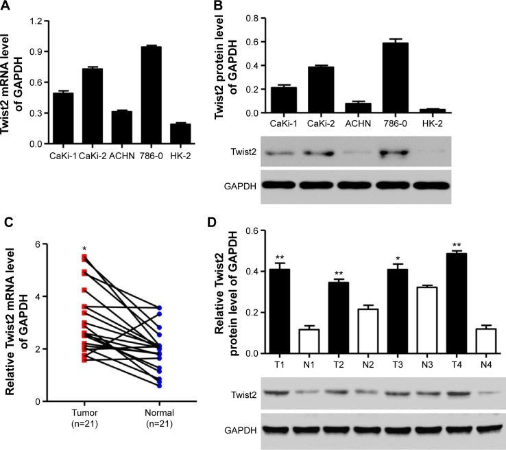
The expression of Twist2 was examined in four kidney cancer cell lines (CaKi-1, CaKi-2, ACHN, and 786-0) and normal kidney cells (HK-2). RT-PCR analysis showed that higher levels of Twist2 expression were found in the four kidney cancer cell lines compared with HK-2 cells, with the highest Twist2 mRNA expression found in 786-0 cells and the lowest Twist2 mRNA expression found in ACHN cells (Figure 1A and B). These two cell lines were therefore used for subsequent experiments. The mRNA expression level of Twist2 was significantly upregulated in kidney cancer tissues when compared with corresponding normal tissues (Figure 1C). Similar Twist2 protein levels were also found in randomly selected four paired normal and kidney cancer tissues (Figure 1D), and the protein levels in other paired normal and kidney cancer tissues are shown in Figure S1. Taken together, these results support the notion that Twist2 may act as an oncogene in kidney cancer.
Figure 1.
Twist2 is upregulated in kidney cancer cell lines and tissues.
Notes: (A, B) The expression of Twist2 in the normal human kidney cell line and four human kidney cancer cell lines was assessed by RT-PCR and Western blot. (C) mRNA expression of Twist2 in 21 paired primary kidney cancer tissues (Tumor) and their corresponding normal tissues (Normal) assessed by RT-PCR. (D) Expression of Twist2 in four primary kidney cancer tissues and their corresponding normal tissues measured by Western blot. *P<0.01, **P<0.001 compared with corresponding normal controls.
Abbreviations: N, normal tissues; T, tumor tissues; RT-PCR, real-time polymerase chain reaction.
Effect of Twist2 on kidney-cancer-derived cell proliferation
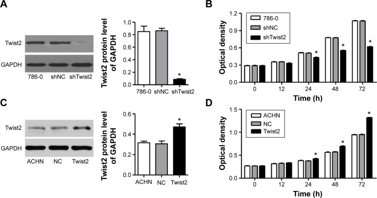
To explore the biological significance of Twist2 in kidney cancer tumorigenesis, we established Twist2 knockdown cell lines of 786-0 cells with a stabilized lentivirus infection. Decreased expression of Twist2 was confirmed by Western blot at 48 hours after transfection (Figure 2A). Forty-eight hours after transfection, cell proliferation was analyzed through a CCK-8 assay. We found that knockdown of Twist2 significantly decreased the proliferation of 786-0 cells compared with the corresponding shNC group (Figure 2B). However, overexpression of Twist2 in ACHN cells showed significant increases in Twist2 protein level (Figure 2C) and cell proliferation (Figure 2D) compared with the ACHN cells without Twist2 overexpression.
Figure 2.
Effect of Twist2 on kidney cancer cell proliferation.
Notes: (A) Successful knockdown of Twist2 was confirmed by Western blot at 48 hours after infection with shTwist2 or shNC control lentivirus. (B) 786-0 cell proliferation was detected using CKK-8 assay at 0, 12, 24, 48, and 72 hours. (C) Successful overexpression of Twist2 was confirmed by Western blot at 48 hours after infection with pWPXL-Twist2 or black pWPXL (NC) control lentivirus. (D) ACHN cell proliferation was detected using CCK-8 assay at 0, 12, 24, 48, and 72 hours. *P<0.001 compared with shNC or NC.
Abbreviations: shNC, recombinant lentivirus pLKO.1-EGFP-scramble shRNA; NC, black pWPXL; CCK, Cell Counting Kit; shTwist2, recombinant lentivirus pLKO.1-EGFP-Twist2.
Effect of Twist2 on kidney-cancer-derived cell cycle and cell apoptosis
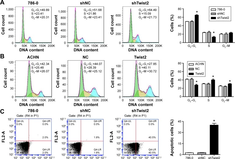
To further validate the effect of Twist2 on cell proliferation, the cell cycle was analyzed in 786-0 cells (Figure 3A). Cell cycle analysis showed that the knockdown of Twist2 through RNAi by Twist2 shRNA notably increased the number of cells in G0–G1 phase and reduced S phase cell population in 786-0 cell lines. However, overexpression of Twist2 in ACHN cells showed a significantly decreased rate of G0–G1 phase cells, and increased S and G2–M phase cell populations (Figure 3B), compared with the ACHN cells without Twist2 overexpression.
Figure 3.
Effect of Twist2 on kidney cancer cell cycle and apoptosis.
Notes: (A, B) 786-0 and ACHN cell cycle profiles were analyzed using flow cytometry. (C, D) 786-0 and ACHN cells were stained with annexin V-fluorescein, and apoptotic rate was analyzed using flow cytometry. *P<0.001 compared with shNC or NC.
Abbreviations: shNC, recombinant lentivirus pLKO.1-EGFP-scramble shRNA; NC, black pWPXL; shTwist2, recombinant lentivirus pLKO.1-EGFP-Twist2.
Then, we evaluated the apoptotic function of Twist2 in 786-0 cells by using an Annexin V–FITC/PI staining assay. As shown in Figure 3C, flow cytometry analysis revealed that the reduction of Twist2 in 786-0 cells significantly induced cell apoptosis by 16-fold compared with the corresponding shNC group. However, overexpression of Twist2 in ACHN cells significantly decreased the apoptotic rate by 55.97% (Figure 3D) compared with the ACHN cells without Twist2 overexpression. Taken together, these data suggest that Twist2 promotes cell proliferation and cell cycle, and suppresses apoptosis of kidney cancer cells in vitro.
Effect of Twist2 on kidney-cancer-derived cell migration and invasion
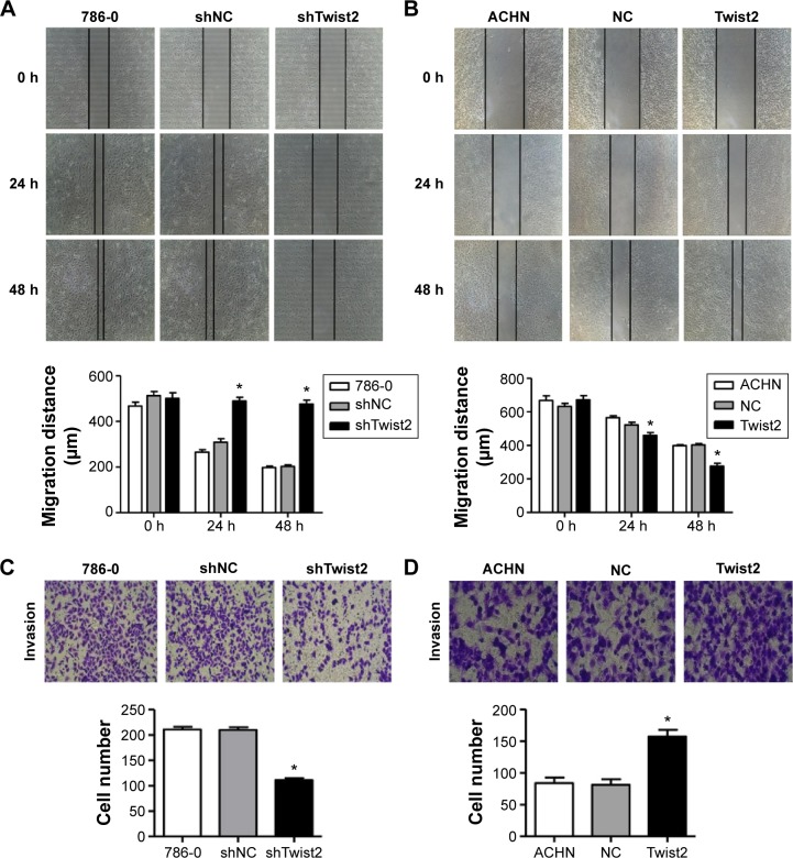
A previous study showed that Twist2 induced cancer cell EMT, which is the first step for metastasis in a cancer cell.28 To investigate the migration-promoting function of Twist2 in kidney-cancer-derived cells, the migration capacity of 786-0 cells was evaluated by a wound healing assay. Knockdown of Twist2 in 786-0 cells significantly reduced the cell migration ability in a time-dependent manner compared with the corresponding shNC group (Figure 4A). However, overexpression of Twist2 in ACHN cells showed significantly increased cell migration (Figure 4B) compared with the ACHN cells without Twist2 overexpression. Also, the Transwell assay showed that the knockdown of Twist2 in 786-0 cells also significantly reduced cell invasion, compared with the corresponding shNC group (Figure 4C). However, overexpression of Twist2 in ACHN cells showed significantly increased cell invasion (Figure 4D), compared with the ACHN cells without Twist2 overexpression. Taken together, these findings demonstrate that Twist2 promotes kidney cancer cell migration and invasion in vitro.
Figure 4.
Effect of Twist2 on kidney cancer cell migration and invasion.
Notes: (A, B) The migration of 786-0 and ACHN cells was performed in in vitro scratch wound healing assay, and photographs were taken 0, 24, and 48 hours after the wound was made. (C, D) The invasion of 786-0 and ACHN cells was performed by Transwell assay, and photographs were taken at 48 hours after incubation in Matrigel precoated Transwell chamber (× 200). *P<0.001 compared with shNC or NC.
Abbreviations: shNC, recombinant lentivirus pLKO.1-EGFP-scramble shRNA; NC, black pWPXL; shTwist2, recombinant lentivirus pLKO.1-EGFP-Twist2.
ITGA6 and CD44 were regulated by Twist2
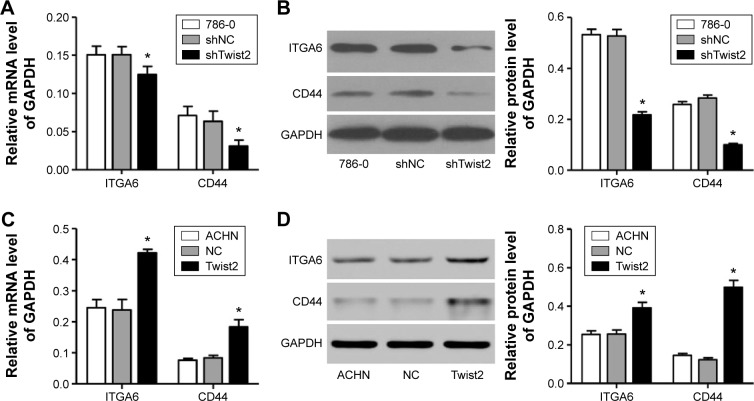
Two core proteins, ITGA6 and CD44, in the extracellular matrix (ECM)-receptor interaction pathway were predicted to be Twist2 targets. To experimentally validate Twist2 regulation of these genes, we decreased the expression of Twist2 in 786-0 cells, and then we detected the mRNA levels of the target genes by RT-PCR. As shown in Figure 5A, the mRNA levels of ITGA6 and CD44 in 786-0 cells were significantly suppressed by Twist2 shRNA, compared with the corresponding shNC group. The same results were found when we performed Western blotting to detect the protein levels of these Twist2 potential targets in the aforementioned cancer cell lines that were transfected with Twist2 shRNA, compared with the corresponding shNC group (Figure 5B). However, overexpression of Twist2 in ACHN cells showed significantly increased expression of ITGA6 and CD44 at both mRNA (Figure 5C) and protein levels (Figure 5D), compared with the ACHN cells without Twist2 overexpression. These results demonstrated that Twist2 promotes cell proliferation, migration, and invasion and inhibits cell apoptosis possibly by regulating the expression of ITGA6 and CD44 in kidney cancer cells.
Figure 5.
Effect of Twist2 on the expression of ITGA6 and CD44.
Notes: The mRNA and protein levels of ITGA6 and CD44 were detected by RT-PCR and Western blot in 786-0 (A, B) and ACHN cells (C, D). *P<0.001 compared with shNC or NC.
Abbreviations: RT-PCR, real-time polymerase chain reaction; shNC, recombinant lentivirus pLKO.1-EGFP-scramble shRNA; NC, black pWPXL; shTwist2, recombinant lentivirus pLKO.1-EGFP-Twist2.
Effect of Twist2 on tumor growth of kidney-cancer-derived cells in nude mice
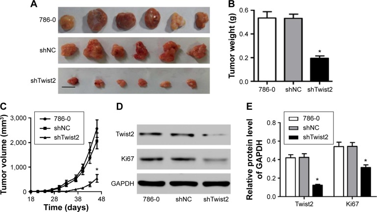
To further examine the effects of Twist2 on in vivo tumor growth, 786-0 cells stably expressing shTwist2 or shNC were injected subcutaneously into the left armpit of nude mice, and tumor size was measured every 3–4 days after a tumor formed. As shown in Figure 6A, at 46 days, tumors of mice with shTwist2 injection were smaller in size than those seen in control and shNC-injected mice. The average tumor weight was also significantly decreased in shTwist2-injected mice, compared with the shNC-injected mice (Figure 6B). Tumor volumes were measured and are shown in Figure 6C. The tumors of shTwist2-injected mice grew slower, whereas tumors of shNC-injected mice grew faster. Moreover, shTwist2-injected mice also showed decreased expression of Twist2 as well as Ki67, suggesting the proliferation inhibitory effect of Twist2 knockdown (Figure 6D and E). Taken together, these data confirm that Twist2 knockdown suppresses the tumorigenesis of kidney-cancer-derived cells in vivo.
Figure 6.
Effect of Twist2 on tumor growth in vivo.
Notes: (A) 786-0 cells transfected with shNC or shTwist2 were subcutaneously injected in athymic nude mice. Tumor growth was shown 46 days after injection (n=6). (B) Tumor weight in shTwist2-treated mice was significantly decreased compared with that in control and shNC mice. (C) shTwist2 decreased tumor growth in mice compared with that in control and shNC mice. (D, E) Expression of Twist2 and Ki67 was detected by Western blot at 46 days after shNC or shTwist2 injection. Scale bars: 10 mm. *P<0.001 compared with shNC.
Abbreviations: shNC, recombinant lentivirus pLKO.1-EGFP-scramble shRNA; NC, black pWPXL; shTwist2, recombinant lentivirus pLKO.1-EGFP-Twist2.
Discussion
Currently, radical nephrectomy is a curative and standard treatment in kidney cancer patients. However, there is a poor prognosis, because about 30% of patients have distant metastases after surgery.6 Therefore, the identification of the molecular mechanisms of kidney cancer development and metastases would provide an opportunity to improve treatment and offer a chance to get a better prognosis.
Twist2 is a member of the bHLH family and plays a critical role in embryogenesis and tumorigenesis. Twist2 is overexpressed in a large number of cancer tissues and cancer-derived cell lines, including in kidney cancer. In this study, Twist2 expression was significantly upregulated in tumor tissues at both mRNA and protein levels. Silencing of Twist2 significantly reduced the expression levels of Twist2 in 786-0 cells, and overexpression of Twist2 significantly increased the expression levels of Twist2 in ACHN cells.
Twist2 inhibits premature senescence of cancer cells and simultaneously promotes EMT, which favors invasion and metastasis of cancer.19,29 In further analysis, we demonstrated that the silencing of Twist2 markedly inhibited the variability of 786-0 cells, while the overexpression of Twist2 significantly increased the variability of ACHN cells, and these were a consequence of the induction of apoptosis, as evidenced by flow cytometry analysis. The data showed that the fundamental event that occurred when 786-0 cells were treated with Twist2 shRNA is a marked decrease in the level of cell proliferation and arrest in G0–G1 phase. In contrast, ACHN cells with Twist2 overexpression revealed the promotion of cell cycle progression and a decrease in cell apoptosis.
Migration and invasion are two of the most important marks of cancer and function as lethal factors for malignant cancer in general and human kidney cancer in particular.30–32 Management of migration and invasion will therefore contribute to the improvement of the prognosis for the human kidney cancer patient. In cultured 786-0 cells, where Twist2 had marked expression, silencing of Twist2 significantly inhibited cell invasion and migration activities, compared with the shNC groups. Interestingly, in cultured ACHN cells where Twist2 had slight expression, overexpression of Twist2 significantly promoted cell invasion and migration activities, compared with the control groups. The strong correlation between Twist2 expression and kidney cancer cell invasion and migration highlights the potential value of Twist2 as a novel biomarker for kidney progression.
Despite improvement in determining the molecular mechanisms of liver cancer tumorigenesis, the specific signal transduction pathways involved have not been fully characterized. The ITGA6 protein belongs to the integrin family, which participates in cell adhesion as well as cell-surface-mediated signaling between the ECM and the cell. It has been reported that ITGA6 promotes cell migration during embryonic development and tumorigenesis.33 CD44 has been reported as one of the most commonly used cancer stem cell (CSC) markers and has prognostic value for patients with cancer in various solid tumors, including colon, lung, and breast cancer.34–36 Moreover, CD44 is found to promote migration and proliferation in gastric cancer cells.37,38 In agreement with these results, our study revealed that Twist2 positively regulated the expression of ITGA6 and CD44 at both mRNA and protein levels.
Further, the proliferative effect of Twist2 was supported by our xenograft experiments in vivo. Nude mice injected with Twist2 shRNA-expressing 786-0 cells showed tumor growth reduction when compared with the NC shRNA-expressing cells over a 46-day time period. Similar to our results that Twist2 promotes tumor growth in vivo, nude mice injected with Twist2-expressed HepG2 cells showed an increase in tumor growth when compared with control.39 Collectively, these results indicate the first demonstration of its crucial function in kidney cancer development.
In conclusion, this study showed that Twist2 was overexpressed in kidney cancer tissues and cell lines, and Twist2 significantly promoted cell proliferation, invasion, migration, and tumor growth and inhibited cell apoptosis. Additionally, Twist2 positively regulated the expression levels of ITGA6 and CD44. These results indicate that Twist2 overexpression may play an important role in cell growth, apoptosis, and motility and that Twist2 may be a potential therapeutic target for the treatment of kidney cancer.
Supplementary material
Twist2 is upregulated in kidney cancer tissues.
Notes: Expression of Twist2 in 17 kidney cancers and their corresponding normal tissues measured by Western blot. *P<0.001 compared with corresponding normal controls.
Abbreviations: N, normal tissues; T, tumor tissues.
Footnotes
Disclosure
The authors report no conflicts of interest in this work.
References
- 1.Quiroga-Garza G, Pina-Oviedo S, Cuevas-Ocampo K, et al. Synchronous clear cell renal cell carcinoma and tubulocystic carcinoma: genetic evidence of independent ontogenesis and implications of chromosomal imbalances in tumor progression. Diagn Pathol. 2012;7:21. doi: 10.1186/1746-1596-7-21. [DOI] [PMC free article] [PubMed] [Google Scholar]
- 2.Jemal A, Bray F, Center MM, Ferlay J, Ward E, Forman D. Global cancer statistics. CA Cancer J Clin. 2011;61:69–90. doi: 10.3322/caac.20107. [DOI] [PubMed] [Google Scholar]
- 3.Bugert P, Kovacs G. Molecular differential diagnosis of renal cell carcinomas by microsatellite analysis. Am J Pathol. 1996;149:2081. [PMC free article] [PubMed] [Google Scholar]
- 4.Kim HJ, Shen SS, Ayala AG, et al. Virtual-karyotyping with SNP microarrays in morphologically challenging renal cell neoplasms: a practical and useful diagnostic modality. Am J Surg Pathol. 2009;33:1276–1286. doi: 10.1097/PAS.0b013e3181a2aa36. [DOI] [PubMed] [Google Scholar]
- 5.Harada K, Miyake H, Kusuda Y, Fujisawa M. Expression of epithelial–mesenchymal transition markers in renal cell carcinoma: impact on prognostic outcomes in patients undergoing radical nephrectomy. BJU Int. 2012;110:E1131–E1137. doi: 10.1111/j.1464-410X.2012.11297.x. [DOI] [PubMed] [Google Scholar]
- 6.Ohba K, Miyata Y, Matsuo T, et al. High expression of Twist is associated with tumor aggressiveness and poor prognosis in patients with renal cell carcinoma. Int J Clin Exp Pathol. 2014;7:3158–3165. [PMC free article] [PubMed] [Google Scholar]
- 7.Franco HL, Casasnovas J, Rodríguez-Medina JR, Cadilla CL. Redundant or separate entities? – roles of Twist1 and Twist2 as molecular switches during gene transcription. Nucleic Acids Res. 2011;39:1177–1186. doi: 10.1093/nar/gkq890. [DOI] [PMC free article] [PubMed] [Google Scholar]
- 8.Wilson-Rawls J, Rhee JM, Rawls A. Paraxis is a basic helix-loop-helix protein that positively regulates transcription through binding to specific E-box elements. J Biol Chem. 2004;279:37685–37692. doi: 10.1074/jbc.M401319200. [DOI] [PubMed] [Google Scholar]
- 9.Murre C, McCaw PS, Vaessin H, et al. Interactions between heterologous helix-loop-helix proteins generate complexes that bind specifically to a common DNA sequence. Cell. 1989;58:537–544. doi: 10.1016/0092-8674(89)90434-0. [DOI] [PubMed] [Google Scholar]
- 10.Ellenberger T, Fass D, Arnaud M, Harrison SC. Crystal structure of transcription factor E47: E-box recognition by a basic region helix-loop-helix dimer. Genes Dev. 1994;8:970–980. doi: 10.1101/gad.8.8.970. [DOI] [PubMed] [Google Scholar]
- 11.Lejard V, Brideau G, Blais F, et al. Scleraxis and NFATc regulate the expression of the pro-α1(I) collagen gene in tendon fibroblasts. J Biol Chem. 2007;282:17665–17675. doi: 10.1074/jbc.M610113200. [DOI] [PubMed] [Google Scholar]
- 12.Firulli BA, Hadzic DB, McDaid JR, Firulli AB. The basic helix-loop-helix transcription factors dHAND and eHAND exhibit dimerization characteristics that suggest complex regulation of function. J Biol Chem. 2000;275:33567–33573. doi: 10.1074/jbc.M005888200. [DOI] [PMC free article] [PubMed] [Google Scholar]
- 13.Firulli BA, Krawchuk D, Centonze VE, et al. Altered Twist1 and Hand2 dimerization is associated with Saethre-Chotzen syndrome and limb abnormalities. Nat Genet. 2005;37:373–381. doi: 10.1038/ng1525. [DOI] [PMC free article] [PubMed] [Google Scholar]
- 14.Wright TM, Brannon AR, Gordan JD, et al. Ror2, a developmentally regulated kinase, promotes tumor growth potential in renal cell carcinoma. Oncogene. 2009;28:2513–2523. doi: 10.1038/onc.2009.116. [DOI] [PMC free article] [PubMed] [Google Scholar]
- 15.Coombs GS, Covey TM, Virshup DM. Wnt signaling in development, disease and translational medicine. Curr Drug Targets. 2008;9:513–531. doi: 10.2174/138945008784911796. [DOI] [PubMed] [Google Scholar]
- 16.Nakagawara A. Trk receptor tyrosine kinases: a bridge between cancer and neural development. Cancer Lett. 2001;169:107–114. doi: 10.1016/s0304-3835(01)00530-4. [DOI] [PubMed] [Google Scholar]
- 17.Sosic D, Richardson JA, Yu K, Ornitz DM, Olson EN. Twist regulates cytokine gene expression through a negative feedback loop that represses NF-kappaB activity. Cell. 2003;112:169–180. doi: 10.1016/s0092-8674(03)00002-3. [DOI] [PubMed] [Google Scholar]
- 18.Ansieau S, Morel AP, Hinkal G, Bastid J, Puisieux A. TWISTing an embryonic transcription factor into an oncoprotein. Oncogene. 2010;29:3173–3184. doi: 10.1038/onc.2010.92. [DOI] [PubMed] [Google Scholar]
- 19.Ansieau S, Bastid J, Doreau A, et al. Induction of EMT by twist proteins as a collateral effect of tumor-promoting inactivation of premature senescence. Cancer Cell. 2008;14(1):79–89. doi: 10.1016/j.ccr.2008.06.005. [DOI] [PubMed] [Google Scholar]
- 20.Hebrok M, Wertz K, Fuchtbauer EM. M-twist is an inhibitor of muscle differentiation. Dev Biol. 1994;165:537–544. doi: 10.1006/dbio.1994.1273. [DOI] [PubMed] [Google Scholar]
- 21.Lee MS, Lowe GN, Strong DD, Wergedal JE, Glackin CA. TWIST, a basic helix-loop-helix transcription factor, can regulate the human osteogenic lineage. J Cell Biochem. 1999;75:566–577. doi: 10.1002/(sici)1097-4644(19991215)75:4<566::aid-jcb3>3.0.co;2-0. [DOI] [PubMed] [Google Scholar]
- 22.Wolf C, Thisse C, Stoetzel C, Thisse B, Gerlinger P, Perrin-Schmitt F. The M-twist gene of Mus is expressed in subsets of mesodermal cells and is closely related to the Xenopus X-twi and the drosophila twist genes. Dev Biol. 1991;143:363–373. doi: 10.1016/0012-1606(91)90086-i. [DOI] [PubMed] [Google Scholar]
- 23.Rourke MP, Tam PP. Twist functions in mouse development. Int J Dev Biol. 2002;46:401–414. [PubMed] [Google Scholar]
- 24.Toji L, Lenchitz T, Kwiatkowski V, Sarama J, Mulivor R. Validation of routine mycoplasma testing by PCR. In Vitro Cell Dev Biol Anim. 1998;34:356–358. doi: 10.1007/s11626-998-0014-0. [DOI] [PubMed] [Google Scholar]
- 25.Tominaga K, Kondo C, Johmura Y, Nishizuka M, Imagawa M. The novel gene fad104, containing a fibronectin type III domain, has a significant role in adipogenesis. FEBS Lett. 2004;577:49–54. doi: 10.1016/j.febslet.2004.09.062. [DOI] [PubMed] [Google Scholar]
- 26.Schmittgen TD, Livak KJ. Analyzing real-time PCR data by the comparative C(T) method. Nat Protoc. 2008;3:1101–1108. doi: 10.1038/nprot.2008.73. [DOI] [PubMed] [Google Scholar]
- 27.Tomayko MM, Reynolds CP. Determination of subcutaneous tumor size in athymic (nude) mice. Cancer Chemother Pharmacol. 1989;24:148–154. doi: 10.1007/BF00300234. [DOI] [PubMed] [Google Scholar]
- 28.Li Y, Wang W, Wang W, et al. Correlation of TWIST2 up-regulation and epithelial-mesenchymal transition during tumorigenesis and progression of cervical carcinoma. Gynecol Oncol. 2012;124:112–118. doi: 10.1016/j.ygyno.2011.09.003. [DOI] [PubMed] [Google Scholar]
- 29.Michaloglou C, Vredeveld LC, Soengas MS, et al. BRAFE600-associated senescence-like cell cycle arrest of human naevi. Nature. 2005;436:720–724. doi: 10.1038/nature03890. [DOI] [PubMed] [Google Scholar]
- 30.Li N, Fu H, Tie Y, et al. miR-34a inhibits migration and invasion by down-regulation of c-Met expression in human hepatocellular carcinoma cells. Cancer Lett. 2009;275:44–53. doi: 10.1016/j.canlet.2008.09.035. [DOI] [PubMed] [Google Scholar]
- 31.Burk U, Schubert J, Wellner U, et al. A reciprocal repression between ZEB1 and members of the miR-200 family promotes EMT and invasion in cancer cells. EMBO Rep. 2008;9:582–589. doi: 10.1038/embor.2008.74. [DOI] [PMC free article] [PubMed] [Google Scholar]
- 32.Martuszewska D, Ljungberg B, Johansson M, et al. Tensin3 is a negative regulator of cell migration and all four Tensin family members are downregulated in human kidney cancer. PLoS One. 2009;4:e4350. doi: 10.1371/journal.pone.0004350. [DOI] [PMC free article] [PubMed] [Google Scholar]
- 33.Golbert DC, Correa-de-Santana E, Ribeiro-Alves M, de Vasconcelos AT, Savino W. ITGA6 gene silencing by RNA interference modulates the expression of a large number of cell migration-related genes in human thymic epithelial cells. BMC Genomics. 2013;14:1–9. doi: 10.1186/1471-2164-14-S6-S3. [DOI] [PMC free article] [PubMed] [Google Scholar]
- 34.Leung EL, Fiscus RR, Tung JW, et al. Non-small cell lung cancer cells expressing CD44 are enriched for stem cell-like properties. PLoS One. 2010;5:e14062. doi: 10.1371/journal.pone.0014062. [DOI] [PMC free article] [PubMed] [Google Scholar]
- 35.Su YJ, Lai HM, Chang YW, Chen GY, Lee JL. Direct reprogramming of stem cell properties in colon cancer cells by CD44. EMBO J. 2011;30:3186–3199. doi: 10.1038/emboj.2011.211. [DOI] [PMC free article] [PubMed] [Google Scholar]
- 36.Brown RL, Reinke LM, Damerow MS, et al. CD44 splice isoform switching in human and mouse epithelium is essential for epithelial–mesenchymal transition and breast cancer progression. J Clin Invest. 2011;121:1064–1074. doi: 10.1172/JCI44540. [DOI] [PMC free article] [PubMed] [Google Scholar]
- 37.Hu Y, Wang J, Qian J, et al. Long noncoding RNA GAPLINC regulates CD44-dependent cell invasiveness and associates with poor prognosis of gastric cancer. Cancer Res. 2014;74:6890–6902. doi: 10.1158/0008-5472.CAN-14-0686. [DOI] [PubMed] [Google Scholar]
- 38.Chai L, Liu H, Zhang Z, et al. CD44 expression is predictive of poor prognosis in pharyngolaryngeal cancer: systematic review and meta-analysis. Tohoku J Exp Med. 2014;232:9–19. doi: 10.1620/tjem.232.9. [DOI] [PubMed] [Google Scholar]
- 39.Liu AY, Cai Y, Mao Y, et al. Twist2 promotes self-renewal of liver cancer stem-like cells by regulating CD24. Carcinogenesis. 2014;35:537–545. doi: 10.1093/carcin/bgt364. [DOI] [PubMed] [Google Scholar]
Associated Data
This section collects any data citations, data availability statements, or supplementary materials included in this article.
Supplementary Materials
Twist2 is upregulated in kidney cancer tissues.
Notes: Expression of Twist2 in 17 kidney cancers and their corresponding normal tissues measured by Western blot. *P<0.001 compared with corresponding normal controls.
Abbreviations: N, normal tissues; T, tumor tissues.


