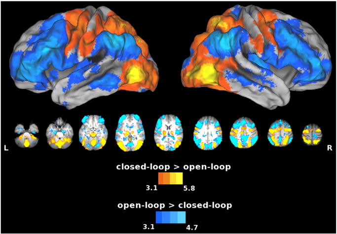
Fig. 5.

Areas showing increased activity for the closed-loop vs. open-loop comparison. Warm colors show the closed-loop > open-loop contrast, and cool colors show the open-loop > closed-loop contrast. Compared to the open-loop condition, the closed-loop condition again showed increased activation in the SPL and cerebellum. Conversely, as was the case for imagery vs. closed-loop execution (cool colors in Fig. 4), open-loop vs. closed-loop execution produced increased activity in the IPL (Ag), MFg, pre-SMA, MTg, and dorsal striatum. When visual feedback is not available, some of these areas might be involved in feedforward processes.
