Colonoscopy has been shown to decrease the incidence of colorectal cancer and cancer death through the removal of adenomatous polyps. To make an accurate diagnosis of colorectal neoplasms, several image-enhanced endoscopic systems, such as narrow band imaging (NBI), flexible spectral imaging color enhancement, and blue laser imaging (BLI), have been developed. However, no significant differences have been reported in the adenoma detection rate between image-enhanced endoscopy and conventional white light endoscopy [1]. NBI and BLI are often criticized for darkening the endoscopy image and, in turn, hampering a wider view of the colon [2]. Moreover, the miss rate for flat lesions was significantly higher than that for pedunculated lesions [3]. We present a case that suggests a possible improvement in adenoma detection rates, especially for flat adenomas.
A 66-year-old woman with a 40-mm laterally spreading tumor of the rectum was referred for endoscopic resection. Before the endoscopic resection, we observed the lesion with an EC-L590ZW endoscope with the LASEREO system (FUJIFILM Co., Tokyo, Japan), with white light (Fig. 1A) and with Linked Color Imaging (LCI) (Fig. 1B). The lesion was clearly seen as a bright reddish area on LCI. Compared with the white light image, the LCI image makes the lesion more easily recognizable, thanks to the striking color contrast between the neoplastic mucosa and the normal mucosa. The lesion was diagnosed as an adenoma on the basis of the magnified BLI images. Successful en bloc endoscopic submucosal dissection was performed for the lesion, and the diagnosis of high-grade adenoma with negative resection margins was confirmed histopathologically (Fig. 1C).
Fig. 1.
Endoscopic images of a laterally spreading tumor (LST). (A) White light endoscopic view of an LST, 40 mm in diameter, in the rectum. (B) The lesion is easily recognizable as a bright reddish area on Linked Color Imaging. (C) Pathological image of the resected lesion (H&E stain, ×4).
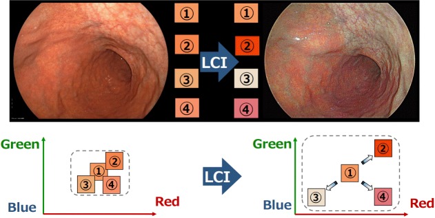
NBI is the most widely used system among several available image enhanced endoscopy systems. NBI modifies the center wavelength and bandwidth of an endoscope’s light into narrow-band illumination at 415±30 nm within the hemoglobin absorption band, thereby facilitating a clearer visualization of vascular structures [4]. However, this technique has limitations such as dark imaging of distant lesions because of narrow-band illumination. BLI was developed to compensate for these inherent limitations of NBI. BLI uses narrow-band laser light combined with white light. This combination results in a bright image of the digestive mucosa, enabling the detailed visualization of both the microstructure and the microvasculature [5]. However, BLI still is not able to obtain sufficient brightness for distant lesions. The newly developed LCI system (FUJIFILM Co.) creates clear and bright endoscopic images by using short-wavelength narrow-band laser light combined with white laser light on the basis of BLI technology. This system can obtain bright endoscopic images even at a distant view because LCI has more intense white light than the short-wavelength narrow-band laser light. Short-wavelength narrow-band laser light enhances the vessels on the mucosal surface and the patterns of the mucosa, which means that BLI enables a clearer visualization of microvascular structures than does LCI. In contrast, LCI enhances differences in hue, in the red region of the spectrum, through digital processing (Fig. 2). This makes red areas appear redder and white areas appear whiter. Thus, it is easier to recognize a slight difference in color of the mucosa. Therefore, LCI may facilitate the detection of flat colorectal neoplasms without magnification. After the detection of lesions, BLI images are easily produced with a push of a button for use in qualitative diagnosis. Further studies are needed to confirm the utility of LCI.
Fig. 2.
Principles of Linked Color Imaging (LCI). LCI enhances differences in hue, in the red region of the spectrum, through digital processing.
Footnotes
Conflicts of Interest: H.Y. has a consultant relationship with FUJIFILM Corporation and has received honoraria, grants, and royalties from the company.
REFERENCES
- 1.Kamiński MF, Hassan C, Bisschops R, et al. Advanced imaging for detection and differentiation of colorectal neoplasia: European Society of Gastrointestinal Endoscopy (ESGE) Guideline. Endoscopy. 2014;46:435–449. doi: 10.1055/s-0034-1365348. [DOI] [PubMed] [Google Scholar]
- 2.Dinesen L, Chua TJ, Kaffes AJ. Meta-analysis of narrow-band imaging versus conventional colonoscopy for adenoma detection. Gastrointest Endosc. 2012;75:604–611. doi: 10.1016/j.gie.2011.10.017. [DOI] [PubMed] [Google Scholar]
- 3.Heresbach D, Barrioz T, Lapalus MG, et al. Miss rate for colorectal neoplastic polyps: a prospective multicenter study of back-to-back video colonoscopies. Endoscopy. 2008;40:284–290. doi: 10.1055/s-2007-995618. [DOI] [PubMed] [Google Scholar]
- 4.Emura F, Saito Y, Ikematsu H. Narrow-band imaging optical chromocolonoscopy: advantages and limitations. World J Gastroenterol. 2008;14:4867–4872. doi: 10.3748/wjg.14.4867. [DOI] [PMC free article] [PubMed] [Google Scholar]
- 5.Osawa H, Yamamoto H. Present and future status of flexible spectral imaging color enhancement and blue laser imaging technology. Dig Endosc. 2014;26 Suppl 1:105–115. doi: 10.1111/den.12205. [DOI] [PubMed] [Google Scholar]


