Abstract
Background
Fluorogenic thrombin generation (TG) assays and turbidity-based fibrin generation (FG)- and fibrinolysis (FL)-resistance assays have been sought to assess bleeding and clotting disorders. Theoretically, TG, FG and FL tests should provide overlapping information because thrombin is responsible for FG and induces protection from FL. The relationships between TG, FG and FL parameters remain poorly investigated, partly because existing experimental systems do not permit simultaneous detection of both TG and FG in the same sample of plasma, and are instead tested in separate experiments.
Objectives and methods
We evaluated the potential benefits of a combined TG/FG/FL assay by testing responses of normal plasma to a wide range of tissue factor (TF) and tissue plasminogen activator (tPA) concentrations. Correlations between multiple parameters extracted from the TG and FG/FL curves were also compared.
Results
Rate of FG correlated well with TG peak height at all TF concentrations, but correlations between TG and FL parameters depended on the TF concentration. Without thrombomodulin, all FG/FL parameters at high TF could be predicted from TG parameters and no FL protection was observed. With thrombomodulin and high TF, TF-dependent FL protection did not correlate with TF-dependent TG. The fluorogenic thrombin substrate did not interfere with optical density readings, and meaningful tPA concentrations did not interfere with TG readings.
Conclusions
In normal plasma, TG, FG and FL parameters may provide interchangeable information. Evaluation of FL-resistance may provide additional data under special assay conditions, but the value of this information should be studied under disease conditions.
Keywords: Thrombin generation, fibrin generation, fibrinolysis, global hemostasis assay, overall hemostasis potential, tissue plasminogen activator, tissue factor, TAFI
Introduction
Thrombin generation assays (TGA) and turbidity-based overall hemostasis assays (OHA) are two global hemostasis methods that are believed to provide a more complete and balanced view on the hemostasis potential under many situations where traditional assays fail. The traditional clotting assays Prothrombin Time (PT) and activated Partial Thromboplastin Time (aPTT) are sensitive to bleeding conditions caused by severe deficiencies in extrinsic and intrinsic coagulation factors. PT and aPTT are less informative for moderate bleeding tendencies caused by mild factor deficiencies (1). Traditional tests are not predictive of prothrombotic tendencies such as those caused by resistance to activated protein C (2). The action of certain anti- and procoagulant therapies such as direct Factor (F) Xa inhibitors (3;4) and recombinant FVIIa (5) are not reflected by the PT and aPTT tests.
In the TGA literature, the inadequacy of clotting tests PT and aPTT is often explained by the theoretical observation that clotting happens very early in the TG response, i.e. when only 5% of thrombin is generated (6). Indeed, while TG is measured using a fluorogenic substrate that is not exhausted over the course of the TGA, FG is measured using fibrinogen which is quickly exhausted and thus measures only the beginning of TG. Furthermore, it is hypothesized that endpoint measurements, e.g. clotting time-based assays, are not reflective of the kinetics of thrombin generation response because peak of thrombin activity (in the TGA) is reached about 10 to 15 minutes after clot formation (7). This TGA-centric view, however, does not appear to be completely justified because strong fibrin clot is a hallmark of normal hemostasis. In principle, Fibrin Generation (FG) should depend on TG. Indeed, abnormal TG has been demonstrated to result in altered FG in various prothrombotic and bleeding coagulopathies (8). Deficient TG in hemophilia leads to formation of weak fibrin clots with increased permeability (9) and poor resistance to fibrinolysis (FL) (10). Increased TG leads to dense clots that are resistant to FL (11;12). In clinical laboratory settings, the importance of FG and FLs observations has long been acknowledged, most notably by the Blombäck group who proposed the OHA, a method for determining the combined effects of coagulation and fibrinolysis based on measurement of the area under the FG curve, as a way to present a global assessment of the hemostasis response (13–16).
Although OHA are speculated to provide better assessment of hemostasis compared to clot-time tests PT and aPTT, their value compared to TGA remains uncertain. Several recent studies employed both fluorogenic thrombin substrate and turbidity-based clotting assessment in an attempt to measure all aspects of the hemostasis response: TG, FG and FL (16–21). However, these studies did not allow direct side-by-side comparison of the TG and FG/FL parameters because simultaneous recording of TG, FG and FL was not implemented. Instead, TG and FG/FL were tested in separate plasma samples under (sometimes, slightly) different conditions, e.g., TG tested without FL activators and FG tested without a fluorogenic substrate for thrombin.
Our study was designed to evaluate the relationships between TG, FG and FL under identical conditions by observations of all three events simultaneously in the same normal plasma sample (evaluation of prothrombotic and other disease conditions was outside the scope of this work). In doing so, we tested two hypotheses: (1) that TG determines FG and FL, i.e., all parameters of FG and FL can be predicted from the TG parameters; and its opposite, (2) that FG or FL may provide information that is different from the TGA, i.e., that some aspects of FG or FL cannot be predicted by the TGA.
In this work we used the simple experiment comprised of normal human plasma spiked with a wide range of tissue factor (TF) concentrations, representing a model of variable coagulation response triggered by vessel wall injury of differing intensity, i.e., propensity of plasma to respond to variable TF stimuli. Increasing TF concentrations induce proportional increases in thrombin generation, allowing us to accurately titrate the response of FG and FL to increased TG. However, our studies were limited to normal plasma and therefore they were not designed to address pathological circumstances (the presence of a direct FXa inhibitor for example) where the relations between the initiation and the propagation mechanisms might be different.
Materials and Methods
Materials
Normal pooled plasma (NPP, VisuCon-F® Normal Control Plasma) and affinity-depleted Factor IX deficient plasma (FIX-DP) were both obtained from Affinity Biologicals (Ancaster, Ontario, Canada). The fluorogenic substrate used in the thrombin generation test, Z-Gly-Gly-Arg-AMC, was from Bachem Americas (King of Prussia, Pennsylvania, USA). Phospholipid vesicles were obtained from Rossix (Molndal, Sweden). Lipidated human recombinant tissue factor (TF, Recombiplastin®) was from Instrumentation Laboratory (Bedford, Massachusetts, USA). Recombinant tissue plasminogen activator (tPA, Cathflo® Activase®) was from Genentech (South San Francisco, USA). Corn trypsin inhibitor (CTI) and rabbit thrombomodulin (TM) were from Haematologic Technologies (Essex Junction, Vermont, USA). Carboxypeptidase inhibitor (CPI) extracted from tubers was from Sigma-Aldrich (St. Louis, USA). Calcium chloride (CaCl2) was from Quality Biologicals (Gaithersburg, USA).
Automated TG/FG/FL assay
NPP or FIX-DP (50% vol/vol in the final reaction) was mixed with fluorogenic substrate Z-Gly-Gly-Arg-AMC (1.25% vol/vol, 800 μM final concentration) and Tris-BSA buffer (pH 7.4, Aniara, West Chester, Ohio, USA). Additional reagents including TF to promote clotting, tPA to induce clot lysis, TM to promote activation of TAFI by thrombin, and CPI to inhibit TAFIa were added to the plasma and the concentration of each varied between individual experiments; however, the volume of all additions to plasma was kept constant. Clotting was initiated by transferring the plasma mixture onto a half-area 96-well flat bottom plate containing CaCl2 (20.0%, 10 mM final concentration) using a 96-channel pipettor (Hydra Liquid Handling System, Thermo Scientific, Pittsburgh, USA) to allow rapid and simultaneous recalcification in all wells of a microplate. The flat bottom plate, containing the activated reaction mixture, was transferred to an Infinite F500 (Tecan, Männedorf, Switzerland) microplate reader regulated at 37°C. Fluoresence (380 nm excitation, 430 nm emission) and absorbance (492 nm) were recorded every 40–50 seconds for at least two hours.
TG/FG/FL curve processing software
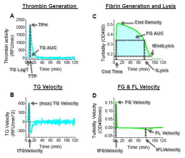
Data processing was performed using a software package designed by Dr. Mikhail Ovanesov using OriginPro (OriginLab, Northampton, MA; the package is available from us upon request). The TGA analysis part of this software was described previously (22). The FG/FL analysis algorithms were developed specifically for this study. The three raw measurements taken by the instrument were relative fluorescence units (RFU), optical density (OD), and time. Fluorogenic substrate conversion measured in RFU was used to monitor TG and clot turbidity measured in OD units was used to monitor FG and FL. All other values were derived from the aforementioned three measurements via calculations performed by our software tool. The software is capable of applying different smoothing algorithms during the conversion of raw fluorescence to the processed TG curve, and computes the final TG curve parameters, such as area under the curve (TG AUC or ETP), thrombin peak height (TPH), time to peak (TTP), and lag time (TG Lag T) (Fig. 1). In addition, the software is capable of converting clot turbidity data to FG/FL curves and computes the FG and FL parameters, such as Clot Density (maximum OD), FG velocity (maximum FG rate), FL velocity (minimum FG rate) and time to lysis (time to half Clot Density). Averaging of multiple wells was performed after processing the TG and FG/FL curves. Although our software can apply the thrombin calibration and thrombin-α2macroglobulin (α2MG) correction algorithms, to simplify the analyses and to avoid introduction of potential mathematical artifacts arising from calibration algorithms, calibration was not performed and thrombin activity was expressed in RFU/min instead of nM.
Figure 1. Parameters used for TG curve and FG/FL curve quantification in this paper.
Parameters quantifying TG, FG, and FL are calculated from the raw fluorescence and absorbance curves and their derivatives. Times and magnitudes where the TG (change in fluorescence over time) and FG/FL (absorbance with respect to time) curves rise from their baselines, reach their maxima, and for FL, fall back toward the starting baseline are analyzed for their response to different TF and tPA concentrations, as are the areas under the TG and FG/FL curves.
Time parameters and power parameters of TG, FG and FL
For the purposes of analyses, all TG and FG/FL curve parameters were divided into two categories. Time parameters were parameters expressed in minutes, e.g., TTP, TG Lag Time, Lysis Time, Time to TG Velocity. The remaining parameters were considered as power parameters. Therefore, time parameters indicate how late or early TG, FL and FL response happens. Power parameters indicate the strength (power) of the response.
Optimization of the assay reagents concentrations
tPA was serially diluted and added at various concentrations (range: 10-0.03 μg/ml and a 0 μg/ml control well) to the reaction wells. This concentration range was selected since the majority of clots exposed to tPA concentrations in this range exhibited lysis within the first 50 minutes of data collection. TF was serially diluted in a range from 20 to 0.14 pM (and a 0 pM control well). The TF range’s upper boundary of 20 pM was selected since it produced a thrombin lag time of 1–2 minutes, which was the minimal time sufficient to transfer the 96-well plate into the microplate reader and initiate data collection. CPI was tested in wide range from 200 to 0.0475 μg/ml. The concentration of TM (6.25 nM) was chosen such that TG parameters were unaffected but TAFI was activated (data not shown). In some experiments, contact activation was inhibited by CTI (130 μg/ml).
Results
1. Trends of TG, FG, and FL parameters as a function of TF trigger concentration in normal plasma
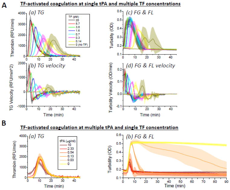
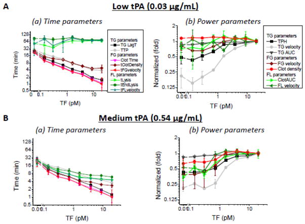
As expected, there was a strong dose-effect of TF concentration on TG, FG, and FL in normal plasma (Fig. 2A). These trends were observed across all tPA concentrations used except the lowest (0.03 μg/ml), where a dose-dependent lysis response was not observed (Figs. 2A and 3). Comparative analysis of various TG, FG, and FL parameters presented in Fig. 4 shows that increasing TF leads to faster TG, FG and FL time parameters (with the exception of lysis parameters at 0.03 μg/mL tPA) in normal plasma. Among the power parameters that characterize magnitude of TG, FG and FL responses, only TPH, TG Velocity, FG velocity and clot AUC demonstrated strong TF-dependence. In contrast, Clot Density, FL Velocity and TG AUC were minimally affected by TF concentration (Fig. 4).
Figure 2. TF- and tPA-dependent TG, FG and FL in normal plasma.
A. TF-dependent TG (a–b) and FG and FL (c–d) at multiple TF concentrations and single tPA concentration. Increase in TF results in higher thrombin peaks, higher TG velocity peaks, and decreased thrombin lag time (TG parameters). Increased TF also results in greater peak clot density and greater peak clotting velocity, and decreased clot time. Curves were generated from experimental data (n = 3) in normal pooled plasma with 0.13 μg/ml tPA and 0 or 0.14–20 pM TF. B. TF-dependent thrombin generation and fibrin generation and lysis at multiple tPA concentrations and single TF concentration. tPA has no effect on thrombin generation (a) but induces faster lysis with increasing concentration (b). Normal pooled plasma activated with 0.71 pM TF and 0 or 0.03–10 μg/ml tPA was used to induce clot lysis. Curves and shaded areas around the curve depict average values and S.D., respectively, for three independent experiments.
Figure 3. Effect of TF and tPA on primary TG, FG and FL parameters in normal plasma.
Only lysis time at low tPA concentrations does not correlate with TF; all other conditions and parameters show strong dose-dependent effects with TF concentration.
Figure 4. Similarity of trends for 15 different TG/FG/FL parameters at low and medium tPA concentrations in normal plasma.
Experiments in normal plasma (n = 3) without TM, CTI, or CPI. TF range spanned from 0.14–20 pM; concentration of tPA was either 0.03 μg/ml (A) or 0.54 μg/ml (B). Power parameters are shown relative to their values at 20 pM TF.
2. Correlation between TG assay parameters and FG/FL assay parameters in normal plasma
To study interconnectedness of TG assay and FG/FL assay parameters in normal plasma, we plotted correlation graphs between the TPH and TTP and all other various TG, FG and FL parameters (Supplemental Figs. S1 and S2). These correlations were quantified using Spearman’s rank correlation coefficient (Table 1 and Supplemental Fig. S3). tPA concentration did not have effect on correlation pattern of most parameters, e.g., higher TPH correlated negatively (inversely) with time parameters TG Lag Time and Clot Time and positively with power parameters TG velocity, TG AUC, FG Velocity, Clot Density and FG AUC. Lysis Time was inversely correlated with TPH at low and moderate tPA concentrations, suggesting that increased TG protects clots from lysis.
Table 1. Correlation coefficients between TPH or TTP and remaining TG, FG and FL parameters for TF-activated coagulation in normal plasma.
Spearman’s rank correlation coefficient for both time and power parameters with respect to thrombin peak height (TPH, top part of the table) and time to thrombin peak (TTP, lower half of the table). Number in parenthesis indicates significance factor. Correlation coefficients with absolute values closer to 1 show better correlation (+1 = positive correlation; −1 = negative [inverse] correlation). Negative correlations are shown in blue.
| TPH vs. | TG parameters | FG parameters | FL parameters | ||||||
|---|---|---|---|---|---|---|---|---|---|
| TG velocity | TG lag time | TG AUC | FG velocity | Clot time | Clot density | FL velocity | Lysis time | FG AUC | |
| tPA: | |||||||||
| 0.03 μg/ml | 0.98 (3.3e-5) | −0.90 (0) | 0.88 (0) | 0.90 (0) | −0.98 (3.3e-5) | 0.90 (0) | 0.98 (3.3e-5) | −0.90 (0) | 0.88 (0) |
| 0.13 μg/ml | 0.90 (0) | −0.98 (3.3e-5) | 0.95 (2.6e-4) | 0.88 (0) | −0.93 (8.6e-4) | 0.76 (0) | 0.71 (0.1) | −0.98 (3.3e-5) | 0.90 (0) |
| 0.54 μg/ml | 1.00 (0) | −0.93 (8.6e-4) | 0.98 (3.3e-5) | 0.90 (0) | −0.98 (3.3e-5) | 0.38 (0.4) | −0.64 (0.1) | −0.48 (0.2) | 0.83 (0) |
| 2.33 μg/ml | 0.98 (3.3e-5) | −0.98 (3.3e-5) | 0.90 (0) | 0.81 (0) | −0.90 (0) | −0.64 (0.1) | 0.21 (0.6) | 0.81 (0) | 0.60 (0.1) |
| TTP vs. | TG parameters | FG parameters | FL parameters | ||||||
|---|---|---|---|---|---|---|---|---|---|
| TG velocity | TG lag time | TG AUC | Time to FG velocity | Clot time | Clot density | FL velocity | Lysis time | FG AUC | |
| tPA: | |||||||||
| 0.03 μg/ml | −0.83 (0) | 1.00 (0) | −0.98 (3.3e-5) | 0.98 (3.3e-5) | 1.00 (0) | −0.86 (0) | 0.24 (0.57) | 0.95 (2.6e-4) | −0.98 (3.3e-5) |
| 0.13 μg/ml | −0.93 (8.6e-4) | 1.00 (0) | −0.98 (3.3e-5) | 1.00 (0) | 1.00 (0) | −0.81 (0) | 0.60 (0.12) | 1.00 (0) | −0.95 (2.6e-4) |
| 0.54 μg/ml | −0.93 (8.6e-4) | 1.00 (0) | −0.98 (3.3e-5) | 1.00 (0) | 1.00 (0) | −0.40 (0.3) | 0.98 (3.3e-5) | 0.95 (2.6e-4) | −0.93 (8.6e-4) |
| 2.33 μg/ml | −0.93 (8.6e-4) | 1.00 (0) | −0.93 (8.6e-4) | 1.00 (0) | 1.00 (0) | 0.62 (0.1) | 0.95 (2.6e-4) | 0.71 (0.1) | −0.62 (0.1) |
Under most tPA concentrations, FL-sensitive parameters FL Rate and Clot Density had correlation coefficients below 0.75 confirming poor agreement with TG parameters TPH or TTP. All other TG, FG and FL parameters correlated very well with TPH and TTP (correlation coefficients close to 1.0). Therefore, our subsequent analysis was focused on TPH, TTP, Lysis Time as representative of the different phenomena and response patterns observed.
3. Analysis of limitations of the TF-activated TG, FG and FL model in normal plasma
Various artifacts such as competition of natural thrombin substrates (e.g., Factor V, Factor XI and Antithrombin III) with the slow fluorogenic substrate Z-GGR-AMC have been previously suspected to diminish the value of the TGA. Since our assay simultaneously records coagulation via two independent channels, fluorescence and absorbance, we can study the effect of substrate on clot formation by observing the absorbance channel with and without fluorogenic substrate addition. In control experiments we showed that substrate does not interfere with FG or FL in our experimental system under most of the TF and tPA conditions of our experiments (Supplemental Figs. S3 and S4).
The slow thrombin substrate used in the TGA was originally developed for the enzyme tPA, suggesting that tPA may have an impact on TGA assay. Indeed, TPH was lower at high tPA (Figs. 2B and 3). We found that this is an artifact caused by increased fluorescence due to fluorogenic substrate consumption by tPA, resulting in inner filter effect (IFE). An algorithm for IFE correction (23) has been used successfully to correct for this artifact in a separate experiment (not shown). The effect was seen only at 10 μg/mL which is not a good condition for analysis of TG/FL because high tPA causes lysis to occur too quickly. In the most informative tPA concentration range of 0.03–0.5 μg/ml, substrate depletion was not reached and IFE artifacts did not require correction for (not shown).
Contact activation is known to cause artificial TG at low TF concentrations (24–28). In our experiments below 0.3 pM of TF, TPH did not decline as TF declines (Fig. 3B). To test if that was caused by spontaneous contact activation, we compared TG, FG and FL parameters with and without CTI, an inhibitor of contact activation. CTI was successful in blocking TG in the absence of TF (not shown). However, even after the addition of CTI, no dose response to TF was observed at low TF concentrations, as evidenced by the good correlation of TPH and TTP in the presence and absence of CTI except at low TF concentrations (not shown).
4. Relationship between TG and FL resistance
Under some conditions of our experiments, FL parameters failed to correlate with TF-dependent TG enhancement. TG was previously suggested to be responsible for FL resistance therefore we investigated the mechanisms behind the good or poor correlation of TG and FL parameters. We considered two previously described mechanisms of FL protection by elevated TG: formation of denser lysis-resistant clots (11;12) or activation of TAFI by thrombin (29). To examine the role of TAFI in our experiments, we used TM to induce TAFI activation and CPI to block TAFIa. Furthermore, we wanted to study correlations between FL and TPHs covering widest range of values in this experiment. Since TPH in normal plasma reached saturation at the lowest TPH level, i.e. no dose response to TF was observed at low TF concentrations, we switched to a plasma with known hypo coagulation status, Factor IX-deficient plasma (FIX-DP). In FIX-DP plasma, no TG or FG was seen in the absence of TF and good dose-response was seen at low TF. The correlations between TG, FG and FL parameters were generally similar to normal plasma (data not shown), however, unlike in normal plasma, TPH was approaching saturation at TF > 5.70 pM (supplemental Figs. S6A and S7A).
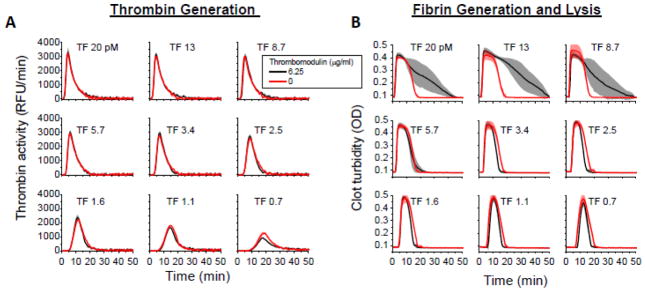
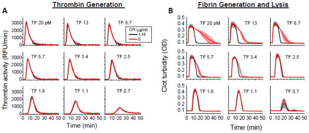
In FIX-DP, addition of TM (chosen at the lowest concentration that would not inhibit the thrombin generation) induced protection from FL (Fig. 5 and supplemental Fig. S6) but only at high TF concentrations. To confirm the role of TAFI in lysis protection, we used a specific inhibitor, CPI, taken at a concentration that inhibited lysis without changing TG or FG (Fig. 6 and supplemental Fig. S7). CPI blocked lysis protection only at high concentrations of TF; lysis protection at low TF remained unchanged with or without CPI (supplemental Fig. S7). This indicates that the observed lysis protection in FIX-DP is TAFI-dependent rather than resulting directly from TG-mediated clot density.
Figure 5. Regulation of clot lysis by TM-dependent activation of TAFI in FIX-DP.
TM promotes inhibition of FL. (A) TG curves demonstrate negligible effect of TM on TG. Black curves: TM, 6.25 nM. Red curves: no TM. (B) FG/FL in the presence of TM (black) are more resilient to lysis than clots in the absence of TM (red). (B) Experiments were performed in FIX-DP supplemented with 0.15 μg/ml tPA, 130 μg/ml CTI, and 0 or 6.25 μg/ml TM. Means and S.D. (2 wells) is shown. Note that lysis resistance in the presence of TM occurs at TF concentrations where TG parameters are constant, see supplemental Fig. S6.
Figure 6. Regulation of clot lysis by TAFI in FIX-DP.
CPI, an inhibitor of TAFIa, promotes FL. (A) The effect of CPI on TG is minimal. (B) Fibrin clots in the presence of CPI (black) lyse more quickly than clots in the absence of CPI (red). Experiments were performed in FIX-DP supplemented with 0.15 μg/ml tPA, 130 μg/ml CTI, 0 or 1.16 μg/ml CPI, and 6.25 nM TM. Note that CPI effects on lysis resistance happen at TF concentrations where TG parameters are largely unchanging; see supplemental Fig. S7.
Discussion
Our study is the first to systematically investigate the correlation between TG, FG and FL parameters determined simultaneously in the same set of normal plasma samples. Simultaneous detection allowed us to show that, within our experimental model, correlation or lack of it between TG, FG and FL parameters is dependent on TF and tPA concentrations. Our studies were intentionally simple in design: we studied TF titration only because we wanted to compare the relationship between different parameters under a well-control trigger-dependent dose-response condition. At low TF, TG determined FG and FL, i.e., all parameters of FG and FL can be predicted from the TG parameters. At high TF, FL provided information that was different from the TG. It is worthy to note that FG parameters correlated remarkably well with the staple TG parameters, TPH and TTP (Table 1). This conclusion contrasts with the common notion that TG is more informative than FG simply because clotting happens earlier. We have found strong correlations within and between TG and FG parameters. At a first glance, this may appear that analyzing a single TG or FG parameter may serve as a proxy for all others. However, our experiments were restricted to normal plasma, suggesting caution against extending the conclusion to abnormal plasmas. Further studies using plasmas from bleeding or prothrombotic patients and various assay conditions may show that this is not always the case.
FL trends did not always track TG or FG, unlike the correlations between TG and FG. At high tPA concentrations lysis time closely tracked parameters such as TG lag time and clot time; however, this could be because FL is activated as quickly as it can under high tPA assay conditions – as lysis time cannot precede clot time, these parameters would correlate even if there are no lysis-specific elements contributing to this trend. FL may be activated before clots are fully formed, resulting in reported TG and FG parameters not reflecting the sample’s intrinsic coaguability or TG potential; however, this might not affect observations within the time resolution of our assay.
Increased TG is thought to protect against FL through production of fast-polymerizing fibrin and thus denser or stronger clots, and, when coupled with TM, activation of thrombin-activated fibrinolysis inhibitor (TAFI). The time sequence of TG, FG and FL is determined by the biochemistry of these reactions and is fairly well reproducible in vitro, e.g. time to thrombin peak is always found after clot time, while lysis time may be much later than both. Therefore, at the time lysis would be occurring, thrombin levels may have already fallen to the baseline, suggesting that protection from lysis at this point is not due to direct involvement of thrombin. This is not to say that lysis time does not depend on TG. We did observe a TF-dependent protection against FL at high TF concentrations. This protection was eliminated when an inhibitor of TAFIa was added to the assay, indicating that protection acts through TG. However, the concentrations of TF where this was observed were beyond the range where there was a TF dose-dependent increase in TG as measured both by peak height (supplemental Figs. S6 and S7) and peak time (not shown). This observation shows that the effects of thrombin persist beyond the TG peak as measured by our assay. Whether this is additional TG or additional activation of proteins activated by thrombin remains to be determined. Nevertheless, FL in the presence of tPA and TM may exhibit phenomena not seen by TG. In theory, observation of FL in conjunction with TG may extend the range of conditions observable by the global hemostasis assay. The differing FL trends between high and low TF concentrations may also reflect the threshold nature of TG- and TAFI-dependent protection from FL.
Robustness of recorded parameters is essential for the practical utility of the assay. So far clot time, lysis time and FG AUC are the only FG/FL parameters we have found to be robust. However, further examination of other FG/FL parameters may reveal phenomena not apparent from clot time or lysis time. Instrumentation may present limitations too. Under most assay conditions in our assays, FG happens so quickly that the majority of the increase in absorbance occurs within one reading cycle of the instrument. If FL is occurring before clot formation is complete, clot time will not detect this due to the limited time resolution of our assay. Only peak absorbance and peak absorbance time, not clot time, sometimes appear to be deflected by possible early lysis seen under high tPA conditions. However, these parameters vary between donor plasmas and between runs and sometimes between duplicate wells (data not shown).
Previously, it has been argued that fluorogenic substrate ZGGR-AMC may interfere with coagulation response because substrate interacts with the many enzymes that protect thrombin from inhibition (30). Combined testing of TG and FG allowed us to test this hypothesis. Otherwise identical conditions run in the presence or absence of the synthetic thrombin substrate show essentially the same FG and FL response over a broad range of TF concentrations. Our findings, in addition to Hemker and de Smedt’s 2007 study using a range of Z-GGR-AMC concentrations (31), show that the effect of the presence of this thrombin substrate on TG, FG, and FL responses is minimal. tPA also cleaves Z-GGR-AMC, but its concentration can be optimized so it does not affect TG while inducing a meaningful level of FL.
While the experiments in this paper are potentially limited because they cover one means of TG activation in two plasmas (normal pooled or Factor IX-deficient), our present study suggests that under some conditions, a single parameter derived from either TG, FG or, possibly, FL assay may serve as predictive readout of a global hemostasis assay. On the other hand, careful selection of assay conditions is needed to increase assay’s sensitivity to different known phenomena. For example, effects of TAFI on FL, TF on TG, TG on FL, TM on TG and TM on FL could be observed under narrow concentration ranges of assay reagents, and the corresponding ranges were oftentimes mutually exclusive. This suggests that a single standardized assay condition, however well optimized, would not possibly cover all known reactions collectively known as global hemostasis. As a result, it should be noted that the current assay may not accurately model the conditions of some thrombotic disorders. Since the results presented in this paper were obtained from normal pooled plasma and affinity-depleted Factor IX deficient plasma, they may have limited validity under pathological conditions that cannot be accurately modeled by these two plasma types.
An alternative to a single global hemostasis assay can be an assay in which multiple assay conditions are tested in parallel, as modeled by the TF and tPA titrations in our study. The simplicity of the TG/FG/FL assay allows analysis of samples at high throughput in a microplate format using nonproprietary commercially available equipment (22). This allows us to detect trends within and between plasma types with ample controls to rule out the bias of run-to-run variations. Future studies in relevant pathological patient populations are needed to reveal how prognostic, if at all, a combined fluorogenic TGA and clot turbidity-based FG/FL assay can be to clinical outcomes compared to existing assay systems.
Supplementary Material
Highlights.
Thrombin generation, clotting and fibrinolysis were measured simultaneously
Correlation between assay parameters was studied at different tissue factor levels
In normal plasma, thrombin and clotting parameters are interchangeable
Fibrinolysis may provide additional data under special assay conditions
The value of this information remains to be studied under disease circumstances
Acknowledgments
This study was supported in part by a Postgraduate and Postbaccalaureate Research Fellowship Award to W.C.C. from the Oak Ridge Institute for Science and Education (ORISE) through an interagency agreement between the U.S. Department of Energy and the U.S. Food and Drug Administration (FDA). This paper is an informal communication and represents authors’ best judgment. These comments do not bind or obligate FDA.
Footnotes
Contribution: K.Z.X. conducted and analyzed experiments. W.C.C. and M.V.O. wrote the manuscript with assistance from K.Z.X. M.V.O. supervised the project and the preparation of the manuscript.
Publisher's Disclaimer: This is a PDF file of an unedited manuscript that has been accepted for publication. As a service to our customers we are providing this early version of the manuscript. The manuscript will undergo copyediting, typesetting, and review of the resulting proof before it is published in its final citable form. Please note that during the production process errors may be discovered which could affect the content, and all legal disclaimers that apply to the journal pertain.
References
- 1.Lippi G, Pasalic L, Favaloro EJ. Detection of mild inherited disorders of blood coagulation: current options and personal recommendations. Expert Rev Hematol. 2015:1–16. doi: 10.1586/17474086.2015.1039978. [DOI] [PubMed] [Google Scholar]
- 2.Kadauke S, Khor B, Van Cott EM. Activated protein C resistance testing for factor V Leiden. Am J Hematol. 2014;89:1147–50. doi: 10.1002/ajh.23867. [DOI] [PubMed] [Google Scholar]
- 3.Lison S, Spannagl M. Monitoring of direct anticoagulants. Wien Med Wochenschr. 2011;161:58–62. doi: 10.1007/s10354-011-0876-8. [DOI] [PubMed] [Google Scholar]
- 4.Hillarp A, Baghaei F, Fagerberg BI, Gustafsson KM, Stigendal L, Sten-Linder M, et al. Effects of the oral, direct factor Xa inhibitor rivaroxaban on commonly used coagulation assays. J Thromb Haemost. 2011;9:133–9. doi: 10.1111/j.1538-7836.2010.04098.x. [DOI] [PubMed] [Google Scholar]
- 5.Qi X, Zhao Y, Li K, Fan L, Hua B. Evaluating and monitoring the efficacy of recombinant activated factor VIIa in patients with haemophilia and inhibitors. Blood Coagul Fibrinolysis. 2014;25:754–60. doi: 10.1097/MBC.0000000000000137. [DOI] [PubMed] [Google Scholar]
- 6.Mann KG, Brummel K, Butenas S. What is all that thrombin for? J Thromb Haemost. 2003;1:1504–14. doi: 10.1046/j.1538-7836.2003.00298.x. [DOI] [PubMed] [Google Scholar]
- 7.Rand MD, Lock JB, van’t Veer C, Gaffney DP, Mann KG. Blood clotting in minimally altered whole blood. Blood. 1996;88:3432–45. [PubMed] [Google Scholar]
- 8.Torbet J. The thrombin activation pathway modulates the assembly, structure and lysis of human plasma clots in vitro. Thromb Haemost. 1995;73:785–92. [PubMed] [Google Scholar]
- 9.He S, Blomback M, Jacobsson EG, Hedner U. The role of recombinant factor VIIa (FVIIa) in fibrin structure in the absence of FVIII/FIX. J Thromb Haemost. 2003;1:1215–9. doi: 10.1046/j.1538-7836.2003.00242.x. [DOI] [PubMed] [Google Scholar]
- 10.Bolliger D, Szlam F, Molinaro RJ, Escobar MA, Levy JH, Tanaka KA. Thrombin generation and fibrinolysis in anti-factor IX treated blood and plasma spiked with factor VIII inhibitor bypassing activity or recombinant factor VIIa. Haemophilia. 2010;16:510–7. doi: 10.1111/j.1365-2516.2009.02164.x. [DOI] [PubMed] [Google Scholar]
- 11.Campbell RA, Overmyer KA, Bagnell CR, Wolberg AS. Cellular procoagulant activity dictates clot structure and stability as a function of distance from the cell surface. Arterioscler Thromb Vasc Biol. 2008;28:2247–54. doi: 10.1161/ATVBAHA.108.176008. [DOI] [PMC free article] [PubMed] [Google Scholar]
- 12.Campbell RA, Overmyer KA, Selzman CH, Sheridan BC, Wolberg AS. Contributions of extravascular and intravascular cells to fibrin network formation, structure, and stability. Blood. 2009;114:4886–96. doi: 10.1182/blood-2009-06-228940. [DOI] [PMC free article] [PubMed] [Google Scholar]
- 13.Antovic A, Blomback M, Sten-Linder M, Petrini P, Holmstrom M, He S. Identifying hypocoagulable states with a modified global assay of overall haemostasis potential in plasma. Blood Coagul Fibrinolysis. 2005;16:585–96. doi: 10.1097/01.mbc.0000188411.94740.d7. [DOI] [PubMed] [Google Scholar]
- 14.Antovic JP, Antovic A, He S, Tengborn L, Blomback M. Overall haemostatic potential can be used for estimation of thrombin-activatable fibrinolysis inhibitor-dependent fibrinolysis in vivo and for possible follow-up of recombinant factor VIIa treatment in patients with inhibitors to factor VIII. Haemophilia. 2002;8:781–6. doi: 10.1046/j.1365-2516.2002.00689.x. [DOI] [PubMed] [Google Scholar]
- 15.He S, Zhu K, Skeppholm M, Vedin J, Svensson J, Egberg N, et al. A global assay of haemostasis which uses recombinant tissue factor and tissue-type plasminogen activator to measure the rate of fibrin formation and fibrin degradation in plasma. Thromb Haemost. 2007;98:871–82. [PubMed] [Google Scholar]
- 16.He S, Wallen H, Bark N, Blomback M. In vitro studies using a global hemostasis assay to examine the anticoagulation effects in plasma by the direct thrombin inhibitors: dabigatran and argatroban. J Thromb Thrombolysis. 2013;35:131–9. doi: 10.1007/s11239-012-0791-x. [DOI] [PubMed] [Google Scholar]
- 17.Nagashima H. Studies on the different modes of action of the anticoagulant protease inhibitors DX-9065a and Argatroban. I. Effects on thrombin generation. J Biol Chem. 2002;277:50439–44. doi: 10.1074/jbc.M208222200. [DOI] [PubMed] [Google Scholar]
- 18.Nagashima H. Studies on the different modes of action of the anticoagulant protease inhibitors DX-9065a and Argatroban. II. Effects on fibrinolysis. J Biol Chem. 2002;277:50445–9. doi: 10.1074/jbc.M208223200. [DOI] [PubMed] [Google Scholar]
- 19.Gebhart J, Laczkovics C, Posch F, Ay C, Reitter-Pfoertner SE, Haslacher H, et al. Plasma clot properties in patients with a mild-to-moderate bleeding tendency of unknown cause. Ann Hematol. 2015 doi: 10.1007/s00277-015-2399-7. [DOI] [PubMed] [Google Scholar]
- 20.Bakhtiari K, Kamphuisen PW, Mancuso ME, Hamulyak K, Schutgens RE, Santagostino E, et al. Clot lysis phenotype and response to recombinant factor VIIa in plasma of haemophilia A inhibitor patients. Br J Haematol. 2013;162:827–35. doi: 10.1111/bjh.12470. [DOI] [PubMed] [Google Scholar]
- 21.McEwen BJ, Morel-Kopp MC, Tofler GH, Ward CM. The effect of omega-3 polyunsaturated fatty acids on fibrin and thrombin generation in healthy subjects and subjects with cardiovascular disease. Semin Thromb Hemost. 2015;41:315–22. doi: 10.1055/s-0034-1395352. [DOI] [PubMed] [Google Scholar]
- 22.Woodle SA, Shibeko AM, Lee TK, Ovanesov MV. Determining the impact of instrument variation and automated software algorithms on the TGT in hemophilia and normalized plasma. Thromb Res. 2013;132:374–80. doi: 10.1016/j.thromres.2013.07.018. [DOI] [PubMed] [Google Scholar]
- 23.Coen Hemker H, Hemker PW, Al Dieri R. The technique of measuring thrombin generation with fluorescent substrates: 4. The H-transform, a mathematical procedure to obtain thrombin concentrations without external calibration. Thromb Haemost. 2009;101:171–7. [PubMed] [Google Scholar]
- 24.Kessels H, Beguin S, Andree H, Hemker HC. Measurement of thrombin generation in whole blood--the effect of heparin and aspirin. Thromb Haemost. 1994;72:78–83. [PubMed] [Google Scholar]
- 25.Dargaud Y, Luddington R, Baglin TP. Elimination of contact factor activation improves measurement of platelet-dependent thrombin generation by calibrated automated thrombography at low-concentration tissue factor. J Thromb Haemost. 2006;4:1160–1. doi: 10.1111/j.1538-7836.2006.01905.x. [DOI] [PubMed] [Google Scholar]
- 26.Luddington R, Baglin T. Clinical measurement of thrombin generation by calibrated automated thrombography requires contact factor inhibition. J Thromb Haemost. 2004;2:1954–9. doi: 10.1111/j.1538-7836.2004.00964.x. [DOI] [PubMed] [Google Scholar]
- 27.Spronk HM, Dielis AW, Panova-Noeva M, van OR, Govers-Riemslag JW, Hamulyak K, et al. Monitoring thrombin generation: is addition of corn trypsin inhibitor needed? Thromb Haemost. 2009;101:1156–62. [PubMed] [Google Scholar]
- 28.van Veen JJ, Gatt A, Cooper PC, Kitchen S, Bowyer AE, Makris M. Corn trypsin inhibitor in fluorogenic thrombin-generation measurements is only necessary at low tissue factor concentrations and influences the relationship between factor VIII coagulant activity and thrombogram parameters. Blood Coagul Fibrinolysis. 2008;19:183–9. doi: 10.1097/MBC.0b013e3282f4bb47. [DOI] [PubMed] [Google Scholar]
- 29.Foley JH, Kim PY, Mutch NJ, Gils A. Insights into thrombin activatable fibrinolysis inhibitor function and regulation. J Thromb Haemost. 2013;11(Suppl 1):306–15. doi: 10.1111/jth.12216. [DOI] [PubMed] [Google Scholar]
- 30.Butenas S, Mann KG. Caution in the interpretation of continuous thrombin generation assays. J Thromb Haemost. 2007;5:1084–5. doi: 10.1111/j.1538-7836.2007.02512.x. [DOI] [PubMed] [Google Scholar]
- 31.Hemker HC, De Smedt E. Caution in the interpretation of continuous thrombin generation assays: a rebuttal. Journal of Thrombosis and Haemostasis. 2007;5:1085–7. doi: 10.1111/j.1538-7836.2007.02512.x. [DOI] [PubMed] [Google Scholar]
Associated Data
This section collects any data citations, data availability statements, or supplementary materials included in this article.


