. 2016 Mar 8;5:e12821. doi: 10.7554/eLife.12821

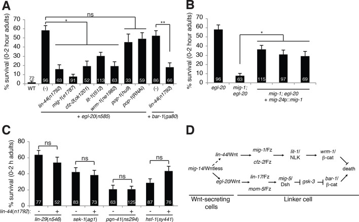
Figure 2. A lin-44/Wnt pathway promotes linker cell survival.

(A) Linker cell survival in indicated genotypes. In (A-C) strains also contain qIs56 and him-5(e1490). *p<10–3; **p <10–4; ns, not significant; Fisher’s exact test. lit-1(t512) is linked to unc-32(e189). (B) Linker cell survival in egl-20(n585) and mig-1(e1787); egl-20(n585) animals harboring a mig-24p::mig-1 transgene. *p<0.001. (C) Linker cell survival in indicated genotypes. ns, not significant; Fisher’s exact test. (D) Model for Wnt pathway interactions in LCD.

DOI: http://dx.doi.org/10.7554/eLife.12821.006

Figure 2.

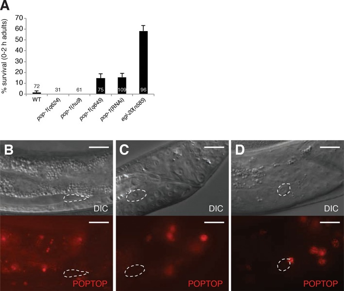
Figure 2—figure supplement 1. pop-1 does not play a significant role in linker cell death.

Figure 2—figure supplement 1.

(A) Linker cell survival in 0-2h adults of the indicated genotypes. All strains also contain the qIs56 reporter transgene to visualize the linker cell and him-5(e1490) to increase the incidence of males. pop-1(RNAi) performed with RNAi-sensitizing rrf-3(pk1426) allele. (B-D) All panels are images of strain unc-119(ed4); him-5(e1490); syIs187[POPTOP::HIS-24-mCherry]. Linker cell outlined in dashed white. (B) Late L3/early L4 male. (C) Mid-L4 male. Note the already-apparent linker cell cytoplasmic changes in the DIC image. (D) Late L4 male. mCherry-staining nucleus at the top right of the linker cell in (D), belongs to a neighboring overlying cell. Scale bars, 10 μm.