Abstract
Stimulation of a single whisker evokes a peak of activity that is centered over the associated barrel in rat primary somatosensory cortex, and yet the evoked local field potential and the intrinsic signal optical imaging response spread symmetrically away from this barrel for over 3.5 mm to cross cytoarchitectonic borders into other “unimodal” sensory cortical areas. To determine whether long horizontal axons have the spatial distribution necessary to underlie this activity spread, we injected adeno-associated viral vectors into barrel cortex and characterized labeled axons extending from the injection site in transverse sections of flattened cortex. A combined qualitative and quantitative analysis revealed labeled axons radiating diffusely in all directions for over 3.5 mm from supragranular injection sites, with density declining over distance. The projection pattern was similar at four different cortical depths, including infragranular laminae. Infragranular vector injections produced patterns similar to the supragranular injections. Long horizontal axons were detected both using a vector with a permissive CMV promoter to label all neuronal subtypes and using a CaMKIIα vector to restrict labeling to excitatory cortical pyramidal neurons. Individual axons were successfully reconstructed from series of supragranular sections, indicating that they traversed gray matter only. Reconstructed axons extended from the injection site, left the barrel field, branched, and sometimes crossed into other sensory cortices identified by cytochrome oxidase staining. Thus, radiations of long horizontal axons indeed have the spatial characteristics necessary to explain horizontal activity spreads. These axons may contribute to multimodal cortical responses and various forms of cortical neural plasticity.
Keywords: Barrel cortex, Horizontal axons, AAV, Somatosensory cortex, Connectivity
Current thinking about the structure and function of neocortex is largely shaped by several underlying principles: parcellation of the cortical sheet into distinct regions containing neurons of similar function (Van Essen 2013; Zilles and Amunts 2010), systematic white matter connections between these discrete functional modules to create larger processing networks recently characterized as the connectome (Sporns 2013; Sepulcre 2014), topography within many of these cortico-cortical projections (Kaas 2012), and vertically organized cortical columns as basic anatomical building blocks (Mountcastle 1997). Whisker representations in the posterior medial barrel subfield (PMBSF) of the rodent primary somatosensory cortex (SI), popularly known as “barrel cortex,” have provided convenient animal models for interventive research into these principles of cortical structure and function (reviewed in Feldmeyer et al. 2013). However, based on functional imaging, neuronal recordings from microelectrode arrays, and histological tract tracing, research from our lab has suggested the existence of an additional, horizontally oriented system in barrel cortex and in other primary sensory cortices that is not clearly evident in current versions of the connectome and that creates a gray matter functional and structural continuum within sensory neocortex (Frostig et al. 2008; Chen-Bee et al. 2012; Stehberg et al. 2014; Johnson and Frostig, 2015).
Our lab has been exploring this horizontal system of connectivity on a systems-based, mesoscopic level rather than by characterizing the responses and anatomy of individual neurons. After a single whisker is stimulated, activity over the rat PMBSF can be detected using intrinsic signal optical imaging (ISOI; Masino et al. 1993). As expected, the peak of ISOI activity evoked by single whisker stimulation is centered over the corresponding whisker barrel; however, evoked intrinsic signal activity spreads a great distance over the cortical surface in all directions, declining in magnitude with increasing distance from the peak of activation (Fig. 1) (Chen-Bee et al. 1996; 2012; Brett-Green et al. 2001). The ISOI signal evoked by whisker stimulation extends beyond the border of PMBSF and even enters the trunk region of somatosensory cortex, visual cortex, or auditory cortex, depending on the location of the stimulated whisker on the rat’s snout (Brett-Green et al. 2001). Similarly, whisker stimulation evokes a spread of activity that crosses borders into other unimodal, primary cortices as seen using voltage-sensitive dye optical imaging (Ferezou et al. 2007, Mohajerani et al. 2013), a technique that directly measures neuronal activity. Finally, a large horizontal spread of activity following whisker stimulation also was measured electrophysiologically in rats as an evoked change in local field potential that extended symmetrically at least 3.5 mm (mean amplitude 11% of peak evoked activity at 3.5 mm away from the activated barrel) to invade other unimodal sensory cortices (Fig. 1) (Frostig et al. 2008; Chen-Bee et al. 2012).
Fig. 1.
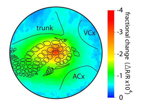
Neuronal activity spreads horizontally for long distances following single whisker stimulation. The intrinsic signal optical imaging response following stimulation of the C2 whisker (the first 500 msec containing the maximal areal extent of the initial dip activity) was averaged across 37 rats as described by Chen-Bee et al. (2012) and was plotted as a false-color image of fractional change relative to prestimulus values. The outer circle has a diameter of 7 mm and represents an extrapolation of the farthest electrode used in the recordings of Frostig et al. (2008), at which an evoked field potential response could be detected in 100% of animals. Black outlines show locations of whisker barrels and cytoarchitectonic areas that were detected by cytochrome oxidase staining in a representative animal. Trunk, trunk region of primary somatosensory cortex; VCx, visual cortex; ACx, auditory cortex
The spread of evoked local field potential outside of the barrel field in rats was disrupted by transection of cortical gray matter between the whisker barrel displaying peak evoked activity and the recording sites, suggesting that the underlying anatomical connection involved shallow axons or axon collaterals running horizontally through cortical gray matter as opposed to either thalamic input or the white matter connections typically considered to underlie the connectome (Frostig et al. 2008). To explore the anatomical basis of the cortical activity spreads further, small, shallow injections of the tracer biotinylated dextran amine were made into PMBSF. Anterograde transport of the tracer revealed cases where long, intrinsic horizontal axons projected through gray matter out from PMBSF and across boundaries into visual and auditory cortical areas (Frostig et al. 2008; Stehberg et al. 2014). Similar long-range horizontal projections also were found following injections of tracers into auditory and visual cortices (Stehberg et al. 2014), which is consistent with the uniform horizontal spread of activation independent of columnar organization that has been observed following optogenetic stimulation of neurons in small regions of tree shrew visual cortex (Huang et al. 2014).
In the present study, we have used a quantitative approach on the mesoscopic scale to determine whether axons project from the PMBSF in a pattern consistent with the pattern of spreading activity evoked by whisker stimulation. Specifically, we asked whether the axons are present in multiple directions from the injection site at distances similar to the 3.5-mm range that has been established electrophysiologically for the spread of the evoked local field potential (Frostig et al. 2008). Labeling at increasing distances from the injection site is expected to become increasingly sparse in concert with the decrease in magnitude of the evoked activity at these distances (Fig. 1), which raises the problem of distinguishing bona fide labeling from spurious features of background staining that might yield false detection of axons. We therefore have analyzed coded images from injected and non-injected brains and have applied a statistical approach to verify the projection pattern.
We have chosen to inject recombinant adeno-associated viruses expressing fluorescent proteins as our anatomical tracers on the basis of reports that they provide predominantly anterograde labeling and high expression levels (Chamberlin et al. 1998; McFarland et al. 2009; White et al. 2011; Harris et al. 2012; Oh et al. 2014). We further have chosen to detect the fluorescent proteins using immunohistochemistry and the ABC-diaminobenzidine method to provide further amplification of the signal and a permanent record of the labeling suitable for repeated quantitative analysis. Our results indicate that long axons coursing through gray matter indeed have a distribution consistent with the horizontal activity spread, that similar patterns of diffuse horizontal projections are present at multiple cortical depths including both supragranular and infragranular laminae, and that axons from excitatory cortical pyramidal neurons contribute to the horizontal projection pattern.
Materials and methods
Virus preparation
All procedures involving adeno-associated virus (AAV) were approved by the UC Irvine Institutional Biosafety Committee and were conducted using biosafety level 2 standards. AAV vectors directing the expression of either enhanced green fluorescent protein (GFP) under a cytomegalovirus (CMV) promoter (AAV2/1.CMV.PI.EGFP.WPRE.bGH, PennVector P0101, 2×10¹³ GC/ml or AAV1.CMV.PI.EGFP.WPRE.bGH, 2.4 ×10¹² GC/ml) or enhanced yellow fluorescent protein under a calcium/calmodulin-dependent protein kinase II α (CaMKIIα) promoter (AAV1.CaMKIIa.ChETA(E123T/H134R)eYFP.WPRE.hGH, AddGene26967P, 8.28×10¹² GC/ml, chosen to label excitatory pyramidal neurons) were obtained from the Penn Vector Core in the School of Medicine Gene Therapy Program at the University of Pennsylvania. Aliquots were stored frozen, and then were thawed and diluted 1:3 in sterile 0.1 M sodium phosphate, 0.9% sodium chloride, 5% glycerol (pH 7.4) or Ringer’s solution immediately prior to loading pipettes.
Capillary micropipettes were pulled from borosilicate glass (1.0 mm outer diameter, 0.25 mm inner diameter) using a Sutter P-97 pipette puller to produce long narrow shafts that were cut to yield beveled tips with internal diameters of between 7 and 9 μm (mean ± sd: 7.5 ± 0.5). The barrel of each pipette was marked at 2-mm intervals to allow for visual identification of volume increments of about 100 nL (measurements of the height of a 3-μm column of fluid in 12 capillary tubes from the same lot indicated 2.05 ± 0.13 mm/100 nL).
Pipettes were back-filled with 1–2 μL of virus. Prior to an injection, each pipette was mounted in a stereotaxic arm and connected to a Picospritzer II pressure injection system (Parker). The patency of the tip was insured by ejecting a small droplet of virus, and the number of pulses needed to deliver 100 nL of virus was determined. The pressure and duration settings of the Picospritzer apparatus then were adjusted to deliver the desired volume of virus (10–100 nL) in 80–100 pulses.
Viral injections
All procedures using rats were consistent with National Institutes of Health guidelines and were approved by the UC Irvine Institutional Animal Care and Use Committee (IACUC). A total of 15 male Sprague-Dawley rats (Charles River Laboratories) between 65 and 98 days of age (mean ± sd: 73 ± 12) were anesthetized using sodium pentobarbital (i.p., 50mg/kg). Anesthesia was maintained by using supplemental doses of sodium pentobarbital (i.p., 30mg/kg) as needed to suppress hindpaw withdrawal and corneal reflexes. Prophylactic injections of ampicillin antibiotic were given intramuscularly (150 mg/kg), hydration was insured by subcutaneous injections of 5% dextrose in physiological saline, and mucous secretions were controlled by intramuscular injections of atropine (0.05 mg/kg). Using sterile technique, the skull over the left somatosensory cortex was exposed and thinned, a small window was excised over the posteromedial barrel subfield, and the dura in this area was removed. The micropipette was angled 45° to contact the brain surface orthogonally and was positioned stereotactically to target the area near the B1 and B2 whisker barrels.
For supragranular injections, the tip was lowered slowly to a depth of 0.2–0.4 mm (cortical layer 2/3). For infragranular injections, the tip was lowered to 1.0 mm (layer 5). Virus was injected using discrete pulses delivered at 15-second intervals. After the final bolus of vector was injected, the pipette was left in place for 10 minutes before being withdrawn slowly. Flunixin meglumine analgesic was injected subcutaneously (1.1 mg/kg). The closed wound was covered with topical antibiotic, after which rats recovered from anesthesia and were housed individually in filter top cages for 12–22 days (mean ± sd: 16.8 ± 3.0).
Histology
Rats previously injected with AAV and non-injected control rats of the same age were anesthetized deeply using sodium pentobarbital, confirmed by absence of hindpaw withdrawal reflexes. They then were perfused transcardially using 0.1 M sodium phosphate, 0.9% sodium chloride (pH 7.4) followed by 4% paraformaldehyde in 0.1 M sodium phosphate (pH7.2). Cerebral cortices were dissected away from deeper structures and then flattened to a thickness of 2 mm between microscope slides. Flattened cortices were stored at 4°C in 0.1 M sodium phosphate, 30% sucrose (pH7.4).
A freezing microtome was used to prepare 40-μm transverse sections, some of which were subjected to histochemical staining for cytochrome oxidase activity to localize cortical barrels as well as other primary sensory cortical regions (Wong-Riley and Welt 1980, Wallace 1987), and others of which were stained using antibodies to GFP in order to identify infected neurons and their processes. Immunohistochemical detection of GFP employed blocking at room temperature in 5% instant milk, 0.3% Triton X-100 in 0.1 M phosphate-buffered saline (pH 7.4) and overnight (typically 21-hour) incubations at 4°C in a 1:2000 dilution of rabbit polyclonal antiserum (Invitrogen, A-6455) in 2% milk, 0.3% Triton X-100 in phosphate-buffered saline. Visualization used a goat anti-rabbit peroxidase Vectastain Elite ABC kit (Vector Laboratories) followed by a 10-minute incubation at room temperature in 0.03% hydrogen peroxide and 0.5 mg/mL diaminobenzidine tetrahydrochloride. In some brains, additional sections were subjected to epifluorescence microscopy to visualize GFP directly, but far fewer fibers were detected using this method, which therefore was abandoned in favor of a more thorough characterization of the horizontal axonal projection pattern using immunohistochemistry.
Microscopy and Image Analysis
For quantitative analysis, immunostaining was visualized using an Olympus BX60 microscope, a 20X objective, an AxioCam MRm monochromatic camera (Zeiss), and AxioVision Rel.4.6 software (Zeiss). Images of each microscopic field (1388 × 1040 pixels, 150 dpi) were saved at two focal depths using microscope brightness and AxioVision software settings adjusted for each section with the goal of utilizing the full grayscale range while allowing the detection of lightly stained, fine axons (see Online Resource 1A, B). The microscope stage was moved between fields to produce images that overlapped sufficiently to reconstruct the entire section as a photomontage (Online Resource 1F). For each brain, four sections were selected for quantitative analysis: these sections were chosen to represent deep layer 1 (depth of 40–120 μm), shallow layer 2/3 (depth of 160–240 μm), deeper layer 2/3 (depth of 280–360 μm), and shallow layer 5 (depth of 800–960 μm).
The two focal planes of each microscopic field were aligned in separate layers of an Adobe Illustrator file (see Online Resource 1E). Copies of the Illustrator files were renamed using a randomly generated code to obscure the identity of the section and the position of the image within the section during fiber tracing. After training, individual researchers traced images from batches of two to five sections that always included sections from both injected and non-injected control brains as well as a set of standard images to allow measurement of inter-observer variance in fiber detection. Tracing of axons was accomplished in an additional layer of the Adobe Illustrator files using a computer mouse or drawing tablet, discrete axons being traced using a 1-point line thickness (Online Resource 1E). Following tracing of all images, the files were decoded and stitched using custom MatLab and Adobe Illustrator scripts to reconstruct the fiber distributions across the sections (see Online Resource 1F).
To generate images of tracings for figures in this paper (e.g., Figs. 3, 7A and 8A), the thicknesses of the tracings were increased to 10 points before reducing the size of the image in order to visualize the sparse fibers at distances from the injection site (see Online Resource 2). For quantitative analyses, the original 1-point tracings were used.
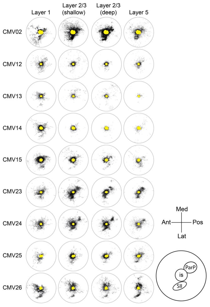
Fig. 3.
Tracings of axons within the 7.2-mm diameter analysis region of four sections from each of nine brains injected supragranularly with AAV-CMV-GFP. The similarity of axonal distributions across different depths in a given brain can be appreciated from this figure, as well as the differences in the absolute levels of labeling across different brains. The yellow area in the center of each tracing represents the extent of the saturated labeling at the core of the injection site in deep layer 2/3. Med, medial; Pos, posterior; Lat, lateral; Ant, anterior; SII, secondary somatosensory cortex; ParP, posterior parietal cortex; is, injection site
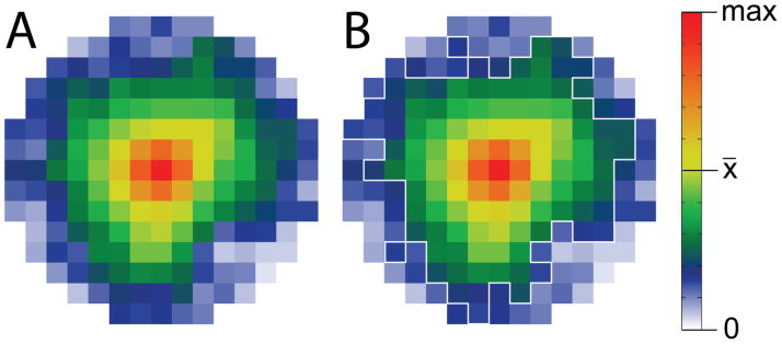
Fig. 7.
Average projection pattern across all brains and section depths for supragranular injections of AAV-CMV-GFP. A. After averaging across four section depths, data arrays corresponding to the 7.2 mm diameter analysis region were re-expressed for each of the nine brains injected with AAV-CMV-GFP in supragranular layers. The re-expressed arrays were then averaged across the nine brains to reveal a largely symmetrical axonal radiation in which axonal density declines with distance from the injection site. B. An outline of the areas displaying differences in one-tailed rank sum tests (p < 0.05) between injected brains and non-injected controls (Fig. 5) is superimposed in white over the average pattern
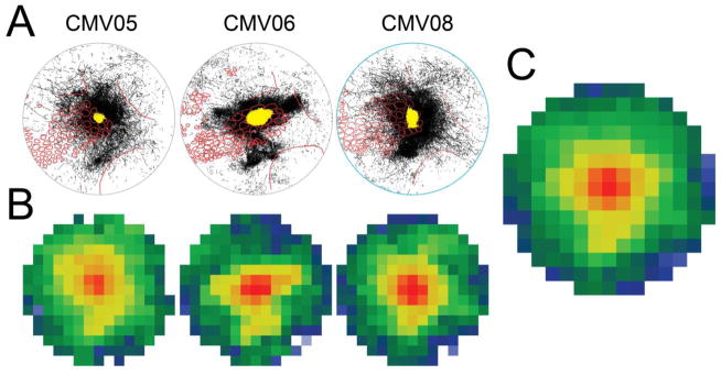
Fig. 8.
Projection patterns following infragranular injection of AAV-CMV-GFP. A. Tracings within the 7.2-mm diameter circular analysis region at four section depths were collapsed together for each of three brains injected infragranularly with AAV-CMV-GFP. These patterns can be compared with the patterns from supragranular injections shown in Fig. 4. The yellow area in the center of each tracing represents the extent of the saturated labeling at the core of the injection site in deep layer 2/3. B. Axonal densities resulting from the three infragranular injections were quantified as described for supragranular injections, averaged across section depth, and re-expressed as in the bottom row of Fig. 6. C. The re-expressed data arrays illustrated in B were averaged to reveal a largely symmetrical axonal radiation, similar to that found for supragranular injections (Fig. 7)
Quantitative analysis of fiber distributions
For each brain, images of sections subjected to cytochrome oxidase staining were copied into separate layers of Adobe Illustrator files and aligned to one other by matching the locations of blood vessels. Barrels and boundaries of other sensory cortices (Wong-Riley and Welt 1980; Wallace 1987) were identified and traced in reference to these sections. Locations of barrels and boundaries of sensory cortices then were transferred to the montages of traced axons by aligning locations of blood vessels detected both in the immunostained sections and in the sections stained for cytochrome oxidase. Then, the traced sections were rotated to provide a best-fit match to the locations of the 27 posterior-most mystacial whisker barrels in a standard barrel map (Brett-Green et al. 2001).
A circle corresponding to a diameter of 7.2 mm was centered on each injection site to define the region of interest for quantitative studies. This circle extended across all of PMBSF and included parts of auditory cortex, visual cortex, secondary somatosensory cortex, and the trunk representation within primary somatosensory cortex (see Fig. 2). For sections from non-injected controls, analysis circles were positioned to give a similar sampling of the cortical surface. Anti-aliased TIFF images of the fiber tracings (10800 pixels in diameter, 144 ppi, grayscale) within the analysis circle were exported for analyses in MatLab R2012a.
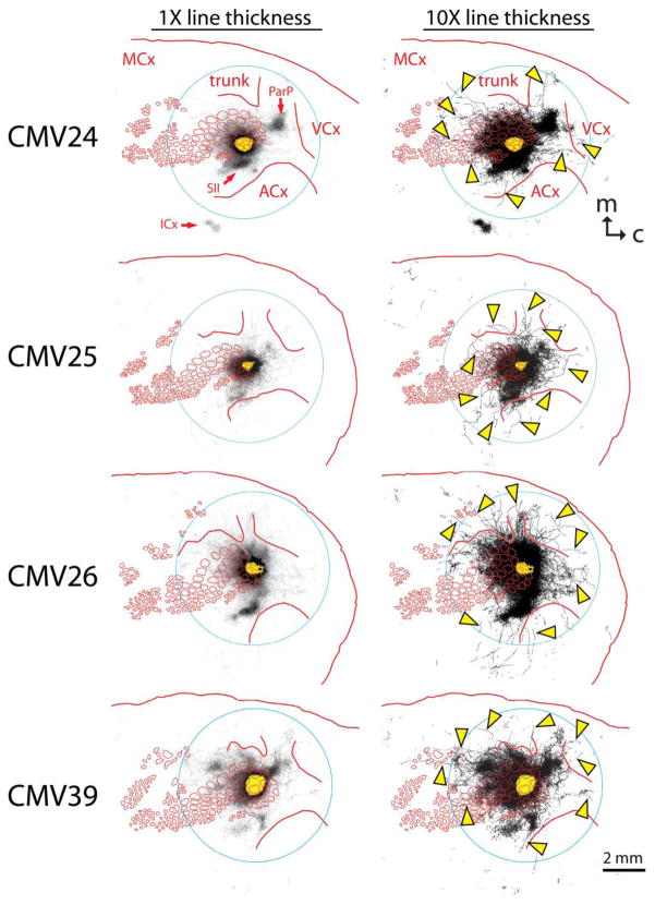
Fig. 2.
Labeled axons detected in the nine most shallow 40-μm sections from four brains injected with an AAV vector directing expression of GFP by way of a non-selective CMV promoter. Red lines indicate the locations of barrels and other cortical regions that were detected by staining layer 4 sections from the same brain for cytochrome oxidase activity. Left panels show tracings rendered using thin lines in order to appreciate the axons found within known specific projection areas including posterior parietal cortex (ParP), secondary somatosensory cortex (SII), insular cortex (ICx), and motor cortex (MCx). Right panels show tracings rendered using thicker lines in order to appreciate more sparsely distributed axons radiating in nearly all directions from the injection site. Several of the longer examples of these axons are indicated with arrowheads. Some of these axons crossed through dysgranular regions of cortex to penetrate the trunk region of somatosensory cortex (trunk), the visual cortex (VCx), and the auditory cortex (ACx). The yellow area in the center of each panel indicates the saturated immunostaining present in a section near layer 4, representing where the majority of the neurons infected by the virus were located (i.e., the “size” of the injection). This figure represents only a sampling of the axons that could have been reconstructed in these brains. Caudal (c) is to the right, and medial (m) is towards the top. The circles (7.2 mm in diameter) represent the area used for quantitative analysis in this study
In the most superficial sections that we analyzed, it was common for the analysis circle to extend past the section edge. Occasionally, the region of interest for quantification in these superficial sections also contained tears or large blood vessels running in the plane of the section. To distinguish between such regions of missing tissue and intact regions lacking axons, we also traced the missing areas in a separate layer of the Illustrator document and exported each of these tracings as a TIFF image to be used as a mask during analyses in MatLab (tan areas in Online Resource 2, column 2).
For each set of sections traced by a given researcher, the standard images that were included to characterize interobserver variability were evaluated for the total amount of tracing. These values were then expressed as a fraction of the mean value across all section sets traced by all researchers. This fraction was used to adjust proportionally the thicknesses of tracings using “erode” and “dilate” functions in MatLab to help remove systematic differences in tracing tendencies across researchers and/or analysis sets.
The TIFF images were converted to 500 × 500 item comma-separated value files in which black was assigned a value of 1 and white (no tracing) a value of 0. After application of the off-section masks and tracer-set normalizations, these arrays were resized to create 15-pixel diameter arrays (Online Resource 2, column 3), each cell representing the density of traced axons within a square area measuring 0.48 mm on a side. For statistical comparisons of the nine supragranular-injected brains and the corresponding six controls, these resized arrays first were averaged across the different section depths for each brain (Online Resource 2). Values from injected brains and non-injected controls then were compared on a cell-by-cell basis using a one-tailed rank sum test in MatLab.
Different sections and different brains were found to possess different levels of GFP immunostaining possibly related to differences in injected volume of vector, batch of vector, time of storage of vector, conditions of immunohistochemical staining, and/or microscopy and imaging parameters. In addition, the range of axonal densities across the analysis region was large, as the very numerous and heavily labeled axons and dendrites at the injection site produced a saturated image using microscopy and imaging settings that were optimal for viewing the sparsely distributed axons at greater distances from the injection site. To visualize patterns of axonal projections independently of the absolute intensity of the staining and to visualize the full range of axonal densities across the analysis region, the arrays were re-expressed further using the equation: y = x^(log(0.5)/log(x̄)), where x represents the original value at each cell of the resized array and x̄ represents the mean value across the analysis region for the same section. This re-expression results in a value of 0.5 at locations within each analysis region that represent the mean staining for the analysis region in that section, while values of 0 continue to represent an absence of staining, and values of 1 continue to represent saturated levels of staining such as usually characterized the injection sites. Arrays re-expressed in this manner were used for the false-color illustrations of staining patterns of individual sections (Online Resource 2, column 4). To visualize the average labeling across different sections within the same brain, values were averaged before re-expression. To visualize the average axonal distributions across different brains at each section depth and to visualize the grand average of axonal distributions across all brains (Fig. 6), averages were calculated after re-expression.
Fig. 6.
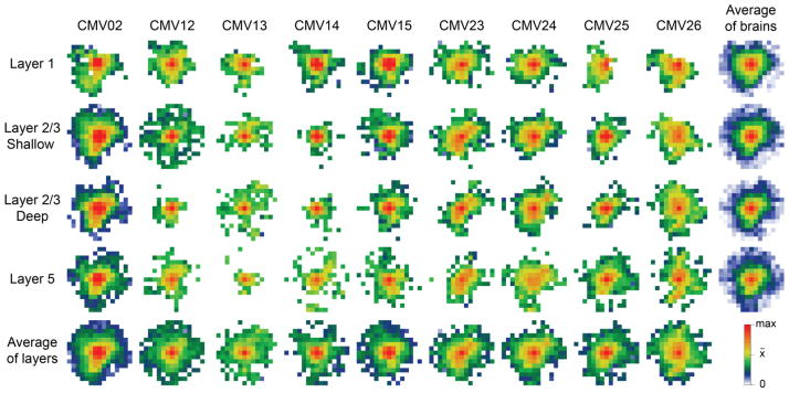
Quantification of relative axonal densities following supragranular injections of AAV-CMV-GFP. To compare projection patterns across brains that exhibited different absolute labeling intensities and to visualize the full range of axonal densities across each 7.2-mm diameter circular analysis region, data arrays measuring 15 pixels across were re-expressed such that the average labeling was assigned a value of 0.5. Values were then visualized by using a pseudocolor look-up table as indicated at bottom right. The bottom row represents re-expressed arrays after averaging across the different section depths within the same brains, revealing the similarity of projection patterns across different brains. The right column represents the averages after re-expression of arrays for different brains at each section depth, revealing the similarity of projection patterns in different cortical laminae
Results
Qualitative description of projection patterns following supragranular injections of AAV-CMV-GFP
Fig. 2 (left panels) illustrates the axon segments detected in the nine most superficial 40-μm transverse sections of flattened cortex from four brains injected with viral vector into layer 2/3 of PMBSF. We observed very heavy labeling for about two barrel diameters around the injection site as well as in areas known to receive specific projections from neurons in PMBSF (Chapin et al. 1987; Koralek et al. 1990; Fabri and Burton 1991; Aronoff et al. 2010). These areas included secondary somatosensory cortex (SII), posterior parietal cortex (ParP), insular cortex (ICx), and motor cortex (MCx). In addition to these well-known specific projections, axon segments also were detected at considerable distances from the injection site in multiple directions not obviously related to the specific projection sites. To better illustrate the locations of these more sparsely distributed axon segments, we have used a thicker line width in the right panels of Fig. 2. The use of thicker lines establishes that the local radiation of axons actually extends more than two barrel diameters from the edges of the injection sites, in most cases leaving the barrel field entirely to impinge on cytochrome oxidase-defined borders of other primary sensory cortices. Segments of sparsely distributed axons (arrowheads in Fig. 2, right panels) were found over anterior whisker barrels and at far distances (>3mm) within regions of secondary sensory cortices (associative dysgranular cortices) located between unimodal sensory cortices. Labeled axon segments also crossed over cytochrome oxidase borders to enter the trunk region of somatosensory cortex, the auditory cortex (ACx) and the visual cortex (VCx). Overall, these sparsely distributed axon segments suggest the presence of a nearly symmetrical, diffuse, horizontal projection radiating for great distances from neurons at the injection site.
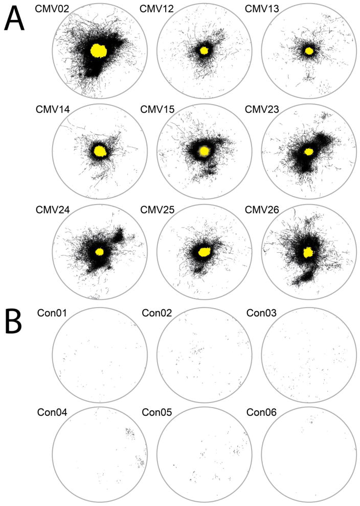
The axon segments contained within the region outlined with the circles in Fig. 2 were of greatest interest in the present study, as this region corresponds to the area displaying changes in evoked local field potential following stimulation of either a single whisker (Frostig et al. 2008) or sets of adjacent whiskers (Chen-Bee et al. 2012)(Fig. 1). To address axon distributions across different animals as well as the potential contribution of falsely detected fibers to the projection patterns contained in this region, we conducted a quantitative analysis involving coded images (see Methods). Fig. 3 shows tracings of the axon segments detected during this blind analysis in the circular region of four transverse sections from each of nine brains injected with the AAV-CMV-GFP vector. The four sections included three supragranular sections corresponding to layer 1, shallow layer 2/3, and deep layer 2/3, as well as one infragranular section from shallow layer 5. In all cases, the injection site was labeled heavily in comparison to other areas of the analysis region. To illustrate the overall distribution of labeled axon segments independently of their depth in the section, the tracings also were collapsed across layers (Fig. 4A).
Fig. 4.
A. Axons shown in Fig. 3 were collapsed across all four sections for each of the nine brains injected supragranularly with AAV-CMV-GFP. B. Falsely detected fibers within the analysis region of six non-injected controls were collapsed across sections at the four depths that were analyzed in injected brains. These fibers were traced in coded images interspersed among those comprising the results shown in A. The yellow area in the center of each tracing represents the extent of the saturated labeling at the core of the injection site in deep layer 2/3. The circles measure 7.2 mm in diameter
As originally reported by Bernardo et al. (1990), and repeatedly confirmed in subsequent reports (e.g., Stehberg et al. 2014; Narayanan et al. 2015), axons immediately surrounding the injection sites within the barrel field tended to extend further along whisker row representations than they did across whisker arcs. This well-known phenomenon was not explored further in the present analyses. Labeling of SII (see inset of Fig. 3 for location within the analysis circle) often could be seen extending laterally from the posterior half of the injection site and then curving anteriorly. A patch of labeling corresponding to ParP also was frequently detected in these sections. Despite these similarities, the pattern of labeling was somewhat different from brain to brain. On the other hand, projection patterns in sections at different depths from a given brain were very similar to one another. All sections of all brains yielded evidence of labeled axon segments distributed throughout the analysis circle in regions other than SII and ParP.
The researchers who detected and traced the axons for our analyses were trained to include short and faintly stained fragments in order to adequately assess the full spatial extent of the projection. Although this training emphasized the identification of features characteristic of axons or axon collaterals and also included samples of non-injected tissue in order to encourage discrimination of real axon segments from distracting features in the background staining that might resemble axon fragments (e.g., capillary walls, chance arrangements of elevated intensity along the cell membranes of adjacent unstained neurons, etc.), we found that some fragments continued to be identified in the non-injected tissue even after extensive experience. We therefore included in the blind analysis coded images of sections from non-injected tissue in order to assess this false discovery rate (see Methods). The distributions of these falsely detected fibers are illustrated in Fig. 4B, where tracings from four sections at different depths from six controls have been collapsed in the same manner used to generate Fig. 4A. Far fewer segments were traced in these non-injected controls than were traced in sections from injected brains, and the traced segments tended to be shorter in the non-injected controls. The falsely detected fibers were for the most part evenly distributed across the analysis region.
Statistical comparison of tracings from supragranularly injected brains and non-injected controls
Due to the false detection of some fibers in our analysis, it was necessary to determine whether the density of traced axon segments at distances from the injection site in injected tissue exceeded the density of tracings resulting from false discovery. As described in greater detail in Methods, TIFF images corresponding to the individual section data in Fig. 3, as well as comparable images from individual sections of non-injected control brains, were converted into data arrays in MatLab, transformed to address systematic differences in tracing among different tracers (as well as among different analyses conducted by the same tracers), and resized to produce arrays that were 15 pixels across (see Online Resource 2). In these resized arrays, each pixel represents the mean value within a square area of the section measuring 0.48 cm on a side. The values from the four sections of the same brain then were averaged together (see Online Resource 2), and the nine average arrays from injected brains were compared to the six corresponding arrays from non-injected controls using cell-by-cell, one-tailed rank sum tests.
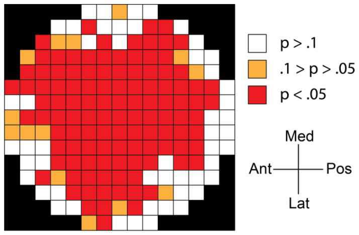
Red squares in Fig. 5 indicate the locations in the circular analysis region where the overlap between fiber densities in injected and non-injected brains was so low as to return a significant (p < 0.05) result in the rank sum test. Differences were found across the great majority of the analysis region, in some cases extending to the pixels furthest (3.12–3.60 mm) from the center of the injection site. These data indicate that, across multiple animals, labeled axons are distributed throughout the majority of the analysis region, with long axons extending nearly to the outer boundaries.
Fig. 5.
Spatial distribution of significant differences in tracing density between brains injected supragranularly with AAV-CMV-GFP and non-injected controls. Images of traced fibers from the 7.2-mm diameter circular analysis region such as shown in Fig. 3 were converted to data arrays measuring 15 pixels across (see Online Resource 2). Arrays from four different section depths of the same brain were averaged, and the nine injected brains were compared to six non-injected controls, applying one-tailed rank sum tests to identify locations where the tracing density in injected brains reliably exceeded the tracing density in non-injected controls. Individual square areas (0.48 cm on a side) where individual rank sum tests yielded p values below 0.05 were colored red to illustrate the large spatial extent of the reliable differences between injected brains and non-injected controls. Med, medial; Ant, anterior; Lat, lateral; Pos, posterior
Quantification of the relative distribution of axons in supragranularly injected brains
Whereas the statistical comparison between injected brains and non-injected controls establishes the presence of axons throughout the region previously shown to be activated by the stimulation of single whiskers or combinations of whiskers (Chen-Bee et al. 2012), it does not give an indication of the overall relationship between the density of axons and the distance and direction from the center of the injection. Axonal density would be expected to decline steadily with distance in all directions if this projection underlies the activity spread (Fig. 1), which itself shows a near-symmetrical diminution in amplitude with increasing distance from the peak of activation (Brett-Green et al. 2001; Frostig et al. 2008; Chen-Bee et al. 2012).
Characterization of the central tendency of the relationship between relative axonal density and location across different brains required a correction for the different absolute levels of labeling that were observed (Fig. 3). Also, the axonal density at the periphery of each analysis region was much lower than in the injection sites and in the specific projection areas, so that a non-linear scaling was required to visualize the full range of axonal density. We chose to re-express the values in the 15-pixel diameter arrays using the equation: y = x^(log(0.5)/log(x̄)), where x represents the original value at each cell of the resized array and x̄ represents the mean value across the same array. Following this transformation, a value of 0.5 represents the mean staining for that array, a value of 0 represents an absence of staining, and a value of 1 represents saturated staining such as present in the core of injection sites. The resulting values were visualized using a heat map where warmer colors indicated higher densities, cooler colors indicated lower densities, and the average value for the array occurred at the transition between yellow and green. Fig. 6 displays the arrays for the four individual sections of the nine injected brains.
For each brain, axonal densities also were averaged across the four section depths and then re-expressed by the same formula used for the individual sections (Fig. 6, bottom row). The similarity in projection patterns across the different brains is clearly apparent. In all brains, axonal density tended to fall off gradually in all directions with increasing distance from the injection site, although the stronger projections to SII and ParP remain evident. For each section depth, arrays were averaged across brains after re-expression (Fig. 6, right column). The projection pattern was very similar at the different depths, with axonal density decreasing in all directions with increasing distance from the centers of the injection sites. It should be noted that the absolute density of axons in layer 5 was lower than in layer 2/3 for most brains, despite the similarity in pattern, as is evident from the original tracings shown in Fig. 3.
Our understanding of the mesoscopic spatial characteristics of the activity spread shown in Fig. 1 is based on averaging results across multiple animals. To determine if the horizontal axonal projection had spatial characteristics predicting this activity spread, we similarly averaged the re-expressed axonal densities across animals. Fig. 7A shows the grand average of projection patterns across all brains (the average of the bottom row of Fig. 6), and Fig. 7B shows this same average overlain with an outline (white) of the areas showing p < 0.05 in the rank sum test (Fig. 5). The overall average density shows a remarkable level of radial symmetry, although there was a lesser density of traced axons at the perimeter of the analysis circle in the direction of the auditory cortex (bottom right portion of the analysis region).
Axonal projection patterns following infragranular injections of AAV-CMV-GFP
In addition to the nine brains injected supragranularly, we also injected AAV-CMV-GFP into layer 5 of three brains (injection depth of 1.0 mm) and analyzed the patterns of projection in the same manner as for the supragranular injections. Axons were found to extend in all directions from the infragranular injection sites (Fig. 8). There was a concentration of axon segments associated with SII, but axons also were distributed in all directions and scattered throughout the 7.2-mm diameter analysis region (Fig. 8A). Quantification of axonal density showed a diminution of axonal density with distance towards the periphery of the analysis region in all three brains (Fig. 8B). The average pattern across the three infragranularly injected brains (Fig. 8C) was remarkably similar to the average pattern resulting from supragranular injections (Fig. 7), displaying a largely symmetrical radiation of axons outward from the injection site. The projection patterns also were similar at the four section depths we investigated.
Reconstruction of individual long horizontal axons
We considered the possibility that fibers detected in our quantitative analyses could include axons that coursed through white matter for varying distances before rising into the sections used for tracing. We therefore wished to determine if we could follow individual axons continuously as they traversed gray matter from the injection site to reach locations and distances comparable to the extent of the previously measured activity spread. We examined the long axon segments in the nine most superficial immunostained sections from two brains (CMV26 and CMV39, illustrated in Fig. 2), and we identified candidate intrinsic axons that appeared to be moving up or down from one section to another from near the injection site to the axon terminus. These segments were imaged again at higher magnification (100X objective) in numerous focal planes, and they were stitched together using blood vessel patterns and other features that were common to the adjacent sections. Although this analysis was limited to the most superficial laminae of gray matter (layers 1, 2, and part of 3), numerous individual intrinsic axons were successfully reconstructed, and although the long fibers were relatively rare compared to the high density of axons immediately surrounding the injection sites, the axons that we reconstructed represent only a subset of the axons present at considerable distances from the injection sites. We also observed continuous axons moving up and down between up to three adjacent infragranular sections, but we have not yet been able to completely reconstruct such axons due to our use of every fourth infragranular section for cytochrome oxidase staining.
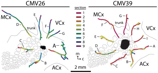
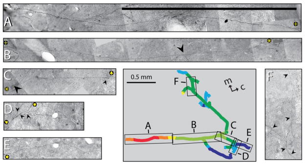
The locations of some of the reconstructed intrinsic supragranular axons are shown in Fig. 9, and some details of the appearance of one of the reconstructed axons are shown in Fig. 10. As shown in Fig. 9, some reconstructed axons extended anteriorly across the PMBSF (CMV26, axon C; CMV39, axon D), and others projected beyond the barrel field to terminate in the somatosensory dysgranular zone between PMBSF and the trunk region (CMV39, axon E), in the trunk region of somatosensory cortex (CMV39, axon G), in dysgranular cortex located between visual cortex (VCx) and somatosensory cortex (CMV26, axon E; CMV39, axons H and I), in the visual cortex (CMV26, axons F and G), in the region of secondary somatosensory cortex (CMV39, axon B), and in dysgranular cortex between somatosensory cortex and auditory cortex (ACx)(CMV26, axon A; CMV39, axon A). One reconstructed supragranular axon in each brain projected through the trunk region of somatosensory cortex to reach motor cortex (CMV26, axon D; CMV39, axon F).
Fig. 9.
Locations of reconstructed supragranular axons from two brains injected with AAV vectors employing CMV promoters. Axon segments were stitched together with segments located in adjacent sections by matching shared blood vessel and other staining patterns, and their locations were superimposed on the pattern of cytochrome oxidase staining obtained in layer 4 sections from the same brain to represent the borders of barrels and other unimodal sensory cortices. Axon segments were colored according to the section in which they were found (see key). The filled shapes around the injection sites represent the saturated labeling in section 9 (closest to the sections used for cytochrome oxidase staining) under imaging conditions that were optimal for the detection of sparse, stained axons at great distances from the injection site. Letters refer to individual axons referenced in the text. ACx, auditory cortex; VCx, visual cortex; MCx, motor cortex; trunk, trunk region of primary somatosensory cortex; m, medial; c, caudal
Fig. 10.
Photomontages detailing the reconstruction of parts of axon A from brain CMV26. Axonal segments were imaged using a 100X objective, and individual images were joined to create the montages. Superimposed rectangles and letters on the tracing in the inset (replicated from Fig. 9) indicate the portions of the axon that are illustrated in the separate smaller panels, and the colors indicate which section contained a given portion of the axon (see key in Fig. 9). Arrowheads indicate branch points, and pairs of yellow symbols on black circles in panels A–E indicate points of overlap between adjacent segments. Scale bars = 500 μm; m, medial; c, caudal
Many of the reconstructed axons extended close to or beyond the established 3.5-mm distance of the activity spread initiated by whisker stimulation. For example, at its furthest point, axon D of CMV26 extended 5.5 mm from the center of the injection site (5.3 mm from its edge), and axon F of CMV39 extended 3.7 mm and 3.2 mm from the center and edge of the injection site, respectively. In most cases, reconstruction of the proximal ends of the axons began around 0.5–1 mm from the edge of the injection site due to a very high density of stained axons in these regions that made it difficult to identify the continuation of axons crossing between adjacent sections. We consider it very likely that these axons would have remained in gray matter for the short distance remaining to the injection sites.
Many of the reconstructed axons branched as they traversed the superficial cortical laminae (Fig. 9, CMV26, axons A, B, C, E, F and G; CMV39, axons A, D, E, G, and H). Although we did not follow all branches to their termini, some of the details of an extensively branched axon (CMV26, axon A) are illustrated in Fig. 10. Over twelve termini were identified for this axon. The most rostral terminus (at the end of the medial branch) was located over 1.8 mm from the most caudal terminus, and the termini also were distributed across multiple depths within the supragranular layers, ranging from section 4 (120–160 μm deep) to section 8 (280–320 μm deep). Given this extensive branching, it seems likely that an individual axon like this one has the potential to affect activity over a large volume of cortex.
Given that we were able to reconstruct continuous supragranular axons projecting in multiple directions from the injection site to distances comparable to the spread of activity, we can conclude that the radiating, diffuse axonal projection likely includes intrinsic horizontal axons (i.e., axons that never enter white matter).
Axonal projection patterns following supragranular injections of AAV directing expression of YFP by way of a CaMKIIα promoter
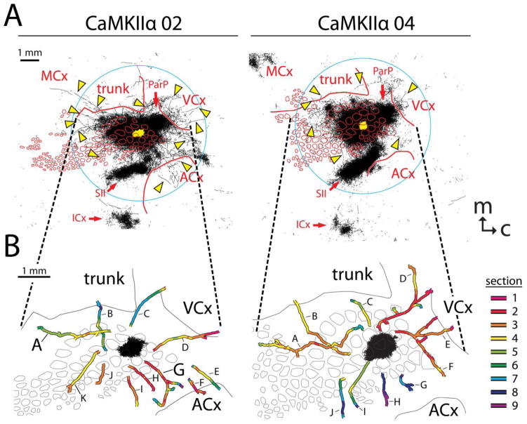
To determine whether the axons contributing to the diffuse, radiating projection pattern involve excitatory cortical pyramidal neurons, we injected additional brains with a vector causing expression of yellow fluorescent protein under the direction of the calcium/calmodulin-dependent protein kinase II alpha (CaMKIIα) promoter. CaMKIIα is limited in its expression primarily to the cortex, where it is found in cortical excitatory neurons and not in inhibitory neurons (Burgin et al. 1990; Wang et al. 2013). As shown in Fig. 11A, supragranular injections of this selective vector into two brains resulted in supragranular projection patterns that were very similar to those produced by injections of the permissive CMV vector (Fig. 2, left panels). Concentrated labeling was observed surrounding the injection site and extending out of the barrel field to impinge upon other primary sensory cortices, as well as within patches representing secondary somatosensory cortex (SII), posterior parietal cortex (ParP), insular cortex (ICx), and motor cortex (MCx). Sparser labeling (arrowheads in Fig. 11A) was found in anterior barrels and in dysgranular cortices between primary sensory cortices, as well as crossing into the trunk region of somatosensory cortex (trunk) and into visual (VCx) and auditory cortices (ACx). Therefore, axons from excitatory cortical pyramidal neurons contribute to the radiating projection pattern.
Fig. 11.
Projection patterns following supragranular injection of an AAV vector directing expression of YFP by way of a CaMKIIα promoter in order to restrict labeling to axons from excitatory cortical neurons. A. Tracings of axons detected in the nine most superficial 40-μm sections of flattened cortices were collapsed together as was shown in Fig. 2 for brains injected with the CMV promoter. As in the right panels of Fig. 2, thicker lines were used to better visualize the sparser axon segments (arrowheads) located outside of major specific projection targets. The yellow area in the center of each panel indicates the saturated immunostaining present in a section near layer 4 (the size of the injection). B. Locations of reconstructed long horizontal axons from the nine most superficial 40-μm transverse sections. Axon segments were stitched together and colored according to the section in which they were located, as described for Fig. 9. Letters refer to individual axons referenced in the text. Axons A and G from brain CaMKIIα 02 are further illustrated in Fig. 12. Axons labeled E and F in brain CaMKIIα 04 may represent a single continuous fiber; however, another axon crossed the proximal end of axon F in section 2 (red segment) at such a similar depth that the continuation of the fiber could not be unambiguously established. This figure represents only a sampling of the axons that could have been reconstructed in these brains. Dashed lines extending between A and B point out identical locations in the two illustrations. ACx, auditory cortex; VCx, visual cortex; MCx, motor cortex; ICx, insular cortex; ParP, posterior parietal cortex; SII, secondary somatosensory cortex; trunk, trunk region of primary somatosensory cortex; m, medial; c, caudal
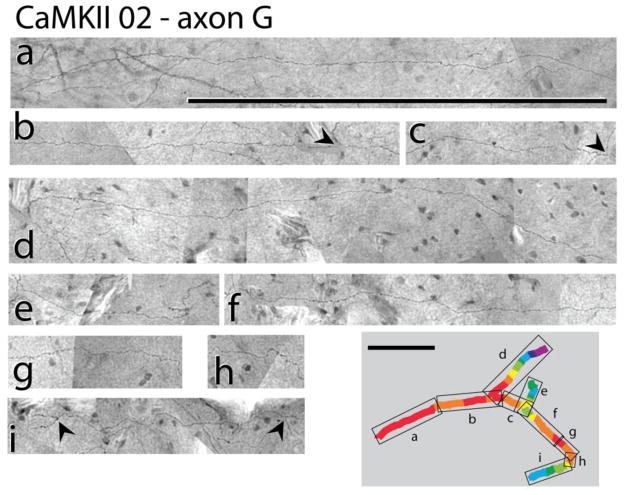
To determine whether supragranular axons labeled using the selective CaMKIIα vector stayed in gray matter throughout their course, we reconstructed some of the axons as was previously described for CMV26 and CMV39 (Fig. 10). As shown in Fig. 11B, numerous intrinsic horizontal axons were successfully reconstructed. Fig. 12 shows the details of the reconstruction of two of these axons. Some of the axons extended anteriorly across the PMBSF (CaMKIIα 02, axons J and K; CaMKIIα 04 axon A), and others were found to project beyond the barrel field to terminate in the somatosensory dysgranular zone between PMBSF and the trunk region (CaMKIIα 02, axon A; CaMKIIα 04, axon C), in the trunk region of somatosensory cortex (CaMKIIα 02, axon B; CaMKIIα 04, axon B), in dysgranular cortex located between visual cortex (VCx) and somatosensory cortex (CaMKIIα 04, axons D, E and F), in the visual cortex (CaMKIIα 02, axons C and D; CaMKIIα 04, axon E), and in dysgranular cortex between somatosensory cortex and auditory cortex (ACx)(CaMKIIα 02, axons G, H and I; CaMKIIα 04, axons E, F, G, H, I and J).
Fig. 12.
Photomontages detailing the reconstruction of two axons from brain CaMKIIα 02 (indicated by large capital letters in Fig. 11B). Axonal segments were imaged using a 100X objective, and individual images were joined to create the montages. Superimposed rectangles and lower case letters on the tracings of the axons in the bottom right panel of each reconstruction indicate the portions of the axons that are illustrated in the separate smaller panels, and the colors indicate which section contained a given portion of the axon (see key in Fig. 11B). Arrowheads indicate branch points, and arrows indicate features resembling boutons in CaMKIIα 02, axon A. Scale bars = 500 μm; m, medial; c, caudal
Reconstruction of the proximal ends of two axons crossing into the auditory cortex in brain CaMKIIα 02 (axons E and F) began in the dysgranular region between somatosensory cortex and auditory cortex. Because the axons dropped precipitously below our range of stained sections at this location, we were unable to trace their full projections, and it is uncertain whether the axons traversed gray matter all the way from the injection site.
Measured as the linear distance from the distal extremity of the axon to the center of the injection site, many of the reconstructed axons reached a distance that was comparable to the extent of the fiber radiation documented in the quantitative analysis (Fig. 7). For example, at its distal extremity, axon A of CaMKIIα 02 was 3.0 mm from the center of the injection site, while axon D of CaMKIIα 02 extended 2.7 mm from the center of the injection site. Axons A and B of CaMKIIα 04 extended 3.9 mm and 3.3 mm from the center of the injection site, respectively. In a few cases, the distal ends of axons rose above the first intact section that we were able to collect on the microtome knife (CaMKIIα 02, axon D; CaMKIIα 04, axons D, E and F). The actual lengths of these fibers therefore may be underestimated.
As was the case for the axons labeled using the permissive CMV vector, many of the reconstructed axons branched multiple times as they traversed the superficial cortical laminae (Fig. 11, especially CaMKIIα 02, axons A and G and CaMKIIα 04, axons A, D, E and F; Fig. 12, arrowheads). There also were sporadic short protuberances from some of the axons that were reminiscent of boutons (Fig. 12: CaMKIIα 02, axon A, arrows). The presence of extensive branching and possible boutons in axons labeled using the CaMKIIα vector suggests that even a single axon might exert excitatory synaptic influence over a large volume of cortex.
Discussion
Technical considerations
Although some researchers have reported that AAV vectors provide anterograde labeling almost exclusively (Chamberlin et al. 1998; McFarland et al. 2009; White et al. 2011; Harris et al. 2012; Oh et al. 2014), others have reported significant retrograde (Burger et al. 2004; Taymans et al. 2007; Cearley and Wolf 2007; Castle et al. 2014) and even trans-synaptic (Provost et al. 2004; Cearley and Wolf 2007; Castle et al. 2014) labeling using AAV vectors such as ours. We also observed sporadic labeling of cell bodies more than 500 μm from the injection sites, which likely reflects either retrograde or trans-synaptic labeling. Labeled cell bodies were evident in regions known to have reciprocal connections with PMBSF such as motor cortex, SII, ParP, and insular cortex, but they also were scattered to a lesser degree throughout the extent of the diffuse horizontal fiber radiation. We therefore cannot rule out the possibility that lightly labeled secondary axons from these cells might have made some contribution to our quantitative analysis of axonal distributions. However, during the reconstruction of axons following injections of the AAV vectors, the distal extremes of axons gave the appearance of terminal branching (e.g., see Fig. 10 and Fig. 12) and never led to a labeled cell body, suggesting that anterograde labeling underlies the axonal radiation visualized by using this approach. The restricted expression of the CaMKIIα vector to excitatory cortical neurons (Burgin et al. 1990; Wang et al. 2013) also argues against a subcortical origin of the reconstructed fibers. A combination of anterograde and retrograde labeling scattered across a more than 3-mm range of the cortical surface is consistent with a generalized presence of widespread, diffuse, horizontal connectivity between multiple neighboring regions of cortex that falls off with distance but that crosses cytoarchitectonic borders in both directions (Stehberg et al. 2014).
We injected virus in small boluses to restrict diffusion of viral particles throughout a large volume of cortex. However, even small volumes of virus would be expected to infect any cell with a dendrite or soma near the micropipette tip. Our injections were into cortical layers 2/3 or 5, and in these layers, pyramidal neurons have dendritic trees that extend horizontally to a radius of up to 0.5 mm (Feldmeyer, 2012). Therefore, cells would be expected to become labeled within 0.5 mm of the injection site in the transverse plane. This volume would span at least one barrel diameter in any direction, minimally labeling neurons associated with several barrels as well as their intervening septa. The cell labeling at the core of our injection sites was fully consistent with these dimensions. (The yellow or black areas around the injection sites in our figures further over-estimate the size of the injections in that they include dense axonal projections emanating from the injection sites.) Because our intention was to characterize horizontal projections contributing to a spread of activity evoked by stimulating a single whisker, the area of our injections was deemed appropriate to address anatomy underlying activity spreads following single whisker stimulation.
Presence and characteristics of a diffuse, radiating, horizontal projection system in rat barrel cortex
By using a combination of descriptive and quantitative approaches following injections of AAV tracers into the PMBSF, we have obtained further evidence that sensory cortex is comprised of two major cortical systems: a specific system that projects through white matter to specific targets and a diffuse horizontal system, a dual pattern found also in primary auditory and visual cortices (Stehberg et al. 2014). Specific projections from rat barrel cortex include well-known targets such as SII and ParP (Chapin et al. 1987; Koralek et al. 1990; Fabri and Burton 1991; Kim and Ebner 1999; Aronoff et al. 2010; Oberlaender et al. 2011; Lee et al. 2011; Kim and Lee 2013), while the diffuse system projects nearly symmetrically (Frostig et al. 2008; Stehberg et al. 2014).
Our analysis following AAV vector injections has shown that the diffuse system involves a radiating network of long, intrinsic horizontal axons running through cortical gray matter with spatial features similar to the horizontal spread of activity initiated by whisker stimulation (Fig. 1). Individual axons were found to extend across and beyond the barrel field in all directions, branching and probably forming synapses with other neurons along their paths. The densities of labeled axon segments were quantitatively significant at distances greater than 3 mm from the center of the injection sites, and individual axons could be followed through superficial cortical laminae farther than 3.5 mm, which is consistent with the subthreshold activity spreading at least 3.5 mm through cortex in various direction (3.5 mm represented the furthest electrode tested, and the average evoked local field potential was still 11% of peak magnitude at that location) (Frostig et al. 2008). The intrinsic horizontal axons radiated in all directions from the injection site, and except within the well-known specific projection regions SII and ParP, axonal density decreased with increasing distance from the injection site, resembling the steady and largely symmetrical decrease in the strength of activity with distance from the peak of activation (Frostig et al. 2008; Chen-Bee et al. 2012).
Alternative pathways for widespread cortical activation upon single whisker stimulation
There are a number of pathways through which activity evoked by a single whisker might diverge en route to the cortex to produce widespread cortical activation following single whisker stimulation. Neurons in barreloids of the ventral posteromedial thalamus that project to barrel columns as part of the lemniscal pathway have been shown to possess multi-whisker receptive fields so that neurons in numerous barreloids can be activated by stimulating a single whisker (Armstrong-James and Callahan 1991; Simons and Carvell 1989; Nicolelis and Chapin 1994). Therefore, numerous barrels would be expected to become activated by the simple relay of this widespread spatial pattern of thalamic activity to the barrel field. Individual ventral posteriomedial thalamic neurons also have been shown to diverge in their projections to multiple cortical barrels (Arnold et al. 2001), which would be expected to lead to additional broadening of the area of activation by a single whisker. Neurons in the medial division of the posterior thalamic complex (POm) also have multi-whisker receptive fields (Chiaia et al. 1991; Diamond et al. 1992), receiving input from multi-whisker responsive neurons in the spinal trigeminal sensory complex (Jacquin et al. 1986; 1989). POm neurons project to septal columns between the cortical barrels as part of the paralemniscal pathway (Koralek et al. 1988; Chmielowska et al. 1989; Lu and Lin 1993; Wimmer et al. 2010), as well as to SII and the dysgranular zone within primary somatosensory cortex (Carvell and Simons 1987; Koralek et al. 1988; Viaene et al. 2011), providing a pathway by which a broad spatial pattern of activity might be relayed directly to barrel cortex and beyond. Finally, multi-whisker receptive neurons in the ventral lateral part of the ventral posteriomedial nucleus of the thalamus also project to septal columns, SII, and the dysgranular zone (Pierret et al. 2000; Bokor et al. 2008), and other thalamic neurons project even more diffusely to the cortex (Rubio-Garrido et al. 2009).
Despite these multiple potential thalamocortical pathways towards widespread cortical activation, transection of gray matter orthogonal to the cortical surface blocks the propagation of activity outside the barrel field (Frostig et al. 2008), which supports the hypothesis that horizontal fibers within cortex are responsible for the spread of activity beyond the barrel field following single whisker stimulation. Relative latencies of responses to “surround” whiskers in cortex and thalamus suggest that the spatially broad responses within the cortical barrel field also involve lateral connections among cortical neurons (Armstrong-James et al. 1991; Armstrong-James and Callahan 1991; Brecht et al. 2003; Katz et al. 2006), and results of pharmacological interventions further support the involvement of lateral connections in generating the surround responses displayed by barrel column neurons (Fox et al. 2003; Wright and Fox 2010).
Although the long, intrinsic horizontal axons or axon collaterals described in the present study might spread activity monosynaptically throughout the area of the activity spread, these data do not rule out the possibility that polysynaptic propagation also contributes to the measured spread of activity. Indeed, a polysynaptic contribution seems likely, as action potentials can be detected from cells an average of 1.5 mm away from the peak of activation, about half the distance of the measured spread of the evoked local field potential (Masino 2003; Frostig et al. 2008). In some animals, action potentials could be measured up to 2.5 mm away from the location of peak activity (Frostig et al. 2008). Postsynaptic activity at the ends of horizontal axons emanating from these secondarily spiking neurons likely contribute to evoked changes in local field potential.
Diffuse, horizontal axonal radiations are observed at multiple cortical depths
Following supragranular vector injections, there was a remarkable similarity in the horizontal axonal projection pattern at different cortical depths, representing axons in both supragranular and infragranular laminae (Johnson and Frostig 2015). This similarity was clearly evident in individual brains, and the average pattern further revealed that the general characteristics of a diffuse radiation of axons occurred at all depths that we investigated. This finding is consistent with the similarity in the symmetrical spread of the local field potential and neuronal spiking measured both supragranularly and infragranularly (Frostig et al. 2008). Supragranular injections of vector result in infection of supragranular neurons as well as infragranular pyramidal neurons that have apical dendrites in these superficial laminae (Feldmeyer 2012), so that it is not possible for us to attribute the diffuse projection pattern to any particular cell type defined by morphology or laminar location revealed using single-cell labeling methods (Narayanan et al. 2015). We found that infragranular injections of vector also resulted in a diffuse radiation of axons at all cortical depths, suggesting that the infragranular neurons make an important contribution to the horizontal axonal radiation. These results are consistent both with electrophysiological data showing an important contribution of infragranular neurons to the horizontal spread of activity in supragranular layers of cortical slices (Wester and Contreras 2012) and with imaging data showing that horizontal spreads of local cortical activity can be initiated optogenetically by point stimulation in transgenic mice expressing channelrhodopsin primarily in pyramidal neurons located in infragranular layer 5A of barrel cortex (Lim et al. 2012; Mohajerani et al. 2013).
It recently has been reported that certain types of individually labeled pyramidal neurons defined by morphology and laminar location differ in the eccentricity of their horizontal projections within the barrel field such that some neurons project axon collaterals more along a whisker row than along a whisker arc in supragranular layers, whereas other neuron types project more along an arc than along a row in infragranular layers, although collaterals extended along both axes for all cell types (Narayanan et al. 2015). These differences were observed for collaterals that extended about one barrel column (~500 μm) away from the labeled soma (Narayanan et al. 2015), which would correspond to the very dense regions within and adjacent to the injection sites in our studies. We have not attempted to address such differences in the eccentricity of local axon projections at different cortical depths following our supragranular versus infragranular injections, as we were more concerned with the horizontal axon segments that extended in parallel with the activity spread (i.e., outside the barrel field or several barrels away within the barrel field). Any such analysis of eccentricity would be complicated for our injections, all of which would be expected to label a mixture of the “rowish” and “arcish” neuron types. Furthermore, the analysis of Narayanan et al. (2015) was based on axons characterized in a sample of 74 individually labeled excitatory neurons (representing 10 cell types), none of which were reported to exhibit axon collaterals that extended as far as the axons we have reconstructed following our viral vector injections. Given the sparsity of the axons contributing to the diffuse projection pattern we have observed, it seems likely that a greater number of neurons would need to be characterized for the morphology and laminar specificity of the cells responsible for the long projections we have characterized to be represented in their data set.
The diffuse horizontal axon radiation includes projections from excitatory neurons
We detected horizontal axonal radiations following injections of two different tracing vectors. One vector expressed GFP under the permissive CMV promoter, which should direct high levels of expression in all infected cells, including excitatory and inhibitory neurons of every subclass (Nathanson et al. 2009), whereas the second vector should restrict expression primarily to excitatory pyramidal neurons by way of the CaMKIIα promoter (Burgin et al. 1990; Wang et al. 2013). Because the latter promoter revealed the presence of long, intrinsic horizontal axons reaching across the extent of the diffuse axonal radiation in all directions, part of the horizontal activity spread recorded by intrinsic signal optical imaging and recording of evoked local field potentials may represent a spread of primary excitation by way of axons or axon collaterals of pyramidal neurons. These data are consistent with the measurement of spiking units up to 2.5 mm from the peak of activation (Frostig et al. 2008).
The present anatomical data do not distinguish whether the excitatory axons contact excitatory or inhibitory postsynaptic neurons; nor can they exclude a contribution of inhibitory axons to either the overall axonal radiation detected using the permissive CMV promoter or the activity spread measured by intrinsic signal optical imaging and recording of evoked local field potentials (Frostig et al. 2008; Chen-Bee et al. 2012). Indeed, some inhibitory neurons in rat somatosensory cortex extend axons horizontally to a distance of several barrel columns from the cell body (Helmstaedter et al. 2008). Although the CaMKIIα promoter is preferentially expressed in pyramidal neurons, the present data also cannot exclude the possibility that the neurons projecting the long horizontal axons represent some uncharacterized subclass of neuron that expresses CaMKIIα.
Apparent novelty of the long, diffusely radiating, horizontal projections from barrel cortex
The extent and near symmetry of the long, diffuse, intrinsic horizontal axonal radiation we report here and elsewhere (Frostig et al. 2008; Stehberg et al 2014; Johnson and Frostig 2015) have not been discussed in the numerous prior studies of cortico-cortical efferent connections of barrel cortex. Long horizontal connections between neurons in other sensory cortices have been established in multiple species (see, for example, Stettler et al. 2002; Martin et al. 2014). In rat barrel cortex, horizontal projections have generally been described as extending only one or two barrel diameters in any given direction for pyramidal neurons in barrel columns, and for two to four barrel diameters in any given direction for neurons in septal columns, although evidence for a sparser projection beyond this extent often has been present in photomicrographs (Chapin et al. 1987; Koralek et al. 1990, Bernardo et al. 1990; Fabri and Burton 1991; Hoeflinger et al. 1995; Gottlieb and Keller 1997; Kim and Ebner 1999; Broser et al. 2008; Oberlaender et al. 2011; Lee et al. 2011; Kim and Lee 2013; Narayanan et al. 2015). Prior studies in rats (Miller and Vogt 1984; Paperna and Malach 1991) and other rodents (Budinger et al. 2006; Larsen et al. 2009; Campi et al. 2007; 2010; Charbonneau et al. 2012; Laramée et al 2013; Henschke et al. 2015) resulted in reports of sparse projections from somatosensory cortex to other primary sensory cortices, but most of these studies employed retrograde neuronal tracers and therefore could not have appreciated that the axons involved coursed diffusely through gray matter, branching along the way. Although neurons in the barrel field are known to have long projections to the dysgranular zone of the somatosensory cortex and to the posterior parietal cortex (Kim and Ebner 1999; Oberlaender et al. 2011; Lee et al. 2011; Kim and Lee 2013), these axons typically are not discussed as being abundant in other associational cortices posterior to PMBSF.
The long axons involved in the diffusely radiating projection that we have described certainly are encountered more rarely than those near the injection site and those involved in clustered specific projections to targets such as the motor cortex, SII, ParP, and insular cortex, and they may have been overlooked or ignored in earlier tract-tracing efforts in favor of describing the denser projections. Although the density of the long, diffusely distributed axons may be low relative to the previously well-known specific projections, the absolute volume of cortex potentially impacted by these horizontal axons is likely to be quite large (Boucsein et al. 2011). Even in our analyses, the extent of the diffuse projection may have been underestimated due to the sampling methods we employed. For example, the axons shown in Fig. 2 and Fig. 11 included only those detected in the nine most superficial sections, and the axons in our quantitative analyses (Fig. 3) represented only four sections chosen to sample different laminae, whereas the entire depth of cortex usually extended through at least 30–32 of the 40-μm transverse sections that we collected through the flattened cortical tissue. Furthermore, our search for axons involved two focal planes imaged through a 20X objective, whereas additional axon segments often became apparent during fiber reconstruction, when we viewed the same tissue using many more focal planes imaged through a 100X objective.
In contrast to many earlier studies, we chose a modern viral tracer and sensitive detection techniques, including amplification by way of avidin-biotin complexes, and we sought explicitly to determine if long, horizontal axons might underlie a previously detected pattern of neural activity. Recently, other researchers working on elucidating the cortical connectome have accumulated data suggesting more profuse efferent projections from the rat (Zakiewicz et al. 2014) and mouse (Zingg et al. 2014; Oh et al. 2014) barrel cortex than were reported in early studies, although the coronal section plane and comprehensive mapping strategies preferred in these reports likely obscure the radiating nature of the mesoscopic projection (Johnson and Frostig 2015). Indeed, the data mining in the analysis of the connectome typically imposes preconceived notions of cortical parcellation that would divide some of the continuous patterns of connectivity we have described into discrete regions before quantitative comparisons are performed (Bota et al. 2012; Zingg et al. 2014; Oh et al. 2014). To distinguish adequately a relatively sparse projection from false detection of fibers, we used a statistical approach involving injections into the same cortical region in multiple animals, whereas fewer injections into each of multiple distinct locations are employed by others to complete the connectome (Zingg et al. 2014; Oh et al. 2014). Likewise, the sparsity of fibers encouraged us to perform axonal reconstructions across a large set of meticulously aligned adjacent histological sections, a detailed strategy not currently exploited in the analysis of the connectome (Johnson and Frostig 2015). A combination of “big data” approaches such as the connectome projects and approaches targeted to answering specific questions such as we have done here is still required for a thorough understanding of cortical connectivity and function.
Functional implications of the diffuse, radiating projection system
As mentioned above, there is evidence that cortico-cortical connections can contribute to the generation of multi-whisker receptive fields in the barrel field, and cortico-cortical projections have been implicated in the facilitation or suppression of responses when multiple whiskers are stimulated simultaneously or sequentially (Brumberg et al. 1996; Shimegi et al. 1999; Mirabella et al. 2001; Hirata and Castro-Alamancos 2008; Chen-Bee et al. 2012; Sachdev et al. 2012). The horizontal axons we have characterized within the barrel field may participate in these phenomena.
The existence of long, horizontal axons radiating diffusely from the barrel field into other sensory cortices may be relevant to cross-modal responses to whisker stimulation that can be recorded in visual and auditory cortices of rats and other rodents (Wallace et al. 2004; Campi et al. 2007; Frostig et al. 2008; Larsen et al. 2009; Vasconcelos et al. 2011) and to the modulatory influence that whisker stimulation can have on responses to visual stimuli in rodent visual cortex (Iurilli et al. 2012). Horizontal projections also may contribute to the ability of stimuli from one modality to evoke activity in cortices corresponding to another modality following associative learning (Cahill et al. 1996). Long, border-crossing horizontal projections from the barrel field may play a role in deprivation-induced neural plasticity, such as when somatosensory stimuli evoke responses in the auditory cortex of congenitally deaf mice (Hunt et al. 2006) or when somatosensory evoked responses invade the visual cortex of rats following neonatal enucleation (Toldi et al. 1988; Wolff et al. 1992). Likewise, horizontal axons within the barrel field may contribute to the phenomenon in which the representations of spared whiskers expand into deprived cortical regions after a subset of whiskers is plucked or trimmed (Diamond et al. 1993; Armstrong-James et al. 1994; Glazewski et al. 1998; Polley et al. 1999; Marik et al. 2010). Finally, the predicted spread of activity carried by the long, horizontal axons diffusely radiating from the barrel field may explain how single whisker stimulation can protect large portions of the cerebral cortex from ischemic damage following blockage of the middle cerebral artery (Lay et al. 2010; 2013).
Supplementary Material
Acknowledgments
This work was supported by the United States National Institute for Neurological Disorders and Stroke (PHS grants NS-055832 and NS-066001). We thank Daniel D. Johnson for designing many of the quantitative approaches used here, and for writing custom MatLab software to facilitate data collection and analysis. We also thank the many students who performed imaging, tracing, and axon reconstruction described here: Joel Ramirez, Theodore Nieblas, Ajitesh Singh, Karishma Patel, Gordon Man, Roblen Guevarra, Raymond Jed Singson, Keli Tahara, Pejman Majd, Jason Louie, Rika Takada, Keith Uyeno, Jeremy Chen, Joel Fong, Velinda Liao, Bahram Rabbani, Marina Gerges, Julie Liang, Camillia Azimi, Maria Najam, Paul Lang, George Khamo, Hilda Zuñiga, Julian Huynh, Tiffany Do, Sean Siguenza, Ambrose Ha, Min Kim, Troy Ruff, Francisco Lee, Justin Hung, and John Hakim. We acknowledge further the creative contributions of Paul Lang, Camillia Azimi, Keli Tahara, Joel Ramirez, Roblen Guevarra, Gordon Man, Pejman Majd, Jason Louie, and Theodore Nieblas. Finally, we thank Cynthia Chen-Bee and Nathan Jacobs for valuable suggestions throughout the project and Dr. Cynthia Woo for comments on the manuscript.
Footnotes
Ethical standards
We declare that all animal studies have been approved by the appropriate ethics committee (see “Materials and methods”) and have, therefore, been performed in accordance with the ethical standards laid down in the 1964 Declaration of Helsinki and its later amendments. The manuscript does not contain clinical studies or patient data.
Conflict of interest
We declare that we have no conflict of interest.
Contributor Information
B.A. Johnson, Email: bajohnso@uci.edu, Department of Neurobiology and Behavior, University of California, Irvine, Irvine, CA 92697-4550
R.D. Frostig, Email: rfrostig@uci.edu, Department of Neurobiology and Behavior, University of California, Irvine, Irvine, CA 92697-4550. Department of Biomedical Engineering and Center for the Neurobiology of Learning and Memory, University of California, Irvine, Irvine, CA 92697
References
- Armstrong-James M, Callahan CA. Thalamo-cortical processing of vibrissal information in the rat. II. Spatiotemporal convergence in the thalamic ventroposterior medial nucleus (VPm) and its relevance to generation of receptive fields of s1 cortical “barrel” neurons. J Comp Neurol. 1991;303:211–224. doi: 10.1002/cne.903030204. [DOI] [PubMed] [Google Scholar]
- Armstrong-James M, Callahan CA, Friedman MA. Thalamo-cortical processing of vibrissal information in the rat. I. Intracortical origins of surround but not centre-receptive fields of layer IV neurones in the rat S1 barrel field cortex. J Comp Neurol. 1991;303:193–210. doi: 10.1002/cne.903030203. [DOI] [PubMed] [Google Scholar]
- Armstrong-James M, Diamond ME, Ebner FF. An innocuous bias in whisker use in adult rats modifies receptive fields of barrel cortex neurons. J Neurosci. 1994;14:6978–6991. doi: 10.1523/JNEUROSCI.14-11-06978.1994. [DOI] [PMC free article] [PubMed] [Google Scholar]
- Arnold PB, Li CX, Waters RS. Thalamocortical arbors extend beyond single cortical barrels: an in vivo intracellular tracing study in rat. Exp Brain Res. 2001;136:152–168. doi: 10.1007/s002210000570. [DOI] [PubMed] [Google Scholar]
- Aronoff R, Matyas F, Mateo C, Ciron C, Schneider B, Petersen CCH. Long-range connectivity of mouse primary somatosensory barrel cortex. Eur J Neurosci. 2010;31:2221–2233. doi: 10.1111/j.1460-9568.2010.07264.x. [DOI] [PubMed] [Google Scholar]
- Bernardo KL, McCasland JS, Woolsey TA, Strominger RN. Local intra- and interlaminar connections in mouse barrel cortex. J Comp Neurol. 1990;291:231–255. doi: 10.1002/cne.902910207. [DOI] [PubMed] [Google Scholar]
- Bokor H, Acsády L, Deschênes M. Vibrissal responses of thalamic cells that project to the septal columns of the barrel cortex and to the second somatosensory area. J Neurosci. 2008;28:5169–5177. doi: 10.1523/JNEUROSCI.0490-08.2008. [DOI] [PMC free article] [PubMed] [Google Scholar]
- Bota M, Dong H-W, Swanson LW. Combining collation and annotation efforts toward completion of the rat and mouse connectomes in BAMS. Front Neuroinform. 2012;6:2. doi: 10.3389/fninf.2012.00002. [DOI] [PMC free article] [PubMed] [Google Scholar]
- Boucsein C, Nawrot MP, Schnepel P, Aetrsen A. Beyond the cortical column: abundance and physiology of horizontal connections imply a strong role for inputs from the surround. Front Neurosci. 2011;5:32. doi: 10.3389/fnins.2011.00032. [DOI] [PMC free article] [PubMed] [Google Scholar]
- Brecht M, Roth A, Sakmann B. Dynamic receptive fields of reconstructed pyramidal cells in layers 3 and 2 of rat somatosensory barrel cortex. J Physiol. 2003;553:243–265. doi: 10.1113/jphysiol.2003.044222. [DOI] [PMC free article] [PubMed] [Google Scholar]
- Brett-Green B, Chen-Bee CH, Frostig RD. Comparing the functional representations of central and border whiskers in the rat primary somatosensory cortex. J Neurosci. 2001;21:9944–9954. doi: 10.1523/JNEUROSCI.21-24-09944.2001. [DOI] [PMC free article] [PubMed] [Google Scholar]
- Broser PJ, Erdogan S, Grinevich V, Osten P, Sakmann B, Wallace DJ. Automated axon length quantification for populations of labelled neurons. J Neurosci Methods. 2008;169:43–54. doi: 10.1016/j.jneumeth.2007.11.027. [DOI] [PubMed] [Google Scholar]
- Brumberg JC, Pinto DJ, Simons DJ. Spatial gradients and inhibitory summation in the rat whisker barrel system. J Neurophysiol. 1996;76:130–140. doi: 10.1152/jn.1996.76.1.130. [DOI] [PubMed] [Google Scholar]
- Budinger E, Heil P, Hess A, Scheich H. Multisensory processing via early cortical stages: connections of the primary auditory cortical field with other sensory sytems. Neurosci. 2006;143:1065–1083. doi: 10.1016/j.neuroscience.2006.08.035. [DOI] [PubMed] [Google Scholar]
- Burger C, Goratyuk OS, Velardo MJ, Peden CS, Williams P, Zolotukhin S, Reier PJ, Mandel RJ, Muzyczka N. Recombinant AAV viral vectors pseudotyped with viral capsids from serotypes 1, 2, and 5 display differential efficiency and cell tropism after delivery to different regions of the central nervous system. Mol Ther. 2004;10:302–317. doi: 10.1016/j.ymthe.2004.05.024. [DOI] [PubMed] [Google Scholar]
- Burgin KE, Waxham MN, Rickling S, Westgate SA, Mobley WC, Kelly PT. In situ hybridization histochemistry of Ca2+/calmodulin-dependent protein kinase in developing rat brain. J Neurosci. 1990;10:1788–1798. doi: 10.1523/JNEUROSCI.10-06-01788.1990. [DOI] [PMC free article] [PubMed] [Google Scholar]
- Cahill L, Ohl F, Scheich H. Alteration of auditory cortex activity with a visual stimulus through conditioning: a 2-deoxyglucose analysis. Neurobiol Learn Mem. 1996;65:213–222. doi: 10.1006/nlme.1996.0026. [DOI] [PubMed] [Google Scholar]
- Campi KL, Bales KL, Grunewald R, Krubitzer L. Connections of auditory and visual cortex in the prairie vole (Microtus ochrogaster): evidence for multisensory processing in primary sensory areas. Cereb Cortex. 2010;20:89–108. doi: 10.1093/cercor/bhp082. [DOI] [PMC free article] [PubMed] [Google Scholar]
- Campi KL, Karlen SJ, Bales KL, Krubitzer L. Organization of sensory neocortex in prairie voles (Microtus ochrogaster) J Comp Neurol. 2007;502:414–426. doi: 10.1002/cne.21314. [DOI] [PubMed] [Google Scholar]
- Carvell GE, Simons DJ. Thalamic and corticocortical connections of the second somatic sensory area of the mouse. J Comp Neurol. 1987;265:409–427. doi: 10.1002/cne.902650309. [DOI] [PubMed] [Google Scholar]
- Castle MJ, Gershenson ZT, Giles AR, Holzbaur ELF, Wolfe JH. AAV serotypes 1, 8, and 9 share conserved mechanisms for anterograde and retrograde axonal transport. Hum Gene Ther. 2014;25:705–720. doi: 10.1089/hum.2013.189. [DOI] [PMC free article] [PubMed] [Google Scholar]
- Cearley CN, Wolfe JH. A single injection of an adeno-associated virus vector into nuclei with divergent connections results in widespread vector distribution in the brain and global correction of a neurogenetic disease. J Neurosci. 2007;27:9928–9940. doi: 10.1523/JNEUROSCI.2185-07.2007. [DOI] [PMC free article] [PubMed] [Google Scholar]
- Chamberlin NL, Du B, de Lacalle S, Saper CB. Recombinant adeno-associated virus vector: use for transgene expression and anterograde tract tracing in the CNS. Brain Res. 1998;793:169–175. doi: 10.1016/s0006-8993(98)00169-3. [DOI] [PMC free article] [PubMed] [Google Scholar]
- Chapin JK, Sadeq M, Guise JL. Corticocortical connections within the primary somatosensory cortex of the rat. J Comp Neurol. 1987;263:326–346. doi: 10.1002/cne.902630303. [DOI] [PubMed] [Google Scholar]
- Charbonneau V, Laramée M-E, Boucher V, Bronchti G, Boire D. Cortical and subcortical projections to primary visual cortex in anophthalmic, enucleated and sighted mice. Eur J Neurosci. 2012;36:2949–2963. doi: 10.1111/j.1460-9568.2012.08215.x. [DOI] [PubMed] [Google Scholar]
- Chen-Bee CH, Kwon M, Masino SA, Frostig RD. Areal extent quantification of functional representations using intrinsic signal optical imaging. J Neurosci Methods. 1996;68:27–37. [PubMed] [Google Scholar]
- Chen-Bee CH, Zhou Y, Jacobs N, Lim B, Frostig RD. Whisker array functional representation in the rat barrel cortex: transcendence of one-to-one topography and its underlying mechanism. Front Neural Circuits. 2012;6:93. doi: 10.3389/fncir.2012.00093. [DOI] [PMC free article] [PubMed] [Google Scholar]
- Chiaia NL, Rhoades RW, Fish SE, Killackey HP. Thalamic processing of vibrissal information in the rat. II. Morphological and functional properties of medial ventral posterior nucleus and posterior nucleus neurons. J Comp Neurol. 1991;314:217–236. doi: 10.1002/cne.903140203. [DOI] [PubMed] [Google Scholar]
- Chmielowska J, Carvell GE, Simons DJ. Spatial organization of thalamocortical and corticothalamic projection systems in the rat SmI barrel cortex. J Comp Neurol. 1989;285:325–338. doi: 10.1002/cne.902850304. [DOI] [PubMed] [Google Scholar]
- Diamond ME, Armstrong-James M, Ebner FF. Somatic sensory responses in the rostral sector of the posterior group (POm) and in the ventral posterior medial nucleus (VPM) of the rat thalamus. J Comp Neurol. 1992;318:462–476. doi: 10.1002/cne.903180410. [DOI] [PubMed] [Google Scholar]
- Diamond ME, Armstrong-James M, Ebner FF. Experience-dependent plasticity in adult rat barrel cortex. Proc Natl Acad Sci USA. 1993;90:2082–2086. doi: 10.1073/pnas.90.5.2082. [DOI] [PMC free article] [PubMed] [Google Scholar]
- Fabri M, Burton H. Ipsilateral cortical connections of primary somatic sensory cortex in rats. J Comp Neurol. 1991;311:405–424. doi: 10.1002/cne.903110310. [DOI] [PubMed] [Google Scholar]
- Feldmeyer D. Excitatory neuronal connectivity in the barrel cortex. Front Neuroanat. 2012;6:24. doi: 10.3389/fnana.2012.00024. [DOI] [PMC free article] [PubMed] [Google Scholar]
- Feldmeyer D, Brecht M, Helmchen F, Petersen CCH, Poulet JFA, Staiger JF, Luhmann, Schwarz C. Barrel cortex function. Prog Neurobiol. 2013;103:3–27. doi: 10.1016/j.pneurobio.2012.11.002. [DOI] [PubMed] [Google Scholar]
- Ferezou I, Haiss F, Gentet LJ, Aronoff R, Weber B, Petersen CC. Spatiotemporal dynamics of cortical sensorimotor integration in behaving mice. Neuron. 2007;56:907–923. doi: 10.1016/j.neuron.2007.10.007. [DOI] [PubMed] [Google Scholar]
- Fox K, Wright N, Wallace H, Glazewski S. The origin of cortical surround receptive fields studied in the barrel cortex. J Neurosci. 2003;23:8380–8391. doi: 10.1523/JNEUROSCI.23-23-08380.2003. [DOI] [PMC free article] [PubMed] [Google Scholar]
- Frostig RD, Xiong Y, Chen-Bee CH, Kvasnák E, Stehberg J. Large-scale organization of rat sensorimotor cortex based on a motif of large activation spreads. Journal of Neuroscience. 2008;28:13274–13284. doi: 10.1523/JNEUROSCI.4074-08.2008. [DOI] [PMC free article] [PubMed] [Google Scholar]
- Glazewski S, McKenna M, Jacquin M, Fox K. Experience-dependent depression of vibrissae responses in adolescent rat barrel cortex. Eur J Neurosci. 1998;10:2107–2116. doi: 10.1046/j.1460-9568.1998.00222.x. [DOI] [PubMed] [Google Scholar]
- Gottlieb JP, Keller A. Intrinsic circuitry and physiological properties of pyramidal neurons in rat barrel cortex. Exp Brain Res. 1997;115:47–60. doi: 10.1007/pl00005684. [DOI] [PubMed] [Google Scholar]
- Harris JA, Oh SW, Zeng H. Adeno-associated viral vectors for anterograde axonal tracing with fluorescent proteins in nontransgenic and Cre driver mice. Curr Protoc Neurosci. 2012;59:1.20.1–1.20.18. doi: 10.1002/0471142301.ns0120s59. [DOI] [PubMed] [Google Scholar]
- Helmstaedter M, Sakmann B, Feldmeyer D. Neuronal correlates of local, lateral, and translaminar inhibition with reference to cortical columns. Cereb Cortex. 2008;19:926–937. doi: 10.1093/cercor/bhn141. [DOI] [PubMed] [Google Scholar]
- Henschke JU, Noesselt T, Scheich H, Budinger E. Possible anatomical pathways for short-latency multisensory integration processes in primary sensory cortices. Brain Struct Funct. 2015;220:955–977. doi: 10.1007/s00429-013-0694-4. [DOI] [PubMed] [Google Scholar]
- Hirata A, Castro-Alamancos MA. Cortical transformation of wide-field (multiwhisker) sensory responses. J Neurophysiol. 2008;100:358–370. doi: 10.1152/jn.90538.2008. [DOI] [PMC free article] [PubMed] [Google Scholar]
- Hoeflinger BF, Bennett-Clarke CA, Chiaia NL, Killackey HP, Rhoades RW. Patterning of local intracortical projections within the vibrissae representation of rat primary somatosensory cortex. J Comp Neurol. 1995;354:551–563. doi: 10.1002/cne.903540406. [DOI] [PubMed] [Google Scholar]
- Huang X, Elyada YM, Bosking WH, Walker T, Fitzpatrick D. Optogenetic assessment of horizontal interactions in primary visual cortex. J Neurosci. 2014;34:4976–4990. doi: 10.1523/JNEUROSCI.4116-13.2014. [DOI] [PMC free article] [PubMed] [Google Scholar]
- Hunt DL, Yamoah EN, Krubitzer L. Multisensory plasticity in congenitally deaf mice: How are cortical areas functionally specified? Neuroscience. 2006;139:1507–1524. doi: 10.1016/j.neuroscience.2006.01.023. [DOI] [PubMed] [Google Scholar]
- Iurilli G, Ghezzi D, Olcese U, Lassi G, Nazzaro C, Tonini R, Tucci V, Benfenati F, Medini P. Sound-driven synaptic inhibition in primary visual cortex. Neuron. 2012;73:814–828. doi: 10.1016/j.neuron.2011.12.026. [DOI] [PMC free article] [PubMed] [Google Scholar]
- Jacquin MF, Mooney RD, Rhoades RW. Morphology, response properties, and collateral projections of trigeminothalamic neurons in brainstem subnucleus interpolaris of rat. Exp Brain Res. 1986;61:457–468. doi: 10.1007/BF00237571. [DOI] [PubMed] [Google Scholar]
- Jacquin MF, Barcia M, Rhoades RW. Structure-function relationships in rat brainstem subnucleus interpolaris: IV. Projection neurons. J Comp Neurol. 1989;282:45–62. doi: 10.1002/cne.902820105. [DOI] [PubMed] [Google Scholar]
- Johnson BA, Frostig RD. Photonics meets connectomics: case of diffuse, long-range horizontal projections in rat cortex. Neurophotonics. 2015 doi: 10.1117/1.NPh.2.4.041403. in press. [DOI] [PMC free article] [PubMed] [Google Scholar]
- Kaas JH. Evolution of columns, modules, and domains in the neocortex of primates. Proc Natl Acad Sci USA. 2012;109:10655–10660. doi: 10.1073/pnas.1201892109. [DOI] [PMC free article] [PubMed] [Google Scholar]
- Katz Y, Heiss JE, Lampl I. Cross-whisker adaptation of neurons in the rat barrel cortex. J Neurosci. 2006;26:13363–13372. doi: 10.1523/JNEUROSCI.4056-06.2006. [DOI] [PMC free article] [PubMed] [Google Scholar]
- Kim U, Ebner FF. Barrels and septa: separate circuits in rat barrel field cortex. J Comp Neurol. 1999;408:489–505. [PubMed] [Google Scholar]
- Kim U, Lee T. Intra-areal and corticocortical circuits arising in the dysgranular zone of rat primary somatosensory cortex that process deep somatic input. J Comp Neurol. 2013;521:2585–2601. doi: 10.1002/cne.23300. [DOI] [PubMed] [Google Scholar]
- Koralek K-A, Jensen KF, Killackey HP. Evidence for two complementary patterns of thalamic input to the rat somatosensory cortex. Brain Res. 1988;463:346–351. doi: 10.1016/0006-8993(88)90408-8. [DOI] [PubMed] [Google Scholar]
- Koralek K-A, Olavarria J, Killackey HP. Areal and laminar organization of corticocortical projections in the rat somatosensory cortex. J Comp Neurol. 1990;299:133–150. doi: 10.1002/cne.902990202. [DOI] [PubMed] [Google Scholar]
- Laramée M-E, Rockland KS, Prince S, Bronchti G, Boire D. Principal component and cluster analysis of layer V pyramidal cells in visual and non-visual cortical areas projecting to the primary visual cortex of the mouse. Cereb Cortex. 2013;23:714–728. doi: 10.1093/cercor/bhs060. [DOI] [PubMed] [Google Scholar]
- Larsen DD, Luu JD, Burns ME, Krubitzer L. What are the effects of severe visual impairment on the cortical organization and connectivity of primary visual cortex? Front Neuroanat. 2009;3:30. doi: 10.3389/neuro.05.030.2009. [DOI] [PMC free article] [PubMed] [Google Scholar]
- Lay CC, Davis MF, Chen-Bee CH, Frostig RD. Mild sensory stimulation completely protects the adult rodent cortex from ischemic stroke. PLoS ONE. 2010;5:e11270. doi: 10.1371/journal.pone.0011270. [DOI] [PMC free article] [PubMed] [Google Scholar]
- Lay CC, Jacobs N, Hancock AM, Zhou Y, Frostig RD. Complete sensory-induced protection from ischemic stroke under isoflurane anesthesia. Eur J Neurosci. 2013;38:2445–2452. doi: 10.1111/ejn.12217. [DOI] [PMC free article] [PubMed] [Google Scholar]
- Lee T, Alloway KD, Kim U. Interconnected cortical networks between primary somatosensory cortex septal columns and posterior parietal cortex in rat. J Comp Neurol. 2011;519:405–419. doi: 10.1002/cne.22505. [DOI] [PubMed] [Google Scholar]
- Lim DH, Mohajerani MH, LeDue J, Boyd J, Chen S, Murphy TH. In vivo large-scale cortical mapping using channelrhodopsin-2 stimulation in transgenic mice reveals asymmetric and reciprocal relationships between cortical areas. Front Neural Circuits. 2012;6:11. doi: 10.3389/fncir.2012.00011. [DOI] [PMC free article] [PubMed] [Google Scholar]
- Lu SM, Lin RC. Thalamic afferents of the rat barrel cortex, a light- and electron-microscopic study using Phaseolus vulgaris leucoagglutinin as an anterograde tracer. Somatosens Mot Res. 1993;10:1–16. doi: 10.3109/08990229309028819. [DOI] [PubMed] [Google Scholar]
- Marik SA, Yamahachi H, McManus JNJ, Szabo G, Gilbert CD. Axonal dynamics of excitatory and inhibitory neurons in somatosensory cortex. PLoS Biol. 2010;8(6):e1000395. doi: 10.1371/journal.pbio.1000395. [DOI] [PMC free article] [PubMed] [Google Scholar]
- Martin KA, Roth S, Rusch ES. Superficial layer pyramidal cells communicate heterogeneously between multiple functional domains of cat primary visual cortex. Nat Commun. 2014;5:5252. doi: 10.1038/ncomms6252. [DOI] [PMC free article] [PubMed] [Google Scholar]
- Masino SA. Quantitative comparison between functional imaging and single-unit spiking in rat somatosensory cortex. J Neurophysiol. 2003;89:1702–1712. doi: 10.1152/jn.00860.2002. [DOI] [PubMed] [Google Scholar]
- Masino SA, Kwon MC, Dory Y, Frostig RD. Characterization of functional organization within rat barrel cortex using intrinsic signal optical imaging through a thinned skull. Proc Natl Acad Sci USA. 1993;90:9998–10002. doi: 10.1073/pnas.90.21.9998. [DOI] [PMC free article] [PubMed] [Google Scholar]
- McFarland N, Lee JS, Hyman B, McLean P. Comparison of transduction efficiency of recombinant AAV serotypes 1, 2, 5, and 8 in the rat nigrostriatal system. J Neurochem. 2009;109:838–845. doi: 10.1111/j.1471-4159.2009.06010.x. [DOI] [PMC free article] [PubMed] [Google Scholar]
- Miller MW, Vogt BA. Direct connections of rat visual cortex with sensory, motor, and association cortices. J Comp Neurol. 1984;226:184–202. doi: 10.1002/cne.902260204. [DOI] [PubMed] [Google Scholar]
- Mirabella G, Battiston S, Diamond ME. Integration of multiple-whisker inputs in rat somatosensory cortex. Cereb Cortex. 2001;11:164–170. doi: 10.1093/cercor/11.2.164. [DOI] [PubMed] [Google Scholar]
- Mohajerani MH, Chan AW, Mohsenvand M, LeDue J, Liu R, McVea DA, Boyd JD, Wang YT, Reimers M, Murphy TH. Spontaneous cortical activity alternates between motifs defined by regional axonal projections. Nat Neurosci. 2013;16:1426–1435. doi: 10.1038/nn.3499. [DOI] [PMC free article] [PubMed] [Google Scholar]
- Mountcastle VB. The columnar organization of the neocortex. Brain. 1997;120:701–722. doi: 10.1093/brain/120.4.701. [DOI] [PubMed] [Google Scholar]
- Narayanan RT, Egger R, Johnson AS, Mansvelder HD, Sakmann B, de Kock CP, Oberlaender M. Beyond columnar organization: cell type- and target layer-specific principles of horizontal axon projection patterns in rat vibrissal cortex. Cereb Cortex. 2015 doi: 10.1093/cercor/bhv053. [DOI] [PMC free article] [PubMed] [Google Scholar]
- Nathanson JL, Yanagawa Y, Obata K, Callaway EM. Preferential labeling of inhibitory and excitatory cortical neurons by endogenous tropism of adeno-associated virus and lentivirus vectors. Neuroscience. 2009;161:441–450. doi: 10.1016/j.neuroscience.2009.03.032. [DOI] [PMC free article] [PubMed] [Google Scholar]
- Nicolelis MAL, Chapin JK. Spatiotemporal structure of somatosensory responses of many-neuron ensembles in the rat ventral posterior medial nucleus of the thalamus. J Neurosci. 1994;14:3511–3532. doi: 10.1523/JNEUROSCI.14-06-03511.1994. [DOI] [PMC free article] [PubMed] [Google Scholar]
- Oberlaender M, Boudewijns ZSRM, Kleele T, Mansvelder HD, Sakmann B, de Kock CPJ. Three-dimensional axon morphologies of individual layer 5 neurons indicate cell type-specific intracortical pathways for whisker motion and touch. Proc Natl Acad Sci USA. 2011;108:4188–4199. doi: 10.1073/pnas.1100647108. [DOI] [PMC free article] [PubMed] [Google Scholar]
- Oh SW, Harris JA, Ng L, Winslow B, Cain N, Mihalas S, Wang Q, Lau C, Kuan L, Henry AM, Mortrud MT, Ouellette B, Nguyen TN, Sorensen SA, Slaughterbeck CR, Wakeman W, Li Y, Feng D, Ho A, Nicholas E, Hirokawa KE, Bohn P, Joines KM, Peng H, Hawrylycz MJ, Phillips JW, Hohmann JG, Wohnoutka P, Gerfen CR, Koch C, Bernard A, Dang C, Jones AR, Zeng H. A mesoscale connectome of the mouse brain. Nature. 2014;508:207–214. doi: 10.1038/nature13186. [DOI] [PMC free article] [PubMed] [Google Scholar]
- Paperna T, Malach R. Patterns of sensory intermodality relationships in the cerebral cortex of the rat. J Comp Neurol. 1991;308:432–456. doi: 10.1002/cne.903080310. [DOI] [PubMed] [Google Scholar]
- Pierret T, Lavallée P, Deschênes M. Parallel streams for the relay of vibrissal information through thalamic barreloids. J Neurosci. 2000;20:7455–7462. doi: 10.1523/JNEUROSCI.20-19-07455.2000. [DOI] [PMC free article] [PubMed] [Google Scholar]
- Polley DB, Chen-Bee CH, Frostig RD. Two directions of plasticity in the sensory-deprived adult cortex. Neuron. 1999;24:623–637. doi: 10.1016/s0896-6273(00)81117-4. [DOI] [PubMed] [Google Scholar]
- Provost N, Le Meur G, Weber M, Mendes-Madeira A, Podevin G, Cherel Y, Colle M-A, Deschamps J-Y, Moullier P, Rolling F. Biodistribution of rAAV vectors following intraocular administration: Evidence for the presence and persistence of vector DNA in the optic nerve and in the brain. Mol Ther. 2004;11:275–283. doi: 10.1016/j.ymthe.2004.09.022. [DOI] [PubMed] [Google Scholar]
- Rubio-Garrido P, Pérez-de-Manzo F, Porrero C, Galazo MJ, Clascá F. Thalamic input to distal apical dendrites in neocortical layer 1 is massive and highly convergent. Cereb Cortex. 2009;19:2380–2395. doi: 10.1093/cercor/bhn259. [DOI] [PubMed] [Google Scholar]
- Sachdev RNS, Krause MR, Mazer JA. Surround suppression and sparse coding in visual and barrel cortices. Front Neural Circuit. 2012;6:43. doi: 10.3389/fncir.2012.00043. [DOI] [PMC free article] [PubMed] [Google Scholar]
- Sepulcre J. Functional streams and cortical integration in the human brain. Neuroscientist. 2014:1–10. doi: 10.1177/1073858414531657. [DOI] [PubMed] [Google Scholar]
- Shimegi S, Ichikawa T, Akasaki T, Sato H. Temporal characteristics of response integration evoked by multiple whisker stimulations in the barrel cortex of rats. J Neurosci. 1999;19:10164–10175. doi: 10.1523/JNEUROSCI.19-22-10164.1999. [DOI] [PMC free article] [PubMed] [Google Scholar]
- Simons DJ, Carvell GE. Thalamocortical response transformation in the rat vibrissa/barrel system. J Neurophysiol. 1989;61:311–330. doi: 10.1152/jn.1989.61.2.311. [DOI] [PubMed] [Google Scholar]
- Sporns O. The human connectome: Origins and challenges. Neuro Image. 2013;80:53–61. doi: 10.1016/j.neuroimage.2013.03.023. [DOI] [PubMed] [Google Scholar]
- Stehberg J, Dang PT, Frostig RD. Unimodal primary sensory cortices are directly connected by long-range horizontal projections in the rat sensory cortex. Front Neuroanat. 2014;8:93. doi: 10.3389/fnana.2014.00093. [DOI] [PMC free article] [PubMed] [Google Scholar]
- Stettler DD, Das A, Bennett J, Gilbert CD. Lateral connectivity and contextual interactions in macaque primary visual cortex. Neuron. 2002;36:739–750. doi: 10.1016/s0896-6273(02)01029-2. [DOI] [PubMed] [Google Scholar]
- Taymans J-M, Vandenberghe LH, van den Haute C, Thiry I, Deroose CM, Mortelmans L, Wilson JM, Debyser Z, Baekelandt V. Comparative analysis of adeno-associated viral vector serotypes 1, 2, 5, 7, and 8 in mouse brain. Human Gene Ther. 2007;18:195–206. doi: 10.1089/hum.2006.178. [DOI] [PubMed] [Google Scholar]
- Toldi J, Joo F, Feher O, Wolff JR. Modified distribution patterns of responses in rat visual cortex induced by monocular enucleation. Neuroscience. 1988;24:59–66. doi: 10.1016/0306-4522(88)90311-9. [DOI] [PubMed] [Google Scholar]
- Van Essen DC. Cartography and connectomes. Neuron. 2013;80:775–790. doi: 10.1016/j.neuron.2013.10.027. [DOI] [PMC free article] [PubMed] [Google Scholar]
- Vasconcelos N, Pantoja J, Belchior H, Caixeta FV, Faber J, Freire MAM, Cota VR, de Macedo EA, Laplagne DA, Gomes HM, Ribiero S. Cross-modal responses in the primary visual cortex encode complex objects and correlate with tactile discrimination. Proc Natl Acad Sci USA. 2011;108:15408–15413. doi: 10.1073/pnas.1102780108. [DOI] [PMC free article] [PubMed] [Google Scholar]
- Viaene AN, Petrof I, Sherman M. Properties of the thalamic projection from the posterior medial nucleus to primary and secondary somatosensory cortices in the mouse. Proc Natl Acad Sci USA. 2011;108:18156–18161. doi: 10.1073/pnas.1114828108. [DOI] [PMC free article] [PubMed] [Google Scholar]
- Wallace MN. Histochemical demonstration of sensory maps in the rat and mouse cerebral cortex. Brain Res. 1987;418:178–182. doi: 10.1016/0006-8993(87)90977-2. [DOI] [PubMed] [Google Scholar]
- Wallace MT, Ramachandran R, Stein BE. A revised view of sensory cortical parcellation. Proc Natl Acad Sci USA. 2004;101:2167–2172. doi: 10.1073/pnas.0305697101. [DOI] [PMC free article] [PubMed] [Google Scholar]
- Wang X, Zhang C, Szábo G, Sun Q-Q. Distribution of CaMKIIα expression in the brain in vivo, studied by CaMKIIα-GFP mice. Brain Res. 2013;1518:9–25. doi: 10.1016/j.brainres.2013.04.042. [DOI] [PMC free article] [PubMed] [Google Scholar]
- Wester JC, Contreras D. Columnar interactions determine horizontal propagation of recurrent network activity in neocortex. J Neurosci. 2012;32:5454–5471. doi: 10.1523/JNEUROSCI.5006-11.2012. [DOI] [PMC free article] [PubMed] [Google Scholar]
- White E, Bienemann A, Sena-Esteves M, Taylor H, Bunnun C, Castrique E, Gill S. Evaluation and optimization of the administration of recombinant adeno-associated viral vectors (serotypes 2/1, 2/2, 2/rh8, 2/9, and 2/rh10) by convection-enhanced delivery to the striatum. Human Gene Ther. 2011;22:237–251. doi: 10.1089/hum.2010.129. [DOI] [PubMed] [Google Scholar]
- Wimmer VC, Bruno RM, de Kock CP, Kuner T, Sakmann B. Dimensions of a projection column and architecture of VPM and POm axons in rat vibrissal cortex. Cereb Cortex. 2010;20:2265–2276. doi: 10.1093/cercor/bhq068. [DOI] [PMC free article] [PubMed] [Google Scholar]
- Wolff JR, Toldi J, Siklós L, Fehér, Joó F. Neonatal enucleation induces correlated modification in sensory responsive areas and pial angioarchitecture of the parietal and occipital cortex of albino rats. J Comp Neurol. 1992;317:187–194. doi: 10.1002/cne.903170207. [DOI] [PubMed] [Google Scholar]
- Wong-Riley MT, Welt C. Histochemical changes in cytochrome oxidase of cortical barrels after vibrissal removal in neonatal and adult mice. Proc Natl Acad Sci USA. 1980;77:2333–2337. doi: 10.1073/pnas.77.4.2333. [DOI] [PMC free article] [PubMed] [Google Scholar]
- Wright N, Fox K. Origins of cortical layer V surround receptive fields in the rat barrel cortex. J Neurophysiol. 2010;103:709–724. doi: 10.1152/jn.00560.2009. [DOI] [PMC free article] [PubMed] [Google Scholar]
- Zakiewicz IM, Bjaalie JG, Leergaard TB. Brain-wide map of efferent projections from rat barrel cortex. Front Neuroinform. 2014;8:5. doi: 10.3389/fninf.2014.00005. [DOI] [PMC free article] [PubMed] [Google Scholar]
- Zilles K, Amunts K. Centenary of Brodmann’s map – conception and fate. Nat Reviews. 2010;11:139–145. doi: 10.1038/nrn2776. [DOI] [PubMed] [Google Scholar]
- Zingg B, Hintiryan H, Gou L, Song MY, Bay M, Bienkowski MS, Foster NN, Yamashita S, Bowman I, Toga AW, Dong H-W. Neural networks of the mouse neocortex. Cell. 2014;156:1096–1111. doi: 10.1016/j.cell.2014.02.023. [DOI] [PMC free article] [PubMed] [Google Scholar]
Associated Data
This section collects any data citations, data availability statements, or supplementary materials included in this article.


