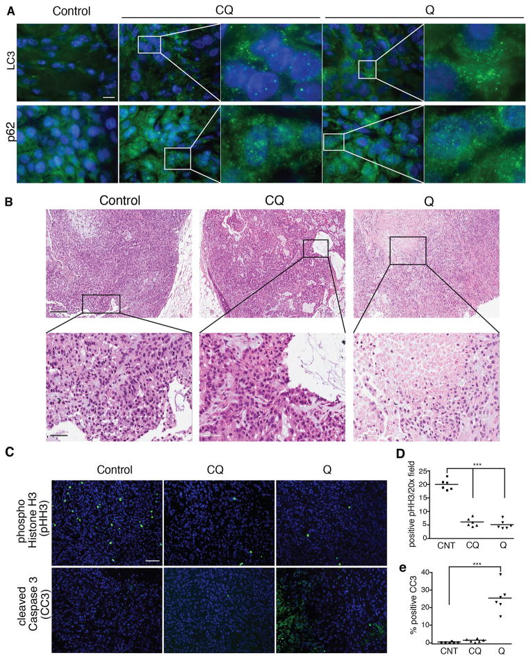
Figure 2. Quinacrine uniquely promotes apoptosis in lung tumors in vivo.
(A) Immunostaining for autophagosome accumulation (punctate LC3) and p62 (SQSTM1) aggregates in A549 xenograft tumors treated with CQ and Q. Bar, 50μm. (B) Increased areas of necrosis and dying cells in Q-treated tumors. Upper panel bar, 200μm; lower panel bar, 50μm. (C) Immunostaining for phospho-histone H3 (pHH3, mitosis marker) and cleaved caspase-3 (CC3, apoptosis marker) of lung tumors treated with CQ and Q. Bar, 100μm. (D, E) Quantification of pHH3 (D) and CC3 (E) positive tumor cells. Bar, 100μm. Data represent mean ± SEM from 6 independent tumors for each cohort. Statistical significance was calculated using ANOVA followed by Tukey’s HSD. ***P≤0.001.

