Abstract
Background & objectives:
The difficulties in diagnosis of neonatal sepsis are due to varied clinical presentation, low sensitivity of blood culture which is considered the gold standard and empirical antibiotic usage affecting the outcome of results. Though polymerase chain reaction (PCR) based detection of bacterial 16S rRNA gene has been reported earlier, this does not provide identification of the causative agent. In this study, we used restriction fragment length polymorphism (RFLP) of amplified 16S rRNA gene to identify the organisms involved in neonatal sepsis and compared the findings with blood culture.
Methods:
Blood samples from 97 neonates were evaluated for diagnosis of neonatal sepsis using BacT/Alert (automated blood culture) and PCR-RFLP.
Results:
Bacterial DNA was detected by 16S rRNA gene PCR in 55 cases, while BacT/Alert culture was positive in 34 cases. Staphylococcus aureus was the most common organism detected with both methods. Klebsiella spp. was isolated from four samples by culture but was detected by PCR-RFLP in five cases while Acinetobacter spp. was isolated from one case but detected in eight cases by PCR-RFLP. The sensitivity of PCR was found to be 82.3 per cent with a negative predictive value of 85.7 per cent. Eighty of the 97 neonates had prior exposure to antibiotics.
Interpretation & conclusions:
The results of our study demonstrate that PCR-RFLP having a rapid turnaround time may be useful for the early diagnosis of culture negative neonatal sepsis.
Keywords: Neonatal sepsis, PCR-RFLP, prior antibiotic therapy, rapid diagnostic
Neonatal septicaemia is a leading cause of morbidity but also in mortality of infants not only in the developing but also in developed countries and is responsible for 30-50 per cent deaths in developing countries1. Neonatal sepsis is the most common cause of neonatal death2 and, therefore, early diagnosis3 and treatment is very important. The clinical signs and symptoms are very subtle and variable and hence cause difficulty in diagnosis4.
The bacteriological profile of the causative agent varies between developed and developing countries5. The source of infection may be vertical being prenatally acquired from the mother or horizontally transmitted from the nosocomial environment6 with the most common agents being Gram negative bacteria such as coliforms (Escherichia coli, Klebsiella spp., Enterobacter spp.), Pseudomonas spp., Acinetobacter spp. and Gram positive bacteria such as Staphylococcus aureus and Group B Streptococcus7,8,9,10,11.
Presently blood culture is considered the gold standard for the diagnosis of neonatal sepsis although new proteomics-based and genomics-based rapid diagnoses have been developed3,12. In spite of the low sensitivity, blood culture has not been replaced by any other technique since isolation of the bacteria is essential for performing antibiogram13. However, there is a justified concern regarding the ability to recover bacteria from the small volume of blood and often, this prompts the clinicians to treat with antibiotics despite a negative blood culture14. Empirical antibiotic treatment is resorted to due to additional concerns such as the intermittent release of organisms in some clinical situations leading to negative results in blood culture. Yet another cause for negative blood culture could be the treatment with antibiotics prior to sample collection. However, the negative results could merely be due to an incomplete course of treatment resulting in the persistence of the bacteria in the blood. Schelonka et al15 reported that in infants with low numbers of bacteria, as much as 60 per cent of culture results may be false negative with blood culture volumes of 0.5 ml. Squire et al16 in a post-mortem study obtained negative blood cultures in 18 per cent of infants who died of bacterial infection. Molecular diagnostic techniques overcome some of the deficiencies of blood culture by their rapidity, sensitivity and specificity17.
Polymerase chain reaction (PCR) has been successfully used to diagnose a wide range of infectious diseases. 16S rDNA based PCR using universal primers that bind to regions that are conserved in all bacteria help in diagnosis of critical conditions such as neonatal sepsis17, meningitis18 and in situations where the organisms are non-culturable due to antibiotic that has been administered prior to sample collection or where culture is negative due to inadequate volume of sample or intermittent release of microorganisms into the blood stream in certain conditions17. However, the limitation of this method is that though positive reactions indicate presence of bacterial DNA, it is not possible to derive any information on the type of organism present. This problem can be circumvented by combining PCR with restriction enzyme treatment. In all such assays following PCR amplification, subtle differences in DNA sequences that arise by enzyme digestion help in distinguishing strains that are phenotypically indistinguishable. The characterization of PCR products based on sequence specific enzymatic cleavage termed restriction fragment length polymorphism (RFLP)19 involves generation of different size and number of DNA fragments that result in characteristic banding patterns. As nucleotide sequence information of several medically important bacteria became available, it became clear that within the 16S rRNA gene, there are variable regions which can be used to discriminate between bacterial species using RFLP technique. This approach has been used to detect and identify bacteria associated with meningitis18,20. This approach has also been termed “amplified rDNA restriction analysis or ARDRA21. The objective of this study was to use PCR-RFLP for identification of bacteria involved in neonatal sepsis and to compare this technique with automated blood culture BacT/Alert.
Material & Methods
Study design: The study was conducted in the department of Microbiology, Kasturba Medical College, Mangalore, India, between 2008 and 2010. The study included neonates less than 28 days old with clinical suspicion of sepsis. A total of 97 cases with suspected neonatal sepsis were chosen consecutively. History of prior antibiotic therapy was collected and prior antibiotic use was not considered as exclusion criterion. The results were correlated with clinical signs and symptoms as well as the C-reactive protein (CRP) level which is well recognized as a useful marker for sepsis especially in neonates22. The study protocol was reviewed and cleared by the institutional ethical committee prior to starting the work.
Reference bacterial cultures: Standard cultures used in the study were from American Type Culture Collection (ATCC); Microbial Type Culture Collection (MTCC), India, or strains of the common pathogens available in our laboratory stock (LSC). The organisms included were Escherichia coli (ATCC 25922), Staphylococcus aureus (ATCC 25923), Enterococcus faecalis (ATCC 29212); Pseudomonas aeruginosa (ATCC 27853), Proteus mirabilis (ATCC 12453), Streptococcus agalactiae (ATCC 13813), Enterobacter aerogenes (ATCC 13048), Klebsiella pneumoniae (MTCC 109), Staphylococcus epidermidis (LSC), and Acinetobacter baumannii (LSC).
Sample collection: Blood sample (1-3 ml) was drawn aseptically from a peripheral site from neonates suspected to have sepsis. About 0.5 to 1.5 ml of the collected blood was inoculated into a BacT/ Alert PF bottle for automated blood culture while the other half was added into a sterile bottle containing 4.5 ml of trypticase soy broth (TSB) for enriching the bacteria for PCR.
Sample processing: The BacT/Alert Microbial Detection system (BacT/ Alert 240, Biomerieux, India) with BacT Alert PF was the system used. A drop from the positive bottles was taken for direct smear for Gram staining and a separate loopful inoculated on 5 per cent sheep blood agar, chocolate agar, MacConkey agar (Fitech systems, India) for culture. Any colony that appeared on the medium was subjected to Gram stain and a battery of biochemical tests for identification of the organism. In some cases, growth was also identified by the Mini API instrument (Database V3.1, Biomerieux). If the bottle recorded negative for growth, incubation was continued until the 7th day before discarding.
PCR assay: The blood samples from patients inoculated into TSB were incubated in a shaker for 5 h followed by DNA extraction using the mini kit (Qiagen, USA). The concentration and purity of DNA was determined using 2 μl of the DNA sample in the NanoDrop ND-1000 spectrophotometer (Thermo, USA). The concentration was measured at OD260 while the ratio of OD260 to OD280 was used to determine the purity. One representative isolate of each of the commonly encountered species in neonatal sepsis was included as positive control. Sterile distilled water and 1x Tris EDTA (TE) (10 mM Tris-Cl; 1 mM EDTA; pH 8.0) buffer (without sample) was used as control each time for DNA extraction procedure.
The 16S rRNA gene was amplified by PCR using universal eubacterial primers (U1: 5’-CCAGCAGCCGCGGTAATACG-3’ and U2 5’-ATCGG(C/T)TACCTTGTTACGACTTC-3’) to generate a 996 bp amplicon18. The reaction was performed in triplicate in 30 μl volumes containing 10 pmol of each primer, dNTP mix which contained 2.5 mM each of dATP, dGTP, dTTP and dCTP (Bangalore Genei, India), 1.5 U Taq DNA polymerase (Bangalore Genei, India), 10× PCR buffer (100 mM Tris-HCl pH 8.3, 20 mM MgCl2, 500 mM KCl, 0.1% gelatin), double distilled Milli Q water and 2-4 μl DNA template. PCR was carried out in a Thermocycler (MJ Research Inc., USA) and included an initial denaturation at 94°C for 5 min followed by 35 cycles of denaturation at 94°C for 1 min, primer annealing at 55°C for 1 min, extension at 72°C for 2 min and a final extension at 72°C for 10 min. Positive controls, extraction controls, negative and reagent controls were included in each run. Reagent controls consisted of all reagent components except template DNA. The electrophoresis of PCR products was performed in 1.2 per cent (w/v) of agarose gel containing ethidium bromide (0.5 μg/ml) and photographed using a gel documentation system (Herolab, Weisloch, Germany).
RFLP of 16SrRNA gene PCR product: The 996 base pair product of 16S rRNA gene was purified (Qiagen, USA) and digested with HaeIII restriction enzyme18 (Fermentas, USA) according to manufacturer's instructions to generate fragments, band patterns of which were characteristic for each organism. The amplified products from all the bacterial strains used as controls were also digested with HaeIII according to manufacturer's instructions. Digestions were carried out in a total volume of 40 μl. The reaction mixture consisted of 10-15 μl of PCR product, 5 U of restriction enzyme and volume adjusted with sterile distilled water. The digest was electrophoresed in 2 per cent agarose with 0.5 μg/ml ethidium bromide and photographed using a gel documentation system (Herolab, Weisloch, Germany). The bands obtained from PCR-RFLP were analyzed using the GelCompare II version 2.5 (Applied Maths, Sint-Martens- Latem, Belgium).
Statistical analysis: PCR results were correlated with culture results using the least square design test (LSDt) and the Wilcoxon signed ranked test. Since the results of RFLP were not normally distributed, comparison with culture results was done using the non-parametric Mann-Whitney U test. The effect of antibiotics on the results of both PCR and culture was analysed using the Z test.
Results
Ninety seven neonates from two secondary neonatal intensive care units (NICU) most having one or more of the risk factors (such as preterm birth, low birth weight, birth asphyxia, intra-partum sepsis, low APGAR, respiratory distress, convulsions, meconium aspiration syndrome, or some surgery) were part of this study. Thirty seven (38%) were preterm and 50 (51.5%) with low birth weight (LBW-less than 2.5 kg) which included both very low birth weight (VLBW) and extremely low birth weight (ELBW). The mean birth weight of all was 2.39 kg; 55 per cent of the neonates investigated were males and 45 per cent females; 55 per cent of the neonates had early onset (<72 h) sepsis and 45 per cent late onset sepsis (>72 h).
All neonates suspected to have sepsis showed one or more of the following signs and symptoms that included respiratory distress, intolerance to feeds, neonatal seizures, apnoea, bilious aspirate, hypoglycaemia, fever and pneumonia. Eight had undergone surgery for conditions like pyloric stenosis, duodenal stenosis, diaphragmatic hernia and anal dysgenesis. CRP levels were estimated and found to be positive in 71 samples (73%). Antibiotics had been administered before the collection of sample in 80 of the 97 (82%) cases. In most of the cases the specimen was collected on the second day after beginning antibiotic therapy. The most common antibiotic prescribed was a combination of piperacillin and tazobactam and in some cases, an aminoglycoside was also added.
BacT/Alert blood culture: Of the 97 samples, 34 were positive for BacT/Alert blood culture. The most common organism was S. aureus which was isolated from 21 neonates followed by K. pneumoniae from four, Citrobacter from two and Enterococcus and Acinetobacter from one case each (Table I). Mixed cultures were observed in five cases. In one case, S. aureus and Pseudomonas spp. were isolated from a five day old infant with Rh incompatibility. This neonate had a negative CRP and was not on any antibiotics. In the second case, Enterococcus spp. with S. aureus were grown from a three day old preterm infant with haemorrhagic disease of the newborn. The third case was that of a 12 day old infant, from whose second sample S. aureus was isolated together with diphtherioids; the neonate had a positive CRP and was preterm with low birth weight. In the fourth case S. aureus was isolated with Acinetobacter spp. and this neonate had a very high CRP. In the fifth case of a neonate with extremely low birth weight and positive CRP, Acinetobacter spp. was isolated with Bacillus spp. Bacillus spp. and diphtherioids were considered contaminants. All these neonates were treated with higher antibiotics to cover both the organisms and showed good clinical response to treatment.
Table I.
Organisms isolated in culture and identified by PCR-RFLP
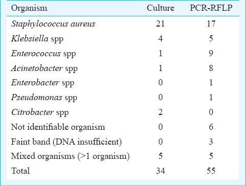
PCR-RFLP analysis: PCR with universal primers for 16S rRNA gene with an amplicon of 996 bp (Fig. 1) was positive in 55 cases (Tables I and II). The specific identification of the organism was by subjecting the amplicon to RFLP. A typical gel with reference cultures and samples is shown in Fig. 2A and 2B, respectively.
Fig. 1.
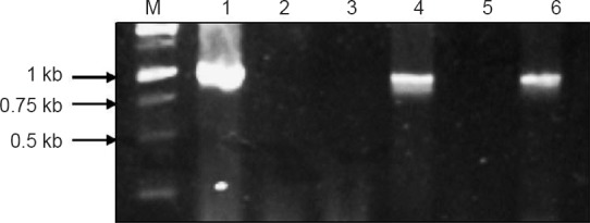
Representative gel picture of PCR amplification of 16S rRNA gene (996 bp) from different samples. M-1 kb DNA ladder. Lane 1-Positive control; Lane 2-Negative control; Lanes 3-6, different blood samples.
Table II.
A comparison of PCR with BacT/Alert culture
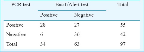
Fig. 2A.
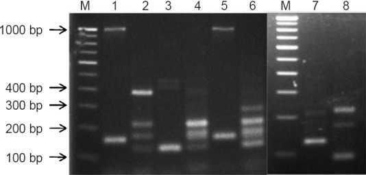
PCR-RFLP photograph of 16S rRNA gene amplicon of reference strains. M: 100 bp DNA ladder Lane 1: Staphylococcus aureus; Lane 2: Pseudomonas aeruginosa; Lane 3: Enterococcus faecalis; Lane 4: Klebsiella pneumoniae; Lane 5: Staphylococcus epidermidis; Lane 6: Enterobacter aerogenes; Lane 7: Acinetobacter baumannii; Lane 8: Group-B Streptococcus.
Fig. 2B.
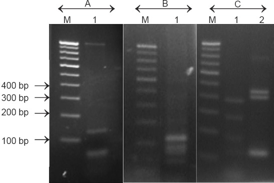
Representative gel picture of RFLP pattern of different samples. M-100 bp DNA ladder. (A) Lane-1, Staphylococcus aureus (B) Lane-1, Klebsiella pneumoniae (C) Lane-1, Pseudomonas aeruginosa (C) Lane-2, Enterococcus faecalis.
The most common organism identified through PCR-RFLP was S. aureus in 17 cases followed by Enterococcus spp. in nine, Acinetobacter spp. in eight, Klebsiella spp. in five and Enterobacter spp. and Pseudomonas spp. in one case each. In seven cases the RFLP pattern did not match any of the known pathogens that were used as reference strain. In two of these cases, Citrobacter was isolated by culture and the panel of standard cultures used in this study for RFLP did not include this organism, while in five cases, mixed organisms were obtained by culture. In seven cases, DNA band was faint and was not adequate for RFLP analysis. No significant association was observed between RFLP results and blood culture.
The results of BacT/Alert blood culture were compared with that of PCR as shown in Table II. There was no significant difference between PCR and blood culture for the detection of neonatal sepsis.
Blood culture has always been considered the gold standard for detection of sepsis and hence PCR when compared to culture yielded a sensitivity of 82.35 per cent. The specificity of PCR was 57.1 per cent with a positive predictive value (PPV) of 50.9 per cent and a negative predictive value (NPV) of 85.71 per cent.
Effect of prior antibiotic treatment on PCR and culture: Antibiotics were used in 80 of the total 97 neonates prior to drawing the sample for PCR and culture. Of the 80 samples, 47 gave a positive PCR result and 27 gave positive blood culture (Table III). There was a significant difference between the results of blood culture and PCR positivity obtained in the presence of antibiotics (P<0.001) suggesting that PCR was a better test to detect neonatal sepsis in cases where antibiotic had been administered.
Table III.
Effect of prior antibiotic treatment on PCR positive results

Discussion
BacT/ Alert, an automated non radiometric system, is a third generation continuous monitoring blood culture system that is used globally and compares with BACTEC 660/730 systems in terms of microbial recovery, providing rapid growth results ensuring maximal recovery of organisms in the shortest possible time with the least contamination23.
Culture was positive in 34 cases in this study. The most common organism isolated was S. aureus from 21 cases. This is in contrast to another study from India that presented Gram negative bacteria as the commonest cause of neonatal sepsis7. Bizzarro and Gallagher24 reported antibiotic-resistant organisms in the NICU and found that the major reservoir for S. aureus in the neonate was the umbilical cord followed by skin, nasopharynx and the gastrointestinal tract with relatively low attack rates (around 2%).
K. pneumoniae isolated from four neonates was extended spectrum beta lactamase (ESBL) producer. Kristof et al25 reported Klebsiella spp. to be a significant pathogen in a NICU setup due to its ability to survive in the environment and transiently in the hands of the health care worker facilitating effective transmission. It was interesting to note that negative blood cultures in 21 samples from neonates showed positive PCR. Several reasons account for negative blood cultures with low blood volume being the most important. Kellogg et al26 found that low-level bacteraemia was common in their setting and recommended drawing 4-4.5 ml of blood for optimal detection of the incriminating agent. However, this is not possible in the neonatal setup. Connell et al27 have concluded that negative blood cultures are inevitable for a large proportion of blood samples analysed due to inadequate volume of blood being submitted, a situation which does not improve even after educational intervention since neonate handling for drawing blood is a challenging task.
The low rate of isolation from blood of neonates administered antibiotics prior to drawing sample demonstrates the inhibitory role of antibiotics in culture based diagnostic technique.
DNA extraction would be a critical step influencing the outcome of PCR and considering the fact that a significant number of culture negative samples were PCR positive in this study, suggests that the column based DNA extraction protocol was quite effective. The total turnaround time for the whole analysis in this study leading to the identification of the specific pathogen was 14 h. The test also required blood volumes as small as 200-500 μl. There was a good agreement between identification of isolates and PCR-RFLP data. The latter was particularly useful for detection of cases due to Acinetobacter and Enterococcus that were missed by culture method. The study demonstrates that PCR-RFLP would be a specific and rapid diagnostic tool for neonatal sepsis, providing reliable identification within a day. Two of the seven unidentified cases by PCR-RFLP were due to non-inclusion of Citrobacter in the control panel. There is a need to expand the control panel to include more organisms encountered in neonatal sepsis. In seven cases, the DNA obtained after PCR as seen from the faint band, was inadequate for RFLP analysis. In such instances, re-amplification of the PCR product would yield sufficient DNA for RFLP analysis28. There were a few cases with mixed isolates, which possibly included skin contaminants. This problem was encountered in BacT/Alert also and would need ample care during sample collection.
In our study, 58.7 per cent neonates who had prior antibiotic exposure, were positive by PCR. Previous investigations also suggested that PCR has the potential to detect bacteria in culture-negative samples even after the initiation of intravenous antibiotics29. Dutta et al30 found universal primer PCR useful in diagnosing sepsis accurately before but not after antibiotic treatment. This is intriguing since PCR detects DNA of both live and dead organisms. In this study, a significant number of culture negative cases being PCR positive showed that DNA of dead/uncultivable bacteria in samples where antibiotic administration might have suppressed/killed them and thus prevented their growth in culture could be detected.
PCR when compared with culture showed a sensitivity of 82.3 per cent and a negative predictive value of 85.7 per cent. The specificity and positive predictive value were low, and this could be due to the positive PCR reaction in culture negative samples. However, the clinical data confirmed them as bacterial sepsis. Jordan and Durso28 compared PCR and BACTEC blood culture system and showed a sensitivity of 96 per cent, specificity of 99.4 per cent, PPV of 88.9 per cent and NPV of 99.8 per cent. In our study, the low values obtained could be due to prior antibiotic exposure in most of the cases and this could be a problem in many developing countries as empirical treatment with antibiotics is undertaken for early management. PCR scored over culture based system as it was not inhibited by the presence of antibiotics that had been administered prior to drawing samples. The study shows that though the PCR-RFLP has almost the same sensitivity as BacT/Alert, it has a rapid turn over. The advantage of PCR-RFLP relates to quick identification of the pathogen, even in some culture negative cases. This facilitates targeted treatment to be given early instead of waiting of the delay for about 30-35 h for culture report. With the cost of consumables and equipment for molecular diagnostics becoming affordable and technical expertise becoming available, PCR- RFLP may be used in routine for early diagnosis of neonatal sepsis.
Footnotes
Conflicts of Interest: None.
References
- 1.Durrene T, Zaidi AKM. Burdern of neonatal infections in developing countries: A review of evidence from community based studies. Padiatr Infect Dis J. 2009;28:S3–9. doi: 10.1097/INF.0b013e3181958755. [DOI] [PubMed] [Google Scholar]
- 2.MacDonald MG, Seshia MMK, Mullett MD, editors. Avery's neonatology: pathophysiology & management of the newborn. 6th ed. Philadelphia, PA: Lippincott Williams & Wilkins; 2005. [Google Scholar]
- 3.Yadav AK, Wilson CG, Prasad PL, Menon PK. Polymerase chain reaction in rapid diagnosis of neonatal sepsis. Indian Pediatr. 2005;42:681–5. [PubMed] [Google Scholar]
- 4.Dunham EC. Septicemia in newborn. Am J Dis Child. 1933;45:229–53. [Google Scholar]
- 5.Mahapatra A, Ghosh SK, Mishra S, Pattnaik D, Pattnaik K, Mohanty SK. Enterobacter cloacae: a predominant pathogen in neonatal septicaemia. Indian J Med Microbiol. 2002;20:110–2. [PubMed] [Google Scholar]
- 6.Kerur BM, Vishnu Bhat B, Harish BN, Habeebullah S, Uday Kumar C. Maternal genital bacteria and surface colonization in early neonatal sepsis. Indian J Pediatr. 2006;73:29–32. doi: 10.1007/BF02758256. [DOI] [PubMed] [Google Scholar]
- 7.Mathur M, Shah H, Dixit K, Khambadkone S, Chakrapani A, Irani S. Bacteriological profile of neonatal septicemia cases (for the year 1990-91) J Postgrad Med. 1994;40:18–20. [PubMed] [Google Scholar]
- 8.Gyawali N, Sanjana RK. Bacteriological profile and antibiogram of neonatal septicemia. Indian J Pediatr. 2013;80:371–4. doi: 10.1007/s12098-012-0911-9. [DOI] [PubMed] [Google Scholar]
- 9.Jyothi P, Basavaraj MC, Basavaraj PV. Bacteriological profile of neonatal septicemia and antibiotic susceptibility pattern of the isolates. J Nat Sci Biol Med. 2013;4:306–9. doi: 10.4103/0976-9668.116981. [DOI] [PMC free article] [PubMed] [Google Scholar]
- 10.Tsering DC, Chanchal L, Pal R, Kar S. Bacteriological profile of septicemia and the risk factors in neonates and infants in Sikkim. J Glob Infect Dis. 2011;3:42–5. doi: 10.4103/0974-777X.77295. [DOI] [PMC free article] [PubMed] [Google Scholar]
- 11.Wu JH, Chen CY, Tsao PN, Hsieh WS, Chou HC. Neonatal sepsis: a 6-year analysis in a neonatal care unit in Taiwan. Pediatr Neonatol. 2009;50:88–95. doi: 10.1016/S1875-9572(09)60042-5. [DOI] [PubMed] [Google Scholar]
- 12.Srinivasan L, Harris MC. New technologies for the rapid diagnosis of neonatal sepsis. Curr Opin Pediatr. 2012;24:165–71. doi: 10.1097/MOP.0b013e3283504df3. [DOI] [PubMed] [Google Scholar]
- 13.Bouza E, Sousa D, Rodríguez-Créixems M, Lechuz JG, Muñoz P. Is the volume of blood cultured still a significant factor in the diagnosis of bloodstream infections? J Clin Microbiol. 2007;45:2765–9. doi: 10.1128/JCM.00140-07. [DOI] [PMC free article] [PubMed] [Google Scholar]
- 14.Piantino JH, Schreiber MD, Alexander K, Hageman J. Culture negative sepsis and systemic inflammatory response syndrome in neonates. Neo Rev. 2013;14:e294–305. [Google Scholar]
- 15.Schelonka RL, Chai MK, Yoder BA, Hensley D, Brockett RM, Ascher DP. Volume of blood required to detect common neonatal pathogens. J Pediatr. 1996;129:275–8. doi: 10.1016/s0022-3476(96)70254-8. [DOI] [PubMed] [Google Scholar]
- 16.Squire E, Favara B, Todd J. Diagnosis of neonatal bacterial infection: hematologic and pathologic findings in fatal and nonfatal cases. Pediatrics. 1979;64:60–4. [PubMed] [Google Scholar]
- 17.Jordan JA, Durso MB, Butchko AR, Jones JG, Bronzanski BS. Evaluating the near-term infant for early onset sepsis: progress and challenges to consider with 16S rDNA polymerase chain reaction testing. J Mol Diagn. 2006;8:357–63. doi: 10.2353/jmoldx.2006.050138. [DOI] [PMC free article] [PubMed] [Google Scholar]
- 18.Lu JJ, Perng CL, Lee SY, Wan CC. Use of PCR with universal primers and restriction endonuclease digestions for detection and identification of common bacterial pathogens in cerebrospinal fluid. J Clin Microbiol. 2000;38:2076–80. doi: 10.1128/jcm.38.6.2076-2080.2000. [DOI] [PMC free article] [PubMed] [Google Scholar]
- 19.Pourzand C, Cerutti P. Genotypic mutation analysis by RFLP/PCR. Mutat Res. 1993;288:113–21. doi: 10.1016/0027-5107(93)90213-y. [DOI] [PubMed] [Google Scholar]
- 20.Pandit L, Kumar S, Karunasagar I, Karunasagar I. Diagnosis of partially treated culture-negative bacterial meningitis using 16S rRNA universal primers and restriction endonuclease digestion. J Med Microbiol. 2005;54:539–42. doi: 10.1099/jmm.0.45599-0. [DOI] [PubMed] [Google Scholar]
- 21.Vaneechoutte M, Rossau R, De Vos P, Gillis M, Janssens D, Paepe N, et al. Rapid identification of bacteria of the Comamonadaceae with amplified ribosomal DNA-restriction analysis (ARDRA) FEMS Microbiol Lett. 1992;72:227–33. doi: 10.1111/j.1574-6968.1992.tb05102.x. [DOI] [PubMed] [Google Scholar]
- 22.Døllner H, Vatten L, Austgulen R. Early diagnostic markers for neonatal sepsis: comparing C-reactive protein, interleukin-6, soluble tumour necrosis factor receptors and soluble adhesion molecules. J Clin Epidemiol. 2001;54:1251–7. doi: 10.1016/s0895-4356(01)00400-0. [DOI] [PubMed] [Google Scholar]
- 23.Wilson ML, Weinstein MP, Reller LB. Automated blood culture systems. Clin Lab Med. 1994;14:149–69. [PubMed] [Google Scholar]
- 24.Bizzarro MJ, Gallagher PG. Antibiotic-resistant organisms in the neonatal intensive care unit. Semin Perinatol. 2007;31:26–32. doi: 10.1053/j.semperi.2007.01.004. [DOI] [PubMed] [Google Scholar]
- 25.Kristof K, Szabo D, Marsh JW, Cser V, Janik L, Rozgonyi F, et al. Extended-spectrum beta-lactamase-producing Klebsiella spp. in a neonatal intensive care unit: risk factors for the infection and the dynamics of the molecular epidemiology. Eur J Clin Microbiol Infect Dis. 2007;26:563–70. doi: 10.1007/s10096-007-0338-9. [DOI] [PubMed] [Google Scholar]
- 26.Kellogg JA, Ferrentino FL, Goodstein MH, Liss J, Shapiro SL, Bankert DA. Frequency of low level bacteremia in infants from birth to two months of age. Pediatr Infect Dis J. 1997;16:381–5. doi: 10.1097/00006454-199704000-00009. [DOI] [PubMed] [Google Scholar]
- 27.Connell TG, Rele M, Cowley D, Buttery JP, Curtis N. How reliable is a negative blood culture result?. Volume of blood submitted for culture in routine practice in a children's hospital. Pediatrics. 2007;119:891–6. doi: 10.1542/peds.2006-0440. [DOI] [PubMed] [Google Scholar]
- 28.Jordan JA, Durso MB. Comparison of 16S rRNA gene PCR and BACTEC 9240 for the detection of neonatal bacteremia. J Clin Microbiol. 2000;38:2574–8. doi: 10.1128/jcm.38.7.2574-2578.2000. [DOI] [PMC free article] [PubMed] [Google Scholar]
- 29.Ohlin A, Bäckman A, Björkqvist M, Mölling P, Jurstrand M, Schollin J. Real-time PCR of the 16S-rRNA gene in the diagnosis of neonatal bacteraemia. Acta Paediatr. 2008;97:1376–80. doi: 10.1111/j.1651-2227.2008.00924.x. [DOI] [PubMed] [Google Scholar]
- 30.Dutta S, Narang A, Chakraborty A, Ray P. Diagnosis of neonatal sepsis using universal primer polymerase chain reaction before and after starting antibiotic drug therapy. Arch Pediatr Adolesc Med. 2009;163:6–11. doi: 10.1001/archpediatrics.2008.513. [DOI] [PubMed] [Google Scholar]


