Abstract
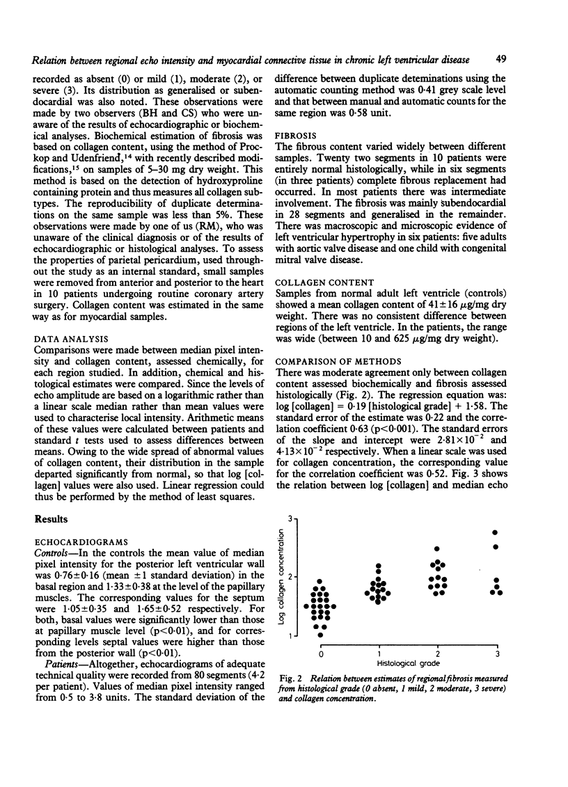
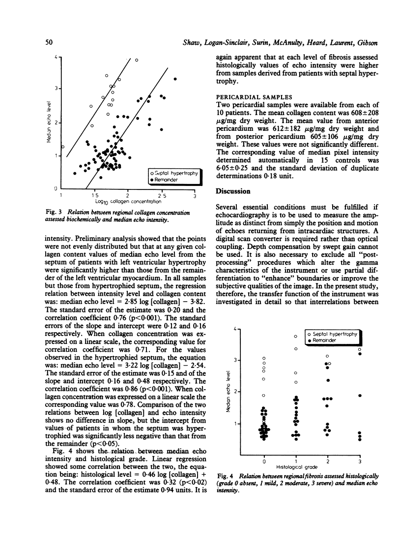
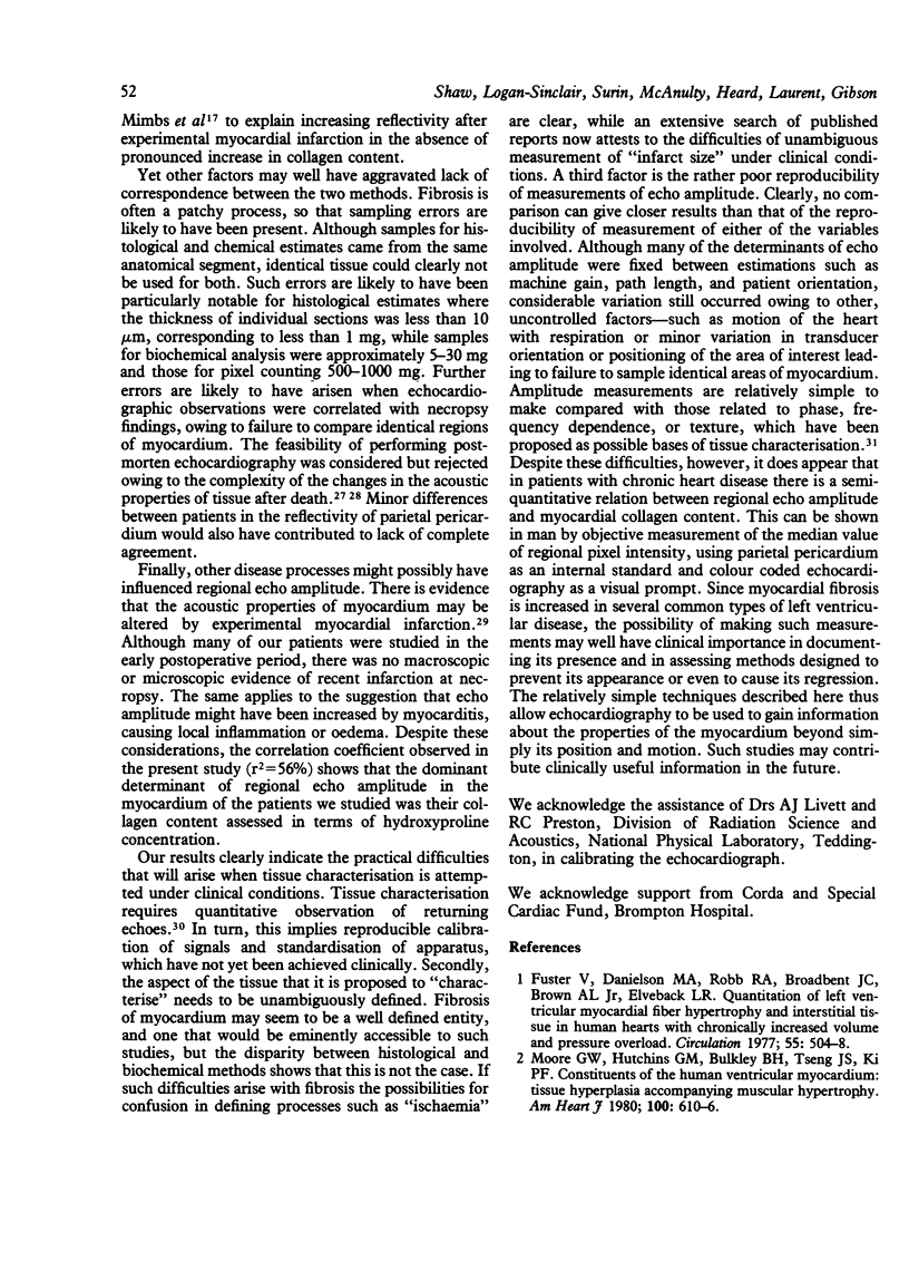
Cross sectional echocardiograms were recorded within one week of death in seven patients with valvular heart disease, four with coronary artery disease, and nine with congenital heart disease. Regional echo amplitude was measured from the cross sectional display by constructing histograms of pixel intensity. Parietal pericardium was used as an internal standard for setting the gain of the instrument. At necropsy myocardium was taken from the free wall of the left ventricle, the papillary muscles, and the septum. Fibrosis was assessed histologically and biochemically as hydroxyproline content. In individual samples histological and biochemical estimates were correlated. In all regions other than the septum in patients with left ventricular hypertrophy, log [collagen] correlated with median pixel intensity. The amplitude of reflected echoes from the hypertrophied septum was significantly higher than that from other samples but was similarly correlated with collagen content. Agreement between echo amplitude and histological grade was significantly less good. Thus in chronic left ventricular disease myocardial collagen content appears to be the major determinant of regional echo intensity. Reproducibility of measurements and more rigorous definition of tissue abnormalities will, however, require further study.
Full text
PDF



Images in this article
Selected References
These references are in PubMed. This may not be the complete list of references from this article.
- Anderson K. R., Sutton M. G., Lie J. T. Histopathological types of cardiac fibrosis in myocardial disease. J Pathol. 1979 Jun;128(2):79–85. doi: 10.1002/path.1711280205. [DOI] [PubMed] [Google Scholar]
- Bornstein P., Sage H. Structurally distinct collagen types. Annu Rev Biochem. 1980;49:957–1003. doi: 10.1146/annurev.bi.49.070180.004521. [DOI] [PubMed] [Google Scholar]
- Caspari P. G., Newcomb M., Gibson K., Harris P. Collagen in the normal and hypertrophied human ventricle. Cardiovasc Res. 1977 Nov;11(6):554–558. doi: 10.1093/cvr/11.6.554. [DOI] [PubMed] [Google Scholar]
- Chivers R. C. Tissue characterization. Ultrasound Med Biol. 1981;7(1):1–20. doi: 10.1016/0301-5629(81)90018-1. [DOI] [PubMed] [Google Scholar]
- Davies J., Gibson D. G., Foale R., Heer K., Spry C. J., Oakley C. M., Goodwin J. F. Echocardiographic features of eosinophilic endomyocardial disease. Br Heart J. 1982 Nov;48(5):434–440. doi: 10.1136/hrt.48.5.434. [DOI] [PMC free article] [PubMed] [Google Scholar]
- Davies M. J., Fulton W. F., Robertson W. B. The relation of coronary thrombosis to ischaemic myocardial necrosis. J Pathol. 1979 Feb;127(2):99–110. doi: 10.1002/path.1711270208. [DOI] [PubMed] [Google Scholar]
- Donaldson R. M., Florio R., Rickards A. F., Bennett J. G., Yacoub M., Ross D. N., Olsen E. Irreversible morphological changes contributing to depressed cardiac function after surgery for chronic aortic regurgitation. Br Heart J. 1982 Dec;48(6):589–597. doi: 10.1136/hrt.48.6.589. [DOI] [PMC free article] [PubMed] [Google Scholar]
- Edwards W. D., Tajik A. J., Seward J. B. Standardized nomenclature and anatomic basis for regional tomographic analysis of the heart. Mayo Clin Proc. 1981 Aug;56(8):479–497. [PubMed] [Google Scholar]
- Fields S., Dunn F. Letter: Correlation of echographic visualizability of tissue with biological composition and physiological state. J Acoust Soc Am. 1973 Sep;54(3):809–812. doi: 10.1121/1.1913668. [DOI] [PubMed] [Google Scholar]
- Frederiksen D. W., Hoffnung J. M., Frederiksen R. T., Williams R. B. The structural proteins of normal and diseased human myocardium. Circ Res. 1978 Apr;42(4):459–466. doi: 10.1161/01.res.42.4.459. [DOI] [PubMed] [Google Scholar]
- Fuster V., Danielson M. A., Robb R. A., Broadbent J. C., Brown A. L., Jr, Elveback L. R. Quantitation of left ventricular myocardial fiber hypertrophy and interstitial tissue in human hearts with chronically increased volume and pressure overload. Circulation. 1977 Mar;55(3):504–508. doi: 10.1161/01.cir.55.3.504. [DOI] [PubMed] [Google Scholar]
- Greenbaum R. A., Ho S. Y., Gibson D. G., Becker A. E., Anderson R. H. Left ventricular fibre architecture in man. Br Heart J. 1981 Mar;45(3):248–263. doi: 10.1136/hrt.45.3.248. [DOI] [PMC free article] [PubMed] [Google Scholar]
- Laurent G. J., Cockerill P., McAnulty R. J., Hastings J. R. A simplified method for quantitation of the relative amounts of type I and type III collagen in small tissue samples. Anal Biochem. 1981 May 15;113(2):301–312. doi: 10.1016/0003-2697(81)90081-6. [DOI] [PubMed] [Google Scholar]
- Laurent G. J., McAnulty R. J., Corrin B., Cockerill P. Biochemical and histological changes in pulmonary fibrosis induced in rabbits with intratracheal bleomycin. Eur J Clin Invest. 1981 Dec;11(6):441–448. doi: 10.1111/j.1365-2362.1981.tb02011.x. [DOI] [PubMed] [Google Scholar]
- Logan-Sinclair R., Wong C. M., Gibson D. G. Clinical application of amplitude processing of echocardiographic images. Br Heart J. 1981 Jun;45(6):621–627. doi: 10.1136/hrt.45.6.621. [DOI] [PMC free article] [PubMed] [Google Scholar]
- Medugorac I., Jacob R. Characterisation of left ventricular collagen in the rat. Cardiovasc Res. 1983 Jan;17(1):15–21. doi: 10.1093/cvr/17.1.15. [DOI] [PubMed] [Google Scholar]
- Mimbs J. W., Bauwens D., Cohen R. D., O'Donnell M., Miller J. G., Sobel B. E. Effects of myocardial ischemia on quantitative ultrasonic backscatter and identification of responsible determinants. Circ Res. 1981 Jul;49(1):89–96. doi: 10.1161/01.res.49.1.89. [DOI] [PubMed] [Google Scholar]
- Mimbs J. W., O'Donnell M., Bauwens D., Miller J. W., Sobel B. E. The dependence of ultrasonic attenuation and backscatter on collagen content in dog and rabbit hearts. Circ Res. 1980 Jul;47(1):49–58. doi: 10.1161/01.res.47.1.49. [DOI] [PubMed] [Google Scholar]
- Mimbs J. W., O'Donnell M., Miller J. G., Sobel B. E. Detection of cardiomyopathic changes induced by doxorubicin based on quantitative analysis of ultrasonic backscatter. Am J Cardiol. 1981 May;47(5):1056–1060. doi: 10.1016/0002-9149(81)90212-5. [DOI] [PubMed] [Google Scholar]
- Moore G. W., Hutchins G. M., Bulkley B. H., Tseng J. S., Ki P. F. Constituents of the human ventricular myocardium: connective tissue hyperplasia accompanying muscular hypertrophy. Am Heart J. 1980 Nov;100(5):610–616. doi: 10.1016/0002-8703(80)90224-0. [DOI] [PubMed] [Google Scholar]
- O'Donnell M., Mimbs J. W., Sobel B. E., Miller J. G. Ultrasonic attenuation of myocardial tissue: dependence on time after excision and on temperature. J Acoust Soc Am. 1977 Oct;62(4):1054–1057. doi: 10.1121/1.381600. [DOI] [PubMed] [Google Scholar]
- Oldershaw P. J., Brooksby I. A., Davies M. J., Coltart D. J., Jenkins B. S., Webb-Peploe M. M. Correlations of fibrosis in endomyocardial biopsies from patients with aortic valve disease. Br Heart J. 1980 Dec;44(6):609–611. doi: 10.1136/hrt.44.6.609. [DOI] [PMC free article] [PubMed] [Google Scholar]
- PROCKOP D. J., UDENFRIEND S. A specific method for the analysis of hydroxyproline in tissues and urine. Anal Biochem. 1960 Nov;1:228–239. doi: 10.1016/0003-2697(60)90050-6. [DOI] [PubMed] [Google Scholar]
- Rasmussen S., Corya B. C., Feigenbaum H., Knoebel S. B. Detection of myocardial scar tissue by M-mode echocardiography. Circulation. 1978 Feb;57(2):230–237. doi: 10.1161/01.cir.57.2.230. [DOI] [PubMed] [Google Scholar]
- Schwarz F., Flameng W., Schaper J., Langebartels F., Sesto M., Hehrlein F., Schlepper M. Myocardial structure and function in patients with aortic valve disease and their relation to postoperative results. Am J Cardiol. 1978 Apr;41(4):661–669. doi: 10.1016/0002-9149(78)90814-7. [DOI] [PubMed] [Google Scholar]
- Sinclair R. B., Oldershaw P. J., Gibson D. G. Computing in echocardiography. Prog Cardiovasc Dis. 1983 May-Jun;25(6):465–486. doi: 10.1016/0033-0620(83)90020-8. [DOI] [PubMed] [Google Scholar]
- Tanaka M., Terasawa Y. Echocardiography. Evaluation of the tissue character in myocardium. Jpn Circ J. 1979 Apr;43(4):367–376. doi: 10.1253/jcj.43.367. [DOI] [PubMed] [Google Scholar]


