Abstract
Objective
To explore the appropriate treatment of malignant germ cell tumor (MGCT) in the female genital system, and to analyze the factors influencing both therapeutic response and survival outcome.
Methods
A cohort of 230-Chinese women diagnosed with MGCT of the genital system was retrospectively reviewed and prospectively followed. The demographic and pathological features, extent of disease and surgery, treatment efficiency, recurrence and survival were analyzed.
Results
MGCTs from different genital origins shared a similar therapeutic strategy and response, except that all eight vaginal cases were infantile yolk sac tumors. The patients’ cure rate following the initial treatment, 5-year overall survival and disease-free survival (DFS) were 85.02%, 95.00%, and 86.00%, respectively. Although more extensive excision could enhance the remission rate; it did not improve the patients’ survival. Instead, the level of the medical institution, extent of surgery and disease were independent prognostic factors for relapse (p<0.05). Approximately 20% of patients had recurrent or refractory disease, more than half of whom were in remission following secondary cytoreductive surgery with salvage chemotherapy.
Conclusion
Fertility-sparing surgery with or without standardized PEB/PVB (cisplatin, etoposide/vincristine, and bleomycin) chemotherapy is applicable for female MGCTs of different origins. Comprehensive staging is not required; nor is excessive debulking suggested. Appropriate cytoreduction by surgery and antineoplastic medicine at an experienced medical institution can bring about an excellent prognosis for these patients.
Keywords: Female Genital System, Malignant Germ Cell Tumor, Prognosis, Therapeutic Uses
INTRODUCTION
Malignant germ cell tumor (MGCT) is a rare entity in the female genital system. Ovarian malignant germ cell tumors (MOGCTs) are highly malignant, rapidly growing, and have a peak incidence in women under the age of 20 years [1]. MOGCTs account for 5% of all ovarian malignancies in Western countries, but may represent approximately 15% of ovarian cancers in Asians and in blacks [2]. However, Surveillance, Epidemiology, and End Results (SEER) data (1973 to 2002) demonstrated lower rates of MOGCTs in blacks than whites and other nonwhites, and an increased incidence for Asian/Pacific Islanders and Hispanics [3]. The relatively low frequency of advanced-stage disease, together with their excellent sensitivity to contemporary platinum-based chemotherapy, has resulted in favorable cure rates for MOGCT patients [4]. Additionally, the predominant unilaterality of these tumors allows for fertility-sparing surgery as initial treatment of choice, and the minimal recommended approach is unilateral oophorectomy [5]. Because this disease affects adolescent and young women of childbearing age and has much less aggressive surgical extent but better curability than epithelial ovarian cancer, the correct evaluation, treatment, and regular follow-ups are of the highest importance for these patients. Tumors arising primarily from the vagina comprise 3% to 8% of all MGCTs [6], and endodermal sinus tumor/ yolk sac tumor is the most common histological subtype at that site [7]. Although the cisplatin, etoposide and bleomycin (PEB) chemotherapy regimen without surgery has achieved complete remission (CR) in some early cases [8], the proper diagnosis and appropriate initial treatment remains unsolved challenges, and long-term surveillance information is limited due to the rarity of this malignancy in the pediatric population [9]. In addition, disorder of sex development (DSD) has been recognized as one of the main risk factors for the development of germ cell tumors [10]. Therefore, we retrospectively analyzed and prospectively followed a cohort of 230 Chinese women diagnosed with MGCT in the genital system, including 17 DSD females (social gender) with concomitant MGCTs. The objective was to summarize our 10-year experience towards the optimal management of MGCTs and to explore the key factors affecting the relapse and prognosis of these tumors.
MATERIALS AND METHODS
We herein collected a total of 230 female patients with MGCTs in the genital system, who were either initially admitted or subsequently transferred to Peking Union Medical College Hospital (PUMCH) between September 2004 and September 2014 (end date of the study). Paraffin-embedded tissue section specimens of the transferred patients were reviewed, and their diagnosis confirmed by experienced pathologists at PUMCH or other tertiary referral hospitals. The histological diagnosis followed the ICD-O-3 (International Classification of Disease for Oncology) criteria that recognize pure dysgerminoma (9060–9061 [n=44]) and non-dysgerminoma (9070–9071 [n=74], 9073 [n=4], 9080–9084 [n=68], 9091 [n=7], and 9100 [n=6]) (Table 1). Moreover, another 27 tumors had at least two malignant components, thus defined as mixed MGCTs. The clinical information was retrieved from the medical record database, including the patient’s age at diagnosis, ethnic group, primary tumor site, histological subtype, extent of disease and operation, International Federation of Gynecology and Obstetrics (FIGO) stage when a comprehensive staging procedure was performed, and chemotherapy regimen. Specifically, the extent of the disease was categorized into local (disease confined to the ovaries) and metastatic disease, the latter comprising regional spread in the pelvis and distant metastases. Fertility-sparing surgery with unilateral salpingo-oophorectomy was the standard practice, and radical cytoreductive strategy was applied for advanced disease based upon the experience with epithelial ovarian cancer, especially for those without child-bearing requirement. The initial treatment, consisting of surgery with or without standardized platinum-based chemotherapy (≤6 courses), was considered to be effective if the neoplasm was excised satisfactorily and the surveillance indicators (tumor markers, imaging, and physical examinations) were all negative for at least 4 weeks after completion of therapy. Potential impact factors for the treatment efficiency were simultaneously analyzed. Long-term surveillance and survival outcomes were obtained through either the Outpatient Surveillance System of PUMCH or telephone interviews with patients and relatives. The overall survival (OS) and disease-free survival (DFS) of patients with germ cell cancer were evaluated, with an emphasis on recurrent cases, and clinical prognostic factors were reviewed. Categorical variables were described with proportions, and continuous variables with median and range. The impact factors for therapeutic response were also analyzed, and differences were compared by chi-square or Fisher exact test. The observation time for OS ranged from the date of diagnosis to the date of death or study end date, whichever occurred first. While the endpoint for DFS was either the date of first recurrence or last follow-up, starting from the end of the first effective treatment. Potential clinical parameters for survival outcome were evaluated univariatly using the Kaplan-Meier method with log-rank tests accessing the differences between groups, and the 95% CI were calculated. Multivariate analysis was performed using Cox regression model. A p<0.050 (two-sided) was considered statistically significant. Statistical analysis was performed using SPSS ver. 20.0 (IBM Co., Armonk, NY, USA). This study was approved by the Institutional Review Board at the PUMCH, Beijing, China (ZS-834). Written informed consent was obtained from all patients at admission to PUMCH.
Table 1. Patients’ characteristics of a 230-Chinese cohort diagnosed with malignant germ cell tumors.
| Characteristic | NO. (%) |
|---|---|
| Age group (yr) | |
| < 1 | 7 (3.04) |
| 1-9 | 16 (6.96) |
| 10-19 | 84 (36.52) |
| 20-29 | 79 (34.35) |
| 30-39 | 32 (13.91) |
| 40-49 | 6 (2.61) |
| 50-59 | 1 (0.44) |
| 60-69 | 2 (0.87) |
| ≥ 70 | 3 (1.30) |
| Ethnic group | |
| Han | 214 (93.04) |
| Minority | 16 (6.96) |
| Extent of disease | |
| Localized | 164 (71.30) |
| Metastatic | 66 (28.70) |
| Primary tumor site | |
| Ovary | 201 (87.39) |
| Pelvis | 4 (1.74) |
| Vagina | 8 (3.48) |
| Dysgenetic gonads | 17 (7.39) |
| Histological type (ICD-O-3) | |
| Dysgerminoma (9060) | 40 (17.39) |
| Seminoma (9061) | 4 (1.74) |
| Embryonal carcinoma (9070) | 1 (0.43) |
| Endodermal sinus tumor (9071) | 73 (31.74) |
| Gonadoblastoma (9073) | 4 (1.74) |
| Immature teratoma (9080) | 62 (26.96) |
| Teratoma, Malignant transformation (9084) | 6 (2.61) |
| Strumal carcinoid (9091) | 7 (3.04) |
| Choriocarcinoma (9100) | 6 (2.61) |
| Mixed germ cell tumor | 27 (11.74) |
| Total | 230 (100.00) |
ICD-O, International Classification of Disease for Oncology.
RESULTS
1. Clinical features
Table 1 shows the clinical characteristics of these 230 Chinese patients enrolled in the study. Within this cohort, 214 patients (93.04%) hold a nationality of Han, and the rest (16 patients, 6.96%) belong to minority groups. Statistical analysis showed that the ethnic attribute was not associated with either the primary tumor site or histological subtype (p=0.403 and p=1.000, respectively).
The age at diagnosis was calculated from birth to the date of surgery when the first MGCT specimen was obtained with subsequent pathological confirmation. The majority of patients (84.78%) were diagnosed between age 10 and their 30s, with a median age of 21 years (range, 7 months to 76 years). There was a significant difference in terms of pathology results (p=0.008) and tumor locations (p<0.001) within the age groups: except seven infants (<1 year) who had their primary tumors in the vagina, the most common site for the remaining age groups was the ovary (p=0.158).
Excluding 27 mixed tumors, approximately 80% of patients had a non-dysgerminoma, with dysgerminoma/seminoma in the other one fifth. Among the 203 patients with a single-component tumor, no dysgerminoma was found in groups <1 year, 30 to 39 years and 50 to 69 years. Fifteen of the mixed tumors contained dysgerminoma, while the other common components included yolk sac tumor (66.67%) and immature teratoma (40.74%).
Overall, 201 MGCTs (87.39%) in this cohort occurred primarily in the ovaries, with another four in the pelvis, eight in the vagina, and 17 at dysgenetic gonads. Tumors of different origins exhibited distinct histological features (p=0.004). Specifically, excluding the mixed tumors, all pelvic and vaginal malignancies were non-dysgerminomas, while more dysgerminomas or seminomas (61.54%) were discovered at abnormal gonads. Moreover, 71.30% of patients had merely local neoplasm (disease confined to the ovaries), manifesting an early-stage pattern at diagnosis.
2. Initial treatment
Eighty-four patients (36.52%) completed their initial diagnostic operations in our hospital, and the remaining cases (146, 63.48%) were transferred to PUMCH, among whom 19 patients received a second surgery (salpingo-oophorectomy or cytoreduction) for suspected residual disease. However, all-around staging was not imposed when tumors were thought to have been removed thoroughly. Overall, conditions were better controlled and the relapses were fewer, if the initial diagnosis had been made at PUMCH (p=0.000).
Without consideration of 17 DSD patients and eight infantile vaginal MGCTs, 165 women expressed interest in childbearing; however, fertility-sparing surgery was implemented merely for 153 of them. That is, the fertility of 12 patients could not be preserved, for 10 of them had metastasis in or beyond the pelvis, and two had involvement of the contralateral ovary. With adjuvant chemotherapy, only four out of these 12 patients achieved CR, and two had a recurrence later. What is worse, the disease progressed in three women, and eventually resulted in their early death in life. Although a yolk sac tumor was detected from eight specimens in this group (66.7%), the tumor histology did not significantly affect the clinical decision whether the uterus was preserved in the whole cohort. Instead, the patient’s age, fertility desire and extent of disease were of importance when preoperative plan is made (p<0.05). Moreover, data from the 56 patients who had comprehensive staging demonstrated that more aggressive operation was not often used for more advanced disease (FIGO stage III or IV).
Overall, 56 patients completed full-scale staging of malignancy with detailed description. 64.29% of the patients presented with FIGO stage I disease, 14.29% with stage II, 19.64% with stage III, but stage IV disease was rare. Sequential surgery and chemotherapy were effective for more than 80% of early-stage (I and II) diseases, whereas 66.67% of patients with metastasis (stage III and IV) also achieved remission as well. In addition, the cure rate of the initial treatment was equivalent whether staging was performed or not (p=0.612).
One hundred and ninety one patients received postoperative chemotherapy, and 176 were administered a combination of PEB/cisplatin, vincristine, and bleomycin (PVB). EMA/CO (etoposide, methotrexate, actinomycin/cyclophosphamide, vincristine) was used for primary choriocarcinoma, and TC (aclitaxel, carboplatin) for teratoma with transformation to squamous cell carcinoma. Unconventional regimens were given to three patients, namely PAC (cisplatin, epirubicin, cyclophosphamide), VAC (vincristine, kasugamycin, cyclophosphamide) and PF (cisplatin, 5-fluorouracil), and their response was less favorable. Additionally, incremental intervals between surgery and chemotherapy reduced significantly the therapeutic efficiency (p=0.002).
Except for three patients who were still under treatment, 193 out of 227 patients (85.02%) achieved CR at once, however, disease persisted for the remaining 34 patients (14.98%). For refractory disease showing inadequate sensitivity to platinum-based chemotherapy, alternative regimens were attempted, such as VIP (etoposide, ifosfamide, cisplatin), TIP (paclitaxel, ifosfamide, cisplatin), VAC (vincristine, kasugamycin, and cyclophosphamide), or EMA-CO (etoposide, methotrexate, actinomycin, cyclophosphamide, and vincristine). Unfortunately, 7 patients with progressive disease failed to have their malignancy under control, and died in the end. Potential impact factors for therapeutic effectiveness were identified, and Table 2 lists the results of both univariate and multivariate analysis. Curability was significantly related to the level of the institution where the primary treatment was rendered (p<0.001), and the surgical pattern whether fertility-sparing surgery or radical procedure was performed (remission rate: 89.29% vs. 61.76%, p<0.001). The correlation may have been the result of the thoroughness of tumor resection. Therefore, the role of optimal debulking was evaluated based upon the data from 82 cases whose residual disease was accurately recorded. After incomplete removal of the neoplasm, those patients had a lower remission rate (75.00% vs. 100.00%, p=0.008) after chemotherapy.
Table 2. Univariate and multivariate analysis of impact factors for therapeutic effect of initial treatment.
| Variable | Univariate analysis | Multivariate analysis | |
|---|---|---|---|
| p-value | OR (95% CI) | p-value | |
| Age at diagnosis | 0.344 | 1.168 (0.762-1.791) | 0.476 |
| Institution for diagnosis | <0.001 | <0.001 | |
| PUMCH | 1 | ||
| Transferred | 28.663 (4.565-179.976) | ||
| Primary tumor site | 0.898 | 0.308 | |
| Ovary | 1 | ||
| Pelvis | 0.281 (0.019-4.267) | ||
| Vagina | 1.436 (0.123-16.798) | ||
| Dysgenetic gonads | 7.371 (0.708-76.715) | ||
| Extent of disease | 0.068 | 0.487 | |
| Localized | 1 | ||
| Metastatic | 1.399 (0.542-3.612) | ||
| Histological subtype | 0.035 | 0.176 | |
| Dysgerminoma/seminoma | 1 | ||
| Non-dysgerminoma/seminoma | 6.189 (0.550-69.607) | ||
| Mixed MGCTs | 8.187 (0.886-75.697) | ||
| Extent of surgery | <0.001 | <0.001 | |
| Fertility-sparing surgery | 1 | ||
| Radical resection | 8.790 (2.731-28.295) | ||
| Performance of staging | 0.680 | 0.917 | |
| Yes | 1 | ||
| No | 1.062 (0.341-3.311) | ||
| Postoperative chemotherapy | 0.090 | 0.959 | |
| No | 1 | ||
| PEB/PVB | 0.851 (0.111-6.497) | ||
| Other regimens | 0.797 (0.169-3.756) | ||
MGCT, malignant germ cell tumor; OR, odds ratio; PEB/PVB, cisplatin, etoposide/vincristine and bleomycin; PUMCH, Peking Union Medical College Hospital.
3. Recurrence and survival
1) Recurrence and salvage therapy
Malignancy was controlled immediately in 193 cases by normative therapy, and 14 of them (7.25%) had a recurrence more than 6 months after completion of the treatment. Most (78.6%) of the relapse occurred within 2 years, and the median interval was 12.1 months (range, 6.1 to 62.2 months). Half of these MGCTs contained yolk sac tumors, and half had immature teratoma. Most primitive diseases were localized in the ovaries, and reoccurred more frequently in the pelvis. Excluding four patients who did not undergo chemotherapy for degraded immature teratoma (grade 0 to 1), only two patients from the recurrent group started initial adjuvant therapy immediately, while 28.30% of the whole cohort’s treatment proceeded within a week. Repeat cytoreductive surgery was preferred for resectable recurrent lesions based on pelvic palpation and imaging. PEB/PVB was also chosen for most cases with prior consideration, since the bleomycin dosage was yet within the individualized lifetime maximal dose (250 mg/m2). Otherwise, radiation therapy or other regimens (e.g., including gemcitabine, VAC, or EMA-CO) was given. With salvage methods, nine recurrent tumors eventually remitted, with one cause-specific death.
2) Overall survival
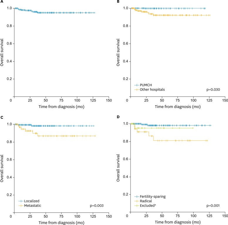
After a median observation time of 3.2 years (range, 0 to 10.5 years), eight patients (3.48%) were deceased, all were due to ovarian cancer. All cause-specific deaths occurred during the first 3 years after diagnosis, and the 5-year OS was predicted to be 95.00% (Fig. 1A).
Fig. 1.
Overall survival (A) in patients stratified by (B) medical institution, (C) extent of disease, and (D) surgery. PUMCH, Peking Union Medical College Hospital. *Twenty-five patients with disorder of sex development or vaginal malignant germ cell tumors were excluded from univariate analysis of fertility-related surgical decisions.
The estimated 5-year OS was 100.00% and 93.60% for patients diagnosed in PUMCH and other hospitals, respectively (p=0.030) (Fig. 1B). Fig. 1C depicts the OS according to local (98.20%) and extensive disease (86.80%, p=0.003). Additionally, the 5-year OS was significantly higher for patients who had opportunities to retain their fertility ability (98.20%) than for those who had to adopt a radical surgery (81.00%, p=0.001) (Fig. 1D).
Although univariate analysis revealed that a higher level of primary medical institution, restricted disease and fertility-sparing operation were predictors for better survival, only the surgical extent was significantly associated with OS in the multivariate model (Table 3).
Table 3. Cox proportional analysis of prognostic factors for recurrence and survival.
| Prognostic factor | Overall survival | Disease-free survival | |
|---|---|---|---|
| p-value | HR (95% CI) | p-value | |
| Age at diagnosis | 0.889 | 1.176 (0.335-4.127) | 0.800 |
| Institution for diagnosis | 0.925 | 0.009 | |
| PUMCH | 1 | ||
| Transferred | 4.307 (1.434-12.939) | ||
| Primary tumor site | >0.999 | >0.999 | |
| Ovary | 1 | ||
| Pelvis | 0 | ||
| Vagina | 0 | ||
| Dysgenetic gonads | 1.007 (0.112-9.078) | ||
| Extent of disease | 0.126 | 0.022 | |
| Localized | 1 | ||
| Metastatic | 2.611 (1.150-5.931) | ||
| Histological subtype | 0.881 | 0.971 | |
| Dysgerminoma/seminoma | 1 | ||
| Non-dysgerminoma/seminoma | 1.199 (0.269-5.352) | ||
| Mixed MGCTs | 1.123 (0.339-3.722) | ||
| Extent of surgery | 0.030 | 0.000 | |
| Fertility-sparing surgery | 1 | ||
| Radical resection | 5.713 (2.323-14.049) | ||
| Performance of staging | 0.265 | 0.891 | |
| Yes | 1 | ||
| No | 1.065 (0.431-2.635) | ||
| Postoperative chemotherapy | 0.997 | 0.945 | |
| None | 1 | ||
| PEB/PVB | 0.803 (0.157-4.120) | ||
| Other regimens | 0.806 (0.225-2.888) | ||
HR, hazard ratio; MGCT, malignant germ cell tumor; PEB/PVB, cisplatin, etoposide/ vincristine and bleomycin; PUMCH, Peking Union Medical College Hospital.
3) Disease-free survival
Although 34 patients failed to remit immediately after surgery combined with standard PEB/PVB (3 to 6 courses) chemotherapy, 24 out of them finally achieved remission with more aggressive operations and/or antineoplastic drugs. Therefore, these patients were grouped as remission cases at the beginning of the long-term surveillance, and 16 had no recurrence until the last follow-up. The 5-year DFS for all patients was 86.00%, with an estimated median period free from MGCTs of 10.1 years (95% CI, 9.4 to 10.7 years).
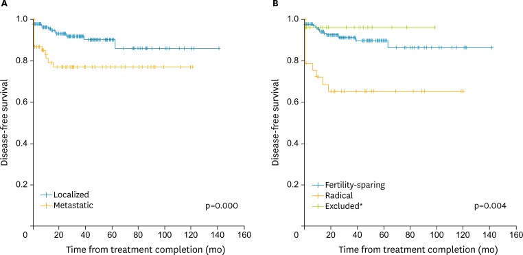
Univariate analysis identified that the extent of disease (p=0.004) and surgery (p<0.001) were factors that influencing progression and recurrence of disease (Fig. 2). In contrast, DFS was not different among the various age groups, primary tumor sites, histological subtypes and adjuvant chemotherapy regimens. Nor did the performance of comprehensive staging prolonged the 5-year DFS (91.3% vs. 96.20%, p=0.550) (Table 3).
Fig. 2.
Disease-free survival stratified by (A) extent of disease and (B) surgical. *Twenty-five patients with disorder of sex development or vaginal malignant germ cell tumors were excluded from univariate analysis of fertility-related surgical decisions.
The Cox regression model adjusted for other potential risk factors revealed the level of the medical center, extent of disease and surgery to be independent predictors of survival. Appropriate removal of tumors, rather than radical resection (hazard ratio, 5.713; 95% CI, 2.323 to 14.049), was sufficient to reduce the risk of relapse (p<0.001). Furthermore, metastatic disease carried a hazards ratio of 2.611 for recurrence (95% CI, 1.150 to 5.931, p=0.022) (Table 3).
DISCUSSION
Without limitation to the ovary, our study included MGCTs of other sites of origin in the female genital system. The results demonstrated that neither short-term therapeutic effect nor long-term survival outcome was different among the groups. Regardless of the primary tumor sites, patients had an excellent prognosis after similar treatment with appropriate surgery combined with platinum-based chemotherapy. Germ cell malignancies are neoplasms derived from primitive germ cells of the embryonic gonad [11], but the exact cause is still unknown. It is worth mentioning that all vaginal MGCTs in our cohort were yolk sac tumors, and occurred in infants. Excluding the infantile population, no significant association of tumor origin sites with histology and age was demonstrated, indicating a probably unique etiology for vaginal MGCTs. Five percent of dysgerminoma occur in phenotypically normal females with dysgenetic gonads, which is linked to Y-chromosome material with karyotypes 46XY (pure gonadal dysgenesis and complete androgen insensitivity) or mosaic 45X/46XY (mixed gonadal dysgenesis) [12]. Because half of the patients with dysgenetic gonads and Y-chromosome in their karyotype will develop malignancy [13], early bilateral gonadectomy was performed for all the DSD patients (7.39%) in our study.
One important finding of this study is the adverse impact of more aggressive surgery on both OS and DFS, with consideration of both the patients’ demographic characteristics and other clinical parameters. Modern principles recommend that conservative fertility-sparing surgery is the initial treatment of choice, and this type of surgery is feasible even with extensive and metastatic disease [14]. In accordance with this, we found that the extent of disease did not affect either the remission or OS rates by multivariate analysis. In particular, though 8 patients with childbearing requirement had their fertility deprived for persistent disease, three of them eventually died of uncontrolled ovarian cancer. Although a retrospective study reported that patients after incomplete resection of the tumor had a higher recurrence rate (68% vs. 28%) and lower DFS (34% vs. 65% at a median of 5 years) compared with completed resection of tumor [15], our study revealed that surgical thoroughness was unrelated to excision extension, OS and DFS, though the disease could be more rapidly under control in the short-term. Hence, although optimal debulking is the standard recommendation in advanced stage disease, judgment must be exercised to weigh the benefit of aggressive cytoreductive surgery against the chemosensitivity of MGCT. Moreover, no pathologically positive lymph note was detected after comprehensive staging in 56 patients, nor did the staging procedure improve the curability or prognosis according to our data. Therefore, staging is not required, especially when debulking is already satisfactorily achieved.
Stage IA dysgerminoma and stage IA, grade 1 immature teratoma are treated adequately with surgery alone, and all other patients are advised to have postoperative chemotherapy. A combination of PEB is the standard regimen [16]. Our results demonstrated that instead of regimen options, timely initiation of chemotherapy (within 7 days after surgery) was the key to enhance the therapeutic effect, but the OS and DFS were not influenced. In other words, anti-neoplastic medicine is of significant value as complementary treatment, and should be applied whenever risk factors are identified. Additionally, institutions lacking experience of treating MGCTs should refer these patients to higher-level hospitals (Chinese Tertiary Medical Centers) where standard chemotherapy can be given. Although three cycles of PEB are adequate for patients with completely resected stage IB–III disease [17], the optimal number of cycles of chemotherapy in incompletely resected or stage IV disease is uncertain. Remission has been realized in many of our patients with four to six cycles of chemotherapy depending on the decline of the serum tumor markers, and the bulkiness of residual disease. Thus, the number of courses may be flexible and individualized.
Approximately 20% of the patients will be resistant to treatment or experience relapse [18], in our study 34 patients (14.78%) had persistent malignancy at first and 14 (6.09%) recurrent tumors at a later stage. Additionally, our study revealed most recurrences occurred within the first 2 years after the end initial treatment, and salvage therapy had made most relapses remitted. Therefore, patients must be followed at gradually prolonged intervals if no abnormality has been detected. Monitoring of tumor markers (e.g., human chorionic gonadotropin, alpha fetal protein, etc), abdominal and pelvic imaging is of importance for timely recognition of recurrence. Although the role of surgery in the management of recurrent and refractory disease is not well defined [19], secondary cytoreductive surgery combined with chemotherapy was preferred in our hospital. As a result, more than half of these patients achieved remission under long-term surveillance. Histological type and stage are the most important prognostic factors, with dysgerminoma having the best survival in other researches [20]. Additionally, a large population-based (2,541 cases) study addressed the prognostic role of age on cause-specific survival in MOGCT [21]. However, our study identified the extent of disease and surgery, together with the level of hospital as the independent impact factor on DFS, while controlling other variables (e.g., age at diagnosis, extent of disease, histological subtype, performance of staging, and postoperative chemotherapy).
We acknowledge that our study has several limitations. Due to the large proportion of transferred patients lacking detailed medical records, some key factors cannot be fully analyzed, such as the extent of residual disease. The median surveillance duration to observe the effect on fertility or teratogenicity after fertility-sparing surgery and chemotherapy for MGCT is relatively short. During follow-up, only 28 patients declared recent desire for fertility, and 14 of them had a healthy baby, with another two patients were diagnosed with definitive infertility. Hence, a longer period of follow-up in a larger multicentral population is needed for more accurate evaluation, and fertility-related quality of life need to be monitored.
In conclusion, our study identified that there was no significant association of the primary tumor sites with the therapeutic response and prognosis in MGCT. The initial surgical option should be chosen with caution, because neither comprehensive staging nor aggressive debulking improves the patients’ DFS or OS. The short-term remission rate and long-term survival outcome should be balanced. Additionally, the preservation of fertility needs to be managed, if a patient so desires and her condition allows the option. Finally, standard but individualized chemotherapy should be supplemented in time, when necessary.
Footnotes
Conflict of Interest: No potential conflict of interest relevant to this article was reported.
References
- 1.Arora RS, Alston RD, Eden TO, Geraci M, Birch JM. Comparative incidence patterns and trends of gonadal and extragonadal germ cell tumors in England, 1979 to 2003. Cancer. 2012;118:4290–4297. doi: 10.1002/cncr.27403. [DOI] [PubMed] [Google Scholar]
- 2.Parkinson CA, Hatcher HM, Ajithkumar TV. Management of malignant ovarian germ cell tumors. Obstet Gynecol Surv. 2011;66:507–514. doi: 10.1097/OGX.0b013e318234ede9. [DOI] [PubMed] [Google Scholar]
- 3.Smith HO, Berwick M, Verschraegen CF, Wiggins C, Lansing L, Muller CY, et al. Incidence and survival rates for female malignant germ cell tumors. Obstet Gynecol. 2006;107:1075–1085. doi: 10.1097/01.AOG.0000216004.22588.ce. [DOI] [PubMed] [Google Scholar]
- 4.Gershenson DM. Management of ovarian germ cell tumors. J Clin Oncol. 2007;25:2938–2943. doi: 10.1200/JCO.2007.10.8738. [DOI] [PubMed] [Google Scholar]
- 5.Low JJ, Perrin LC, Crandon AJ, Hacker NF. Conservative surgery to preserve ovarian function in patients with malignant ovarian germ cell tumors. A review of 74 cases. Cancer. 2000;89:391–398. [PubMed] [Google Scholar]
- 6.Rescorla F, Billmire D, Vinocur C, Colombani P, London W, Giller R, et al. The effect of neoadjuvant chemotherapy and surgery in children with malignant germ cell tumors of the genital region: a pediatric intergroup trial. J Pediatr Surg. 2003;38:910–912. doi: 10.1016/s0022-3468(03)00121-0. [DOI] [PubMed] [Google Scholar]
- 7.Terenziani M, Spreafico F, Collini P, Meazza C, Massimino M, Piva L. Endodermal sinus tumor of the vagina. Pediatr Blood Cancer. 2007;48:577–578. doi: 10.1002/pbc.20636. [DOI] [PubMed] [Google Scholar]
- 8.Tao T, Yang J, Cao D, Guo L, Chen J, Lang J, et al. Conservative treatment and long-term follow up of endodermal sinus tumor of the vagina. Gynecol Oncol. 2012;125:358–361. doi: 10.1016/j.ygyno.2011.12.430. [DOI] [PubMed] [Google Scholar]
- 9.Tang QL, Jiang XF, Yuan XP, Liu Y, Zhang L, Tang XF, et al. Prognosis of eight Chinese cases of primary vaginal yolk sac tumor with a review of the literature. Asian Pac J Cancer Prev. 2014;15:9395–9404. doi: 10.7314/apjcp.2014.15.21.9395. [DOI] [PubMed] [Google Scholar]
- 10.Poynter JN, Amatruda JF, Ross JA. Trends in incidence and survival of pediatric and adolescent patients with germ cell tumors in the United States, 1975 to 2006. Cancer. 2010;116:4882–4891. doi: 10.1002/cncr.25454. [DOI] [PMC free article] [PubMed] [Google Scholar]
- 11.Parkinson CA, Hatcher HM, Ajithkumar TV. Management of malignant ovarian germ cell tumors. Obstet Gynecol Surv. 2011;66:507–514. doi: 10.1097/OGX.0b013e318234ede9. [DOI] [PubMed] [Google Scholar]
- 12.Obata NH, Nakashima N, Kawai M, Kikkawa F, Mamba S, Tomoda Y. Gonadoblastoma with dysgerminoma in one ovary and gonadoblastoma with dysgerminoma and yolk sac tumor in the contralateral ovary in a girl with 46XX karyotype. Gynecol Oncol. 1995;58:124–128. doi: 10.1006/gyno.1995.1195. [DOI] [PubMed] [Google Scholar]
- 13.Bremer GL, Land JA, Tiebosch A, van der Putten HW. Five different histological subtypes of germ cell malignancies in an XY female. Gynecol Oncol. 1993;50:247–248. doi: 10.1006/gyno.1993.1201. [DOI] [PubMed] [Google Scholar]
- 14.Kanazawa K, Suzuki T, Sakumoto K. Treatment of malignant ovarian germ cell tumors with preservation of fertility: reproductive performance after persistent remission. Am J Clin Oncol. 2000;23:244–248. doi: 10.1097/00000421-200006000-00007. [DOI] [PubMed] [Google Scholar]
- 15.Slayton RE, Park RC, Silverberg SG, Shingleton H, Creasman WT, Blessing JA. Vincristine, dactinomycin, and cyclophosphamide in the treatment of malignant germ cell tumors of the ovary. A Gynecologic Oncology Group Study (a final report) Cancer. 1985;56:243–248. doi: 10.1002/1097-0142(19850715)56:2<243::aid-cncr2820560206>3.0.co;2-t. [DOI] [PubMed] [Google Scholar]
- 16.Murugaesu N, Schmid P, Dancey G, Agarwal R, Holden L, McNeish I, et al. Malignant ovarian germ cell tumors: identification of novel prognostic markers and long-term outcome after multimodality treatment. J Clin Oncol. 2006;24:4862–4864. doi: 10.1200/JCO.2006.06.2489. [DOI] [PubMed] [Google Scholar]
- 17.Williams S, Blessing JA, Liao SY, Ball H, Hanjani P. Adjuvant therapy of ovarian germ cell tumors with cisplatin, etoposide, and bleomycin: a trial of the Gynecologic Oncology Group. J Clin Oncol. 1994;12:701–706. doi: 10.1200/JCO.1994.12.4.701. [DOI] [PubMed] [Google Scholar]
- 18.Dimopoulos MA, Papadimitriou C, Hamilos G, Efstathiou E, Vlahos G, Rodolakis A, et al. Treatment of ovarian germ cell tumors with a 3-day bleomycin, etoposide, and cisplatin regimen: a prospective multicenter study. Gynecol Oncol. 2004;95:695–700. doi: 10.1016/j.ygyno.2004.08.018. [DOI] [PubMed] [Google Scholar]
- 19.Einhorn LH, Brames MJ, Juliar B, Williams SD. Phase II study of paclitaxel plus gemcitabine salvage chemotherapy for germ cell tumors after progression following high-dose chemotherapy with tandem transplant. J Clin Oncol. 2007;25:513–516. doi: 10.1200/JCO.2006.07.7271. [DOI] [PubMed] [Google Scholar]
- 20.Yilmaz F, Gül T, Uzunlar AK. Malignant ovarian germ cell tumors: analysis of 32 cases. Eur J Gynaecol Oncol. 2003;24:569–573. [PubMed] [Google Scholar]
- 21.Solheim O, Gershenson DM, Tropé CG, Rokkones E, Sun CC, Weedon-Fekjaer H, et al. Prognostic factors in malignant ovarian germ cell tumours (The Surveillance, Epidemiology and End Results experience 1978-2010) Eur J Cancer. 2014;50:1942–1950. doi: 10.1016/j.ejca.2014.03.288. [DOI] [PubMed] [Google Scholar]


