Abstract
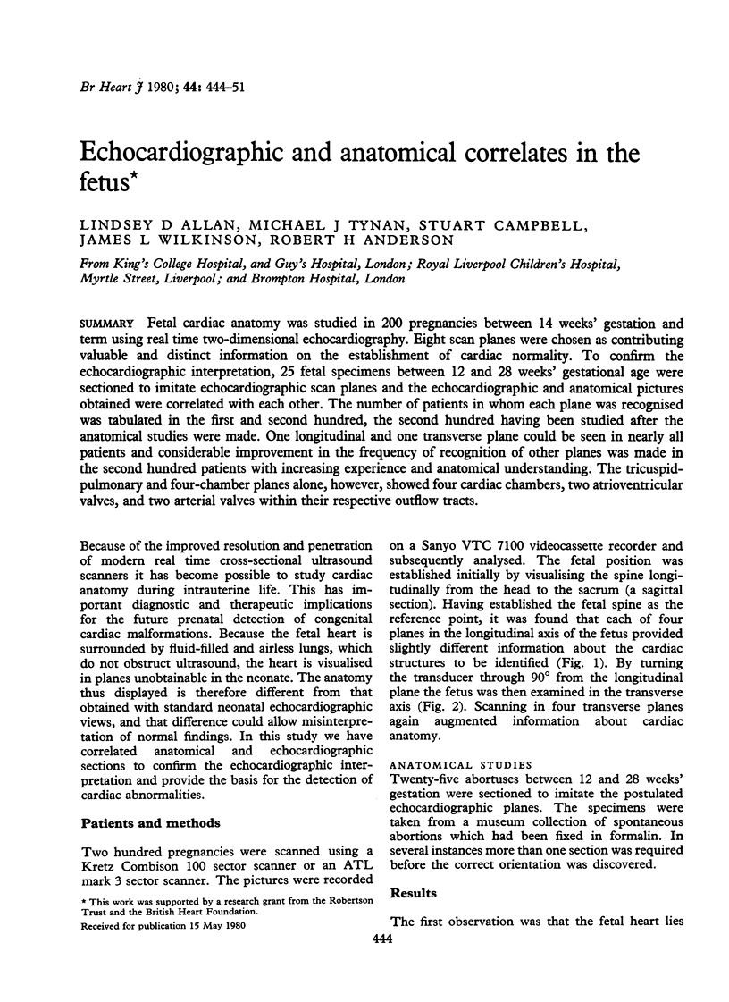
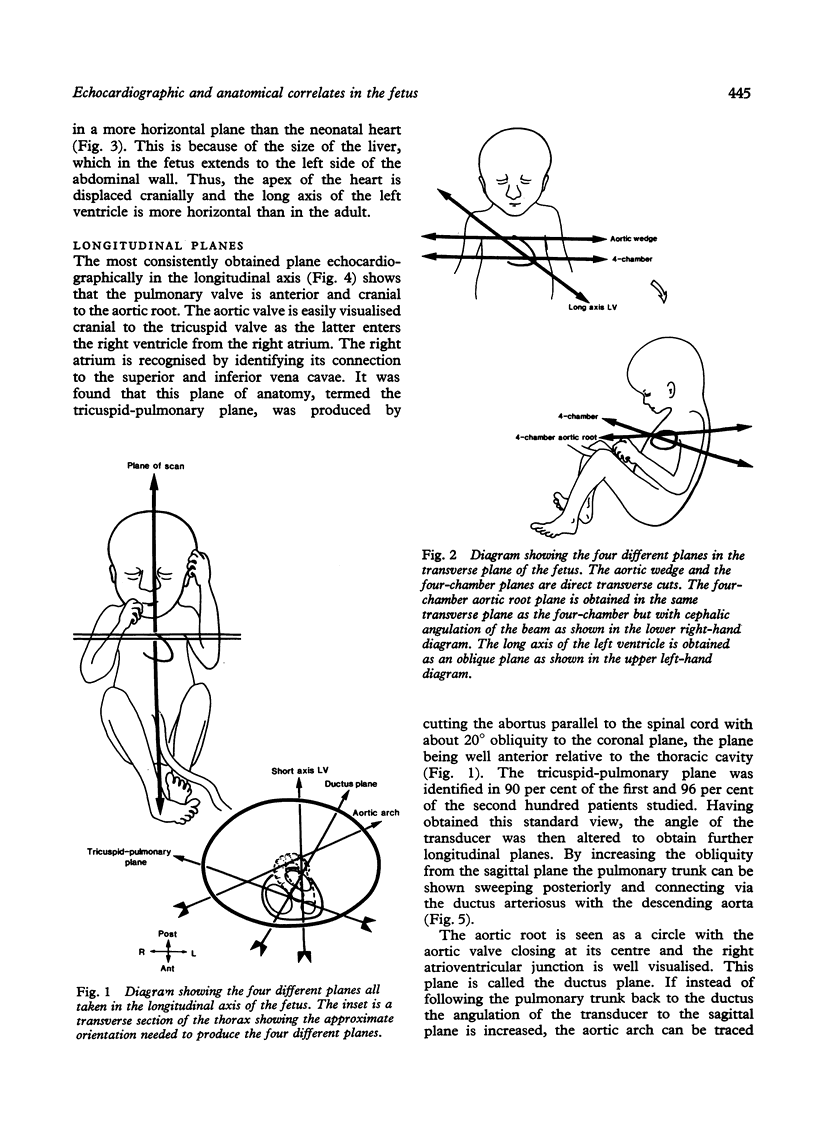
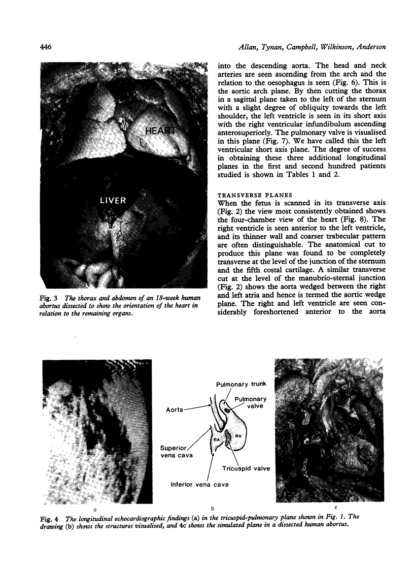
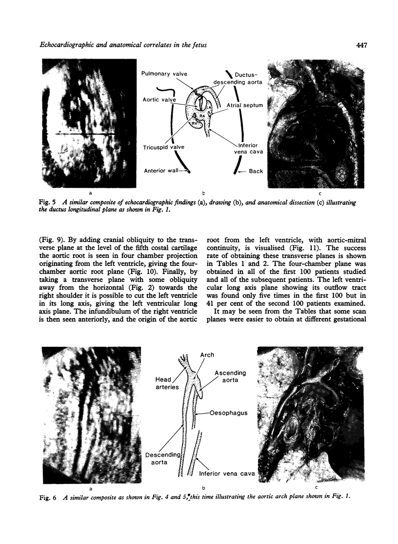
Fetal cardiac anatomy was studied in 200 pregnancies between 14 weeks' gestation and term using real time two-dimensional echocardiography. Eight scan planes were chosen as contributing valuable and distinct information on the establishment of cardiac normality. To confirm the echocardiographic interpretation, 25 fetal specimens between 12 and 28 weeks' gestational age were sectioned to imitate echocardiographic scan planes and the echocardiographic and anatomical pictures obtained were correlated with each other. The number of patients in whom each plane was recognized was tabulated in the first and second hundred, the second hundred having been studied after the anatomical studies were made. One longitudinal and one transverse plane could be seen in nearly all patients and considerable improvement in the frequency of recognition of other planes was made in the second hundred patients with increasing experience and anatomical understanding. The tricuspid-pulmonary and four-chamber planes alone, however, showed four cardiac chambers, two atrioventricular valves, and two arterial valves within their respective outflow tracts.
Full text
PDF