Abstract
Legionella pneumophila, the major causative agent of Legionnaires’ disease, is found in freshwater environments in close association with free-living amoebae and multispecies biofilms, leading to persistence, spread, biocide resistance, and elevated virulence of the bacterium. Indeed, legionellosis outbreaks are mainly due to the ability of this bacterium to colonize and persist in water facilities, despite harsh physical and chemical treatments. However, these treatments are not totally efficient and, after a lag period, L. pneumophila may be able to quickly re-colonize these systems. Several natural compounds (biosurfactants, antimicrobial peptides…) with anti-Legionella properties have recently been described in the literature, highlighting their specific activities against this pathogen. In this review, we first consider this hallmark of Legionella to resist killing, in regard to its biofilm or host-associated life style. Then, we focus more accurately on natural anti-Legionella molecules described so far, which could provide new eco-friendly and alternative ways to struggle against this important pathogen in plumbing.
Keywords: Legionella pneumophila, biofilms, amoebae, biocides, natural compounds, antimicrobial peptides, essential oils, biosurfactants
Introduction
Legionella pneumophila is a Gram-negative opportunistic intracellular human pathogen that is responsible for severe pneumonia called Legionnaires’ disease (LD; Fields et al., 2002). The case fatality rate of LD associated with outbreaks is lower than that of sporadic cases, generally around 8–15%, but it can be higher, particularly for hospital-acquired infections, acquired immune-deficiency syndrome (AIDS) patients, transplant patients, and those undergoing aggressive chemotherapy (Dominguez et al., 2009). Among 60 Legionella species1, L. pneumophila is the leading cause of LD and L. pneumophila serogroup 1 is associated with almost 85–90% of the cases worldwide (Fields et al., 2002; Yu et al., 2002; Campese et al., 2011; Beaute et al., 2013). Since the first outbreak of pneumonia in 1976 (Fraser et al., 1977; McDade et al., 1977), many LD outbreaks have been linked to various sources of contaminated water in hospitals, hotels, cruise ships, industrial facilities, and family residences (Falkinham et al., 2015). Generally, the economic cost of waterborne diseases including LD is elevated with over $430 million per year in the United States considering only hospitalized patients (Collier et al., 2012). Thus, L. pneumophila has a high epidemiological and economical significance, being considered as an opportunistic plumbing pathogen. The bacterium is currently on the United States Environmental Protection Agency (USEPA) candidate contaminant list 42. Transmission to humans occurs after inhalation of contaminated water droplets. L. pneumophila reaches the alveolar mucosa and, thanks to its ability to resist phagocytosis, multiplies inside macrophages (Prashar and Terebiznik, 2015). These latter are considered as the primary target of L. pneumophila although various data indicate that L. pneumophila can also invade epithelial cells, in which it can replicate (Cirillo et al., 1994; Mallegol et al., 2014). Its resistance mechanisms to phagocytosis have been thoroughly described and several key steps are highly studied, among which the delivery of effectors into the host cytosol through the Dot/Icm type IV secretion system and the formation of the Legionella containing vacuole, which is known as the intracellular replicative niche for the bacterium (Isberg et al., 2009; Hubber and Roy, 2010; Xu and Luo, 2013).
Within freshwater environments, L. pneumophila bacteria are ubiquitous organisms, mostly found as parasites of various free-living protozoa such as amoebae, their natural hosts (Steinert et al., 2002). Free-living amoebae are not solely responsible for L. pneumophila spreading, but they are also considered as biological shields as they protect intracellular bacteria from adverse conditions or biocide treatments (Loret and Greub, 2010). Thus, amoebae play a key role in the life cycle and pathogenesis of L. pneumophila, and its ability to infect human macrophages is thought to be a consequence of prior adaptation to intracellular growth within various primitive eukaryotic hosts such as protozoa (Franco et al., 2009; Al-Quadan et al., 2012). Moreover, upon transfer from natural freshwater habitats into anthropogenic systems, generally at higher temperature than ambient, L. pneumophila colonizes existing multispecies biofilms (Rogers et al., 1994). Colonization of these naturally occurring biofilms by L. pneumophila can be influenced by several other microorganisms among which protozoa are arguably of particular importance, as they constitute an ecological niche for the pathogen to replicate and to persist (Abdel-Nour et al., 2013). Co-evolution with multiple species of protozoa has resulted in the development of mechanisms that allow L. pneumophila to occupy a very broad host range (Abu Kwaik et al., 1998). Biofilms and free-living amoebae are thus considered to serve as main environmental reservoirs for L. pneumophila and represent a potential source of drinking water contamination, resulting in a potential health risk for humans (Dupuy et al., 2011; Wingender and Flemming, 2011). Thus, it is of primary importance to find new antibacterial agents to control L. pneumophila environmental spread.
This paper presents an overview of the literature regarding the discovery of potential anti-Legionella control agents and their mechanisms of action, if known. First, various elements that allow L. pneumophila to resist to biocides in its environment are reviewed. Then, the high sensitivity of this bacterium to a diversity of biomolecules that could become of interest in the control of environmental pathogens in water systems is discussed.
Persistence of L. pneumophila in Its Microenvironment
Resistance of L. pneumophila within Biofilms
Legionella pneumophila is ubiquitous in natural and anthropogenic water systems, in which it is able to survive for long periods within biofilms (Rogers et al., 1994). Biofilms are defined as complex microbial communities characterized by cells that are attached to a substrate or phase boundary and to each other, and embedded into a matrix of self-produced extracellular polymeric substances (Donlan and Costerton, 2002). Biofilms provide shelter and nutrients, exhibit a remarkable resistance to many stress factors, thus representing an interesting ecological niche for Legionella persistence. L. pneumophila has also the ability to parasitize protozoa, which commonly graze on biofilm communities (Declerck et al., 2005, 2007). Due to the intracellular lifestyle of L. pneumophila within protozoa, it is difficult to tease out whether the resistance of L. pneumophila in environmental biofilms is due to the biofilm structure, its association with amoebae or both (Abdel-Nour et al., 2013).
In artificial water systems as in drinking water distribution, Legionella growth is detected almost exclusively in biofilms covering the interior of pipe walls, ventilation, and air-conditioning systems, for example (Lau and Ashbolt, 2009). In addition to Legionella, these biofilms can become transient or long-term habitats for hygienically relevant microorganisms among which fecal indicator bacteria (Escherichia coli), obligate bacterial pathogens of fecal origin (Campylobacter sp.), opportunistic bacteria of environmental origin (Pseudomonas aeruginosa, Mycobacterium sp., Aeromonas sp.), enteric viruses (adenoviruses, rotaviruses, noroviruses), and parasitic protozoa (Cryptosporidium parvum). These organisms can attach to preexisting biofilms, where they become integrated and survive for days to weeks or even longer, depending on the biology and ecology of the organism and the environmental conditions (Blasco et al., 2008; Buse et al., 2014; Richards et al., 2015).
In order to restrain L. pneumophila growth, various treatments are used (e.g., physical, thermal, and chemical) in water systems (Jjemba et al., 2015). However, they are not fully efficient, and after a lag period, L. pneumophila may be able to quickly re-colonize the system (Thomas et al., 2004; Cooper et al., 2008; Cooper and Hanlon, 2010). Environmental L. pneumophila found in biofilms are extremely resilient to treatment with biocides (Kim et al., 2002; Emtiazi et al., 2004; Borella et al., 2005; Saby et al., 2005). When this bacterium is exposed to environmental stresses including biocides and/or found within biofilms, it can enter in a viable but non-culturable state (Giao et al., 2009). The most common biocides used to control waterborne pathogens are generally chlorine derivatives (Walker et al., 1995; Liu et al., 1998; Schwartz et al., 2003; Cervero-Arago et al., 2015). While hyperchlorination of potable water has been shown appropriate for treatment and removal of planktonic cultures of L. pneumophila, it remains ineffective against sessile communities (Cooper and Hanlon, 2010; Simoes et al., 2010). Exposure to chlorine at regular intervals has also been shown to facilitate a higher tolerance to disinfectant, thus promoting bacterial resistance (Cooper and Hanlon, 2010). Chlorine dioxide is probably more effective than chlorine because of its superior oxidative power and effect on biofilms (Walker et al., 1995; Hamilton et al., 1996). Chloramine, a powerful chlorine derivative biocide, is a recommended commercial formulation for disinfecting cooling towers. Yet, it has been shown to not completely eradicate L. pneumophila from biofilms (Sanli-Yurudu et al., 2007). Recent inquiries into the microbial ecology of distribution systems have shown that pathogen resistance to chlorination is affected by microbial community diversity and interspecies relationships. Multispecies biofilms are generally more resistant to chloramine disinfection than single-species biofilms. One of the reasons may be the presence of nitrifying bacteria leading to depletion of chloramine disinfectant residuals (Berry et al., 2006).
Resistance of L. pneumophila in Association with Its Eukaryotic Hosts
According to the recommendations of the World Health Organization, water disinfection with chlorine has to be performed using a concentration of chlorine between 0.2 and 0.5 mg/l. However, it appears that Legionella is recovered if the treatment is not continuous. The persistence of L. pneumophila is due, at least partly, to its intra-amoeba lifestyle (Steinert et al., 1998; Hilbi et al., 2011) since these protozoa act as biological shields, protecting bacteria from biocides (Loret and Greub, 2010). Amoeba-grown L. pneumophila are thus more resistant than planktonic cells to chemical disinfectants, biocides (Barker et al., 1992; Dupuy et al., 2011), and antibiotics (Barker et al., 1995). It has been indeed reported that intracellular L. pneumophila are released from amoebae within vesicles containing several hundreds of resistant bacteria to biocides such as the isothiazolone-derivative minimum bactericidal concentration (MBC215; a mixture of 5-chloro-2-methyl-4-isothiazolin-3-one and 2-methyl-4-isothiazolin) and the quaternary ammonium compound poly(oxyethylene) (dimethylimino) ethylene (dimethylimino) ethylene dichloride (Berk et al., 1998). In addition, bacteria released through these vesicles were viable up to 6 months (Bouyer et al., 2007). In the same way, it has been demonstrated that amoebae promote resuscitation of viable but non-culturable Legionella, enhancing in parallel their resistance to sodium hypochlorite (Garcia et al., 2007). The level of Legionella resistance into amoebae is also dependent of the disinfectant used. For example, monochloramine displays the same efficiency against planktonic bacteria in the presence or not of amoebae whereas chlorine and chlorine dioxide are less active against Legionella co-cultured with amoebae (Dupuy et al., 2011). The understanding, at the molecular level, of the intra-protozoa acquired Legionella resistance (Garduno et al., 2002; Koubar et al., 2011) is obviously an important information in order to develop strategies to limit or eradicate this phenomenon. Interestingly, it was also shown that L. pneumophila resistance against chlorine acquired into amoebae is host-dependent (Chang et al., 2009), suggesting the involvement of specific molecular mechanisms currently unknown.
In conclusion, the use of biocides such as chlorine or chloramine to disinfect water appears to only limit the development of L. pneumophila without being able to eradicate this pathogen (Wang et al., 2012). More critically, intracellularly grown L. pneumophila become more resistant when exposed to biocides (Cervero-Arago et al., 2015), suggesting that it is necessary to control, in water, both bacterial pathogens and their natural hosts like amoebae. In this way, studies of the direct impact of biocides on amoebae, alone or infected with a pathogen, would be very helpful (Srikanth and Berk, 1994; Mogoa et al., 2010, 2011; Fouque et al., 2015). Noticeably, infected amoebae were shown to become more pathogenic than uninfected amoebae (Brieland et al., 1997), suggesting that the two partners (i.e., amoebae and L. pneumophila) enhance synergistically their pathogenesis. Therefore, studies dealing with treatments against Legionella should be performed on bacteria associated with their natural host in order to (i) determine how the bacteria are protected during their intracellular life cycle, (ii) if the passage inside host cells modifies the Legionella resistance after its escape, and (iii) how the host responds to treatments (Thomas et al., 2004; Donlan et al., 2005; Dupuy et al., 2011).
Natural Biocides: An Alternative Way to Control L. pneumophila Spread in the Environment?
For a few years now, some studies have highlighted natural compounds with anti-Legionella properties. Why such an interest? Probably because (i) L. pneumophila is a waterborne bacterium ubiquitously found in freshwater environments, (ii) LD is a severe and sometimes fatal multisystem illness involving atypical pneumonia, (iii) the development of man-made water systems such as air-conditioners and cooling towers has expanded the environmental niche of L. pneumophila in association with amoebae, (iv) emerging pathogens such as L. pneumophila or spore-forming bacteria such as Bacillus are able to resist to currently used water disinfection procedures, and (v) more efforts are needed to control disinfection by-products and minimize people exposure to potentially hazardous chemicals (Trihalomethanes, Haloacetic acids…) while maintaining adequate disinfection and control of targeted pathogens while respecting the environment. Subsequently, we review in this chapter recent advances in finding natural compounds exhibiting direct or indirect anti-Legionella activity and discuss, if known, their mode of action.
Proteins
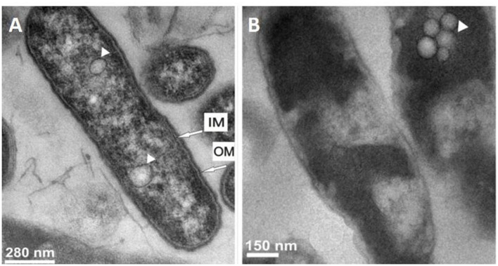
To date, only two proteins have been found to be directly active against Legionella cells: the greater wax moth Galleria mellonella apolipophorin III (ApoLp-III) and the human lactoferrin. The ApoLp-III protein family is composed of low-molecular weight apolipoproteins (161–166 amino acid residues) characterized by a globular amphipathic α-helix bundle conformation (Weers and Ryan, 2006). ApoLp-III has been shown to be an important component of hemolymph of numerous insect species of Orthoptera, Leptidoptera, Coleoptera, and Hemiptera genus. In addition, ApoLp-III is involved in lipid transport and immunity. Recently, the protein was recovered by methanol extraction, purified and evaluated against three Legionella species: L. dumoffii, L. pneumophila, and L. gormanii (Palusinska-Szysz et al., 2012; Chmiel et al., 2014; Zdybicka-Barabas et al., 2014). Antimicrobial assays demonstrated a difference in susceptibility among Legionella species. An 1-hincubation time of cells with 0.1 mg/ml of protein induced a moderate mortality rate of 55% for L. pneumophila vs. 40% for L. gormanii. The highest protein concentration tested in the studies, 0.4 mg/ml for L. dumoffii, 1.6 mg/ml for L. pneumophila, and 0.2 mg/ml for L. gormanii decreased the survival rate of 30, 100, and 50%, respectively. The effect of the protein on L. dumoffii was also investigated by transmission electron microscopy (Palusinska-Szysz et al., 2012). This study highlighted cell wall damages and strong intracellular alterations, such as increased vacuolization and condensation in the cytoplasm (Figure 1). Interestingly, cell envelope damages appeared greater for bacteria cultured on medium with choline supplementation (Palusinska-Szysz et al., 2012). Consistently, the sensitivity toward ApoLp-III of this species was threefold increased when cells were grown in the presence of choline. Extracellular choline is known to be used by some Legionella species for the synthesis of phosphatidylcholine (PC) that are phospholipids commonly found as component of eukaryotic membranes while only encountered in the envelope of about 15% of bacteria (Martinez-Morales et al., 2003; Geiger et al., 2013; Sohlenkamp and Geiger, 2016). Based on these observations as well as atomic force microscopy (AFM), Fourier transform infrared spectroscopy (FTIR), and lipopolysaccharide (LPS) binding studies, the authors assumed that ApoLp-III interacts with lipid components of Legionella cell membrane (Zdybicka-Barabas et al., 2014). Indeed, the protein most probably interacts with phospholipids (especially PC) of L. dumoffii while the anti-L. pneumophila effect is rather driven by interaction with LPS and other lipid components of its membrane. ApoLp-III shares homology with the 22-kDa N-terminal domain of the human apolipoprotein E (ApoE). This 37-kDa apolipoprotein has similar roles as ApoLp-III including lipid transport, host immunity as well as immunomodulatory properties. As for its insect homolog, SDS-PAGE and FTIR analysis revealed that ApoE strongly interacts with LPS of L. pneumophila outer membrane. However, although AFM analysis demonstrated alterations in the cell surface topography and properties, 0.8 mg/ml of protein did not reduce viability of L. pneumophila cells after an 1-h treatment (Palusinska-Szysz et al., 2015).
FIGURE 1.
Ultrastructural changes in Legionella dumoffii cells after treatment with Galleria mellonella apolipophorin III. Transmission electron micrographs of (A) untreated bacteria and (B) treated bacteria with 0.4 mg/ml ApoLp-III. The presence of vacuoles is indicated by arrowhead. IM, Inner membrane; OM, Outer membrane (Source: Palusinska-Szysz et al., 2012).
Lactoferrin is a glycoprotein of the transferrin family found at high levels in milk. The molecule, thanks to its ferric ions binding capacity, presents multiple biological functions. Indeed, lactoferrin is known to interact with the molecular and cellular components of hosts and pathogens (Siqueiros-Cendon et al., 2014). Bortner et al. (1986, 1989) studied the anti-Legionella activity of the human lactoferrin under both its iron-free and iron-saturated states. The study reveals a difference in terms of activity between the two states of the protein. Indeed, 0.09 mg/ml of apolactoferrin (the iron-free state) was able to kill 99.99% of exponentially grown Legionella cells after 2 h of incubation, while the iron-saturated form was unable to reduce viability of the bacteria in the same conditions (Bortner et al., 1986). The activity of apolactoferrin was abrogated at temperatures below 22°C and by the addition of MgCl2, CaCl2, or Mg(NO3)2 but not NaCl (Bortner et al., 1986, 1989). The physiological state of Legionella also played a role in the sensitivity toward apolactoferrin since stationary phase cells became more resistant to the protein. The mechanism by which apolactoferrin kills Legionella is currently unknown. Previously, the bactericidal activity of lactoferrin against other Gram-negative bacteria was shown to be mediated through the binding of the protein to receptors on the bacterial surface inducing cell-death due to a disruption in the cell wall (Jenssen and Hancock, 2009). The bactericidal activity against Gram-positive bacteria is mediated by electrostatic interactions between the positively charged protein and the bacterial membrane leading to its permeabilization (Jenssen and Hancock, 2009).
Protein-Derived Peptides
Regarding protein-derived peptides, two synthetic fragments of protein were shown to have anti-Legionella activity. The first one, C18G (ALYKKLLKKLLKSAKKLG; 2043 Da), is based on the antimicrobial peptide C13 corresponding to the last 13 amino acids of the carboxyl terminus of human platelet factor IV. The peptide was designed to improve its antibacterial potency by increasing the length of C13 and substituting a negative charge with a positive charge (Darveau et al., 1992). The activity of the synthetic amphipathic α-helical cationic peptide has been evaluated against L. pneumophila (Robey et al., 2001). Minimal bactericidal concentration was determined on logarithmic-phase bacteria as 32 to 128 μg/ml, depending on Mg2+ concentration. Deletion of rcp gene, encoding a protein with homology to the lipid A palmitoyltransferase PagP of Salmonella serovar Typhimurium and E. coli, led to a slight increase in susceptibility of the bacteria to the peptide indicating that this gene is involved in the resistance to cationic antimicrobial peptides by a Mg2+ mediated pathway. This latter is linked to the addition of palmitate on LPS, leading to a decrease of membrane fluidity thus preventing peptide insertion (Guo et al., 1998).
When compared to C18G, another synthetic protein fragment named NK-2 demonstrated its efficacy in Legionella killing. NK-2 (KILRGVCKKIMRTFLRRISKDILTGKK; 3203 Da) was designed as the partial sequence of the porcine lymphatic effector protein NK-lysin corresponding to the core region of the protein (residues 39–65) (Leippe, 1995). Various studies highlighted the very high potency of the peptide in the killing of cancer cells and various pathogens including Gram-negative and Gram-positive bacteria, the yeast Candida albicans as well as the intracellular parasites Trypanosoma cruzi and Plasmodium falciparum (Andra and Leippe, 1999; Jacobs et al., 2003; Schroder-Borm et al., 2003; Gelhaus et al., 2008; Jena et al., 2011). Interestingly, NK-2 appears to be non-hemolytic with no cytotoxicity toward normal mammalian cells such as keratinocytes, lymphocytes, macrophages, and glioblastoma cells at bactericidal concentrations (Andra and Leippe, 1999; Jacobs et al., 2003; Schroder-Borm et al., 2003; Jena et al., 2011). Recently, the knowledge about the potency of the synthetic peptide was extended to Legionella. The minimal bactericidal concentration was determined on exponentially grown L. pneumophila (Schlusselhuber et al., 2015). The study revealed that a concentration of 1.6 μM was able to kill the bacteria. NK-2 is a cationic peptide that adopts an amphipathic alpha helical secondary structure upon membrane interaction. A previous study revealed the possible mode of action of the peptide (Willumeit et al., 2005). Indeed, NK-2 was shown to bind and permeabilize membranes containing negatively charged phosphatidylglycerol (PG; found in the cytoplasmic membranes of bacteria) whereas no effects were observed with pure zwitterionic PC model membranes (as a mimetic for human cell membranes). Regarding phosphatidylethanolamine (PE) model membranes (a major phospholipid component of bacterial cell membranes), NK-2 binds and slightly inserts into the model membrane. The direct interaction with lipids leads to an increase of the membrane stiffness, thus favoring the formation of inverted lipid structures promoting the intrinsic negative membrane curvature. Enhancement of this effect results in membrane tension and disruption (Willumeit et al., 2005).
Antimicrobial Peptides (AMPs)
Historically, the first antimicrobial peptides tested against L. pneumophila were some apidaecin-type peptides, consisting in proline-rich molecules isolated from various hymenopteran insects (Casteels et al., 1994). Interestingly, in the same study the most active anti-Legionella peptide tested was Cecropin P1, a 31 amino acids long peptide isolated from the pig small intestine (Lee et al., 1989). However, the antimicrobial activities of these peptides were only estimated from the diameter of the inhibition zones observed on agar plates.
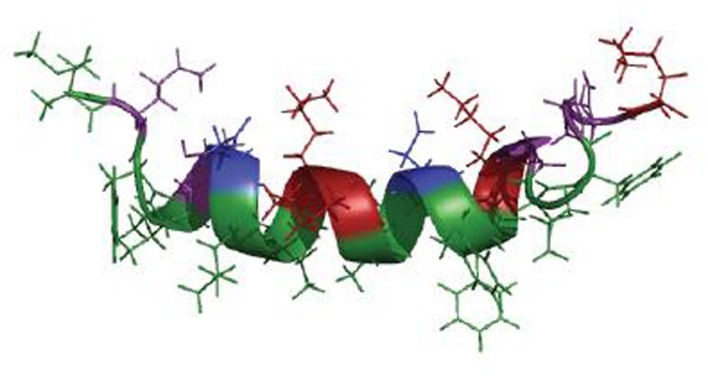
The first anti-Legionella peptides, produced by bacteria, were purified and characterized from the culture supernatant of a Staphylococcus warneri strain. This strain, S. warneri RK, was first detected as a contaminant colony on a L. pneumophila culture surrounded by a characteristic inhibition zone (Hechard et al., 2005). This activity was assigned to a molecule secreted by S. warneri RK. This molecule displayed a high heat-stability and its activity was lost after protease treatments, indicating that it might be an antimicrobial peptide. Finally, three anti-Legionella peptides produced by S. warneri RK were characterized (Verdon et al., 2008). One peptide, warnericin RK, is original, while the two others are delta-lysin I and delta-lysin II, encoded by genes previously described (Tegmark et al., 1998). They are close to S. aureus delta-hemolysin which was known for its action on red blood cells and was deemed to be devoid of antibacterial activity (Verdon et al., 2009b). The S. warneri peptides share similar biochemical characteristics as they are short (22 amino acids), cationic and highly hydrophobic. They display the same antibacterial spectrum, which is almost restricted to the Legionella genus. However, the amino acids sequence alignment (Table 1) shows that no high similarity exists between the three peptides even if they were predicted to adopt an α-helical structure (Verdon et al., 2008). This structure was further confirmed for warnericin RK by circular dichroism (CD) and nuclear magnetic resonance (NMR) spectroscopy analyses (Verdon et al., 2009a). CD spectroscopy showed that the peptide did not have a defined secondary structure in aqueous solution. However, in a membrane-like environment that is mimicked by the addition of dimyristoylphosphatidylcholine vesicles or 8% trifluoroethanol (TFE), a defined α-helical secondary structure was formed. From the 2D-NMR analysis, performed in 8% TFE, NOESY spectra revealed a well-defined α-helix extending from residue 4 to residue 16 (Figure 2). This central part of the peptide forms a nearly perfect amphiphilic helix, which is also observed in the S. aureus delta-hemolysin (Lee et al., 1987).
Table 1.
Anti-Legionella antimicrobial peptides (AMPs) produced by Staphylococci (Adapted from Marchand et al., 2011).
| Peptide | Producing bacteria | Amino acids sequence (Nter–Ctter) | MIC (μM) | |
|---|---|---|---|---|
| Group 1 | Warnericin RK | S. warneri | MQFITDLIKKAVDFFKGLFGNK | 0.3 |
| δ-Lysin I* | S. warneri | MAADIISTIGDLVKLIINTVKKFQK | 1.08 | |
| δ-Lysin II | S. warneri | MTADIISTIGDFVKWILDTVKKFTK | 0.54 | |
| δ-Hemolysin | S. aureus | MAQDIISTIGDLVKWIIDTVNKFTKK | 1.05 | |
| Ggi I | S. haemolyticus | MQKLAEAIAAAVSAGQDKDWGKMGTSIVGIVENGITVLGKIFGF | 4.15 | |
| SLUSH C | S. lugdunensis | MDGIFEAISKAVQAGLDKDWATMGTSIAEALAKGVDFIIGLFH | 5.16 | |
| SLUSH A | S. lugdunensis | MSGIVDAITKAVQAGLDKDWATMATSIADAIAKGVDFIAGFFN | 11.28 | |
| Group 2 | PSMα | S. epidermidis | MADVIAKIVEIVKGLIDQFTQK | 0.63 |
| δ-Hemolysin | S. epidermidis | MMAADIISTIGDLVKWIIDTVNKFKK | 1.59 | |
| PSMβ | S. epidermidis | MSKLAEAIANTVKAAQDQDWTKLGTSIVDIVESGVSVLGKIFGF | 2.69 | |
| H2U* | S. cohnii | MDFIIDIIKKIVGLFTGK | 3.04 | |
| Ggi II | S. haemolyticus | MEKIANAVKSAIEAGQNQDWTKLGTSILDIVSNGVTELSKIFGF | 13.23 | |
| Haemo 3 | S. haemolyticus | n.d. | 1.38 |
Minimum inhibitory concentrations were determined against L. pneumophila Lens and for the formylated forms of the peptides except for the peptides indicated by *.
FIGURE 2.
Structural model of warnericin RK in a membrane-like environment. Green: hydrophobic residues; Purple: hydrophilic and neutral residues; Blue: negatively charged residues; Red: positively charged residues (Source: Adapted from Verdon et al., 2009a).
Several anti-Legionella peptides have been found so far, mainly in bacteria belonging to the Staphylococcus genus (Marchand et al., 2011). Indeed, nine strains representing nine different species of staphylococci were found to secrete anti-Legionella compounds. All the purified compounds (Table 1), except one (Haemo 3 from S. haemolyticus), corresponded to previously described hemolytic peptides and were not known for their anti-Legionella activity. It should be noted that, beside the non-substituted peptides, the N-formylated forms (N-formylmethionine) of these compounds have been isolated and are active against Legionella (Verdon et al., 2008; Marchand et al., 2011). Moreover, the formylated forms of warnericin RK, δ-hemolysin II from S. warneri, and PSMα from S. epidermidis showed a higher inhibitory activity (MIC < 0.6 μM) than the corresponding non-substituted forms (MIC > 1.1 μM) (Marchand et al., 2011). These three peptides were found active against all the Legionella tested, corresponding to 6 L. pneumophila strains belonging to 4 different serogroups (1, 3, 5, and 6) and 6 non-pneumophila species. They appear to be very specific of the Legionella genus. Nevertheless, all the 12 anti-Legionella peptides described to date (Verdon et al., 2008; Marchand et al., 2011) display hemolytic activity.
On the basis of their antimicrobial [minimum inhibitory concentration (MIC), minimum permeabilization concentration, decrease of bacterial cultivability] and hemolytic activities, the purified peptides were separated into two groups (Table 1). The first group, including warnericin RK, corresponds to highly hemolytic and bactericidal peptides. The peptides of the second group, including PSMα from Staphylococcus epidermidis, are bacteriostatic and poorly hemolytic. Thus, a structure/activity relationships study was performed on the archetypes of each group of anti-Legionella peptides, warnericin RK and PSMα, in order to determine key amino acids (Marchand et al., 2015). Firstly, it was shown that the predicted helical wheel projections of these two peptides, assuming that the whole sequences were in an ideal α-helical structure, appeared similar when one of the sequence was reversed. Consequently, the authors designed a library of variants by replacing selected amino acids from one sequence by the corresponding of the reverse sequence of the other. Comparison of the anti-Legionella and hemolytic activities of these variants with these of the parent peptides succeeded in determining specific amino acid residues in warnericin RK and PSMα sequences that are critical. Surprisingly, the residue in the 14th position in both sequences (Phenylalanine for warnericin RK and Glycine for PSMα) appeared crucial for hemolytic activity but not for antibacterial activity (Table 1). However, as expected, the authors showed that the antibacterial activity of such peptides was correlated with their global positive charge.
Only the mode of action of warnericin RK has been studied in detail so far. A concentration of this peptide equals or superior to 3.12 μM was shown to fully suppress the growth ability of L. pneumophila (Verdon et al., 2009a). By using planar lipid bilayer studies and osmotic protection experiments, it was suggested that warnericin RK is membrane active. More precisely, results indicated that warnericin RK forms large channels of various sizes in erythrocytes as well as in model lipid membranes (Verdon et al., 2009a). This means that warnericin RK is likely to have a detergent-like mode of action, as detailed for several others AMPs (Bechinger and Lohner, 2006; Haney et al., 2009). The peptides indeed self-associate and transiently destabilize the membrane. At higher concentrations, this destabilization could lead to cell lysis. Furthermore, it was demonstrated that Legionella is particularly sensitive to detergents (by 10- to 1000-fold) in comparison to other tested bacteria (Verdon et al., 2009a), which is fully consistent with a putative detergent-like mode of action for warnericin RK.
The specific sensitivity of Legionella to warnericin RK, and probably to detergents, seems to be related to the lipid composition of its membrane and not to the presence of a dedicated proteinaceous receptor. This was confirmed by Verdon et al. (2011) who tried unsuccessfully to obtain a Legionella mutant resistant to warnericin RK by screening a collection of mutants obtained by transposition mutagenesis. However, in the same study, the authors isolated an adapted strain which was able to grow at a concentration 33-fold higher than the MIC of the wild type strain. Therefore, the comparison of the fatty acids content of the wild type and adapted strains cell membranes indicated that the increase in branched-chain fatty acids and the decrease in fatty acid chain length in cell membranes were correlated with an increase in resistance to warnericin RK. Therefore, the fatty acids profile seems to play a critical role in the sensitivity of L. pneumophila to warnericin RK (Verdon et al., 2011). The other characteristic of the lipid composition of the Legionella membrane consists in its high level (30%) of PC which is mainly considered as an eukaryotic phospholipid (Hindahl and Iglewski, 1984; Conover et al., 2008).
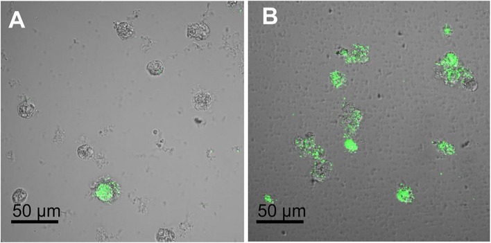
Anti-Legionella activity of AMPs from a non-prokaryotic source was also described in the literature. To date only three peptides, among which two were derived from natural AMPs of the marine organism Ciona intestinalis and one was purified from the greater wax moth Galleria mellonella, have been studied. Ci-MAM-A and Ci-PAP-A are naturally present in the ascidian tunic as well as in granulocytes of inflamed tissues of C. intestinalis, thus constituting a chemical protection to microbial invasion for this organism (Fedders and Leippe, 2008; Di Bella et al., 2011). The authors assumed, based on the knowledge about the processing of AMPs precursors, that sequences of Ci-MAM-A and Ci-PAP-A may represent the prepropeptides of two mature cationic peptides. Therefore, two synthetic peptides, named Ci-PAP-A22 and Ci-MAM-A24, were designed, and represent the cationic amphipathic regions of these two precursors (Fedders and Leippe, 2008; Fedders et al., 2008). Both share a similar size (22–24 amino acid residues) and the propensity to adopt an amphipathic alpha-helical structure. Synthetic peptides were shown to be microbicidal at low micromolar concentrations (below 12.5 μM) against various Gram-positive and Gram-negative bacteria as well as the fungus Candida albicans (Fedders and Leippe, 2008; Fedders et al., 2008). Moreover, Ci-MAM-A24 is extraordinarily salt tolerant and was also found to be remarkably effective against mycobacteria as well as multi-resistant clinically important aerobic and anaerobic strains (Fedders et al., 2008; Jena et al., 2011). Recently the anti-Legionella and the anti-Acanthamoebae activities of these two highly potent peptides were evaluated (Schlusselhuber et al., 2015). The EC50 (concentration that kills 50% of Legionella planctonic cells) was very low for both peptides, below 0.5 μM. However, when considering the minimal bactericidal concentration, Ci-MAM-A24 was found to be much more effective against Legionella cells (1.6 μM) compared to Ci-PAP-A22 (25 μM). The anti-Acanthamoeaba activity of peptides was also determined. The highest concentration tested (25 μM) led to 85% permeabilization of cells by Ci-MAM-A24, but showed no significant effect below this concentration. In comparison, Ci-PAP-A22 did not induce significant permeabilization at these concentrations. Interestingly, the most effective peptide, Ci-MAM-A24, was also found to reduce the intra-amoebae Legionella cell number at a non-toxic concentration for the host cell (12.5 μM), as illustrated in Figure 3. In the frame of elaborating anti-Legionella surfaces, the peptide was then immobilized on gold surfaces to assess its antimicrobial activity. The study revealed that the potent bactericidal activity of the peptide was conserved after its immobilization (Schlusselhuber et al., 2015). CD measurements clearly showed that Ci-PAP-A22 and Ci-MAM-A24 undergo a distinct conformational change upon interaction with some liposomal membranes (Fedders et al., 2008). After mixing with anionic phospholipids (especially PG, L-α-phosphatidyl-DL-glycerol and phosphatidylserine (PS), L-α-phosphatidylserine), both peptides adopted an α-helical structure. Indeed, the CD spectra exhibited the typical shape of a linear peptide in the absence of liposomes, while the typical minima at 222 and 208 nm appeared after mixing with PG or PS. Moreover, with those lipids, peptides adopted a parallel orientation to the membrane surface. The killing activity of these peptides was found to be due to membrane permeabilization. However, a minimalistic system using the depolarization of liposomes revealed a weak pore-forming activity. These data suggested that Ci-MAM-A24 and Ci-PAP-A22 act more likely via a carpet or toroidal-type mechanism, leading to transient pore formation (Fedders et al., 2008).
FIGURE 3.
Activity of Ci-MAM-A24 against intra-amoebic L. pneumophila observed by confocal microscopy. A. castellanii cells infected with GFP expressing L. pneumophila Lens were incubated 6 h post-infection with (A) 12.5 μM of Ci-MAM-A24 or (B) peptide solvent during 42 h (48 h post-infection) (Source: Schlusselhuber et al., 2015).
The Galleria defensin, a 43 aminoacids long peptide (Cytrynska et al., 2007), isolated from greater wax moth Galleria mellonella, was showed to be active against Legionella dumoffii (Palusinska-Szysz et al., 2012). Interestingly, it was shown that the bacteria grown on choline supplemented medium were more sensitive to the peptide than those grown on non-supplemented medium. Like other Legionella species (L. pneumophila, L. bozemanae, L. lytica), L. dumoffii can use extracellular choline for the synthesis of PC. As a consequence, it could be postulated that there is a direct relationship between the level of PC in the Legionella membrane and its sensitivity to the Galleria defensin. Moreover, the lytic activity of δ-hemolysin from S. aureus toward di-palmitoyl-PC vesicles was described (Laabei et al., 2014). This hemolytic peptide was also shown to display an antimicrobial activity restricted to the Legionella genus (Marchand et al., 2011). Taken together, these data suggest that the peculiar sensitivity of bacteria from the Legionella genus to specific AMPs could be related to the high content of PC in its membrane. However, it was shown that, contrary to L. dumoffii, choline supplementation did not induce higher sensitivity of L. pneumophila to ApoLp-III (Zdybicka-Barabas et al., 2014), and the authors suggested that the sensitivity of the bacteria was related to the interaction of the antimicrobial protein and the LPS.
Essential Oils (EOs)
Essential oils are aromatic oily liquids obtained from plant material such as flowers, buds, seeds, leaves, twigs, bark, herbs fruits, or roots, and are mainly composed of a mixture of terpenoïds and aromatic compounds. Among terpenes, monoterpenes, diterpenes, and sesquiterpenes are the most currently found (Dorman and Deans, 2000; Bakkali et al., 2008; Mkaddem et al., 2009). EOs are classified according to the chemical nature of their main active components (Burt, 2004; Bakkali et al., 2008). EOs are well known to possess a wide spectrum of antagonistic activities like antibacterial (Burt, 2004; Solorzano-Santos and Miranda-Novales, 2012; Silva et al., 2013; Seow et al., 2014), antiviral (Reichling et al., 2005; Saddi et al., 2007), antifungal (Penicillium expansum, Botrytis cinerea, and Candida oleophila) (Mari et al., 2003), antitoxigenic (Mycotoxin) (Juglal et al., 2002), antiparasitic (Tariku et al., 2011; Rodrigues et al., 2013) or acaricidal (Neves and da Camara, 2011), and insecticidal activities (Drosophila) (Karpouhtsis et al., 1998).
Investigations were performed by Chang et al. (2008b) to assess the antibacterial activity of EOs against L. pneumophila. Indeed, the authors determined the anti-L. pneumophila activity of EOs extracted from Cinnamomun osmophloeum leaves and from different tissues of Cryptomeria japonica. Among the ten kinds of EOs tested, those extracted from C. osmophloeum leaves exhibited a stronger anti-Legionella activity than those extracted from C. japonica. More precisely, the highest bactericidal effect was obtained with the C. osmophloeum leaf EO of cinnamaldehyde type (characterized by its major constituent, cinnamaldehyde, accounting for 91.3% of EO) (Table 2). The great bioactivity of cinnamon oil appears to be a promising candidate for controlling Legionella growth in recreational spring water and possibly other environments generally at basic pH, i.e., cooling towers (Chang et al., 2008a).
Table 2.
Major components and minimum bactericidal concentration (MBC) or MIC of EOs that exhibit anti-Legionella pneumophila properties.
| Common name of EO | Latin name of plant source | Major components | Approximate concentration (%) | *MBC100 (MIC) | Reference |
|---|---|---|---|---|---|
| Cinnamon | Cinnamomum osmophloeum | Trans-Cinnamaldehyde Benzenpropanal 4-allylanisole | 91.32 3.18 1.42 |
1000 μg/ml | Chang et al., 2008b |
| Tea tree | Melaleuca alternifolia | Terpinen-4-ol 1,8-Cineole |
42.35 3.57 |
0.5% v/v | Mondello et al., 2009 |
| Juniper | Juniperus phoenicea | Isoborneol 1S-α-Pinene | 20.91 18.30 |
(0.03 mg/ml) | Chaftar et al., 2015b |
| Thyme | Thymus vulgaris | Carvacrol | 88.50 | (0.07 mg/ml) | Chaftar et al., 2015b |
*MBC100: The minimum bactericidal concentration of EO that inactivated at least 99.9% of the bacteria. MICs values are indicated in brackets.
In Mondello et al. (2009), a study was conducted to determine the antimicrobial activity of Melaleuca alternifolia cheel (tea tree) oil (TTO) against 22 strains of L. pneumophila of various serogroups and sources of isolation. Results showed that L. pneumophila, quite irrespectively of serogroups and sources of isolation, is highly sensitive to TTO, with MICs ranging from 0.125 to 0.5% v/v, and a minimum bactericidal concentration (MBC100) at 0.5% v/v (Table 2). Therefore, TTO could be used as an anti-Legionella disinfectant for the control of water system contamination, specifically in spa, small waterlines, or in respiratory medical devices.
Recently, the effects of Citrus EOs vapors were tested on different strains of Legionella in water and soil systems (Laird et al., 2014). Among all the tested strains, an antagonistic effect was observed on L. pneumophila, L. longbeachae, L. bozemanii, and on intra-amoebae cultured L. pneumophila with an acute susceptibility for L. pneumophila in water. Different systems of vapors production (passive and active sintering of the vapor) were tested. EOs vapors components were identified (linalool, β-pinene, and citral) and their antimicrobial efficacy was determined. There was up to a 5-log cells/ml reduction in Legionella sp. in soil after exposure to the citrus EO vapors (15 mg/l air). Moreover, data showed that sintering the vapor through water increased the presence of antimicrobial components, including an increase of linalool (57.17 mg/l) compared to the passive system (35.43 mg/l). Thus, the appropriate method for delivering Citrus EO vapor may go some way in controlling Legionella spp. from environmental sources (Laird et al., 2014).
More recently, results obtained by Chaftar et al., 2015a,b), have highlighted the anti-Legionella activity of essential oils extracted from Tunisian plants. Oils extracted from Juniperus phoenicea and Thymus vulgaris exhibited the highest anti-L. pneumophila activity, with MICs lower than 0.03 mg/ml and lower than 0.07 mg/ml, respectively. J. phoenicea oil is mainly composed of isoborneol (20.91%), (1S) α-pinene (18.30%), β-phellandrene (8.08%), α-campholenal (7.91%), and α- phellandrene (7.58%). Concerning the T. vulgaris oil, carvacrol (88.50%) and p-cymene (7.86%) were the major components (Table 2).
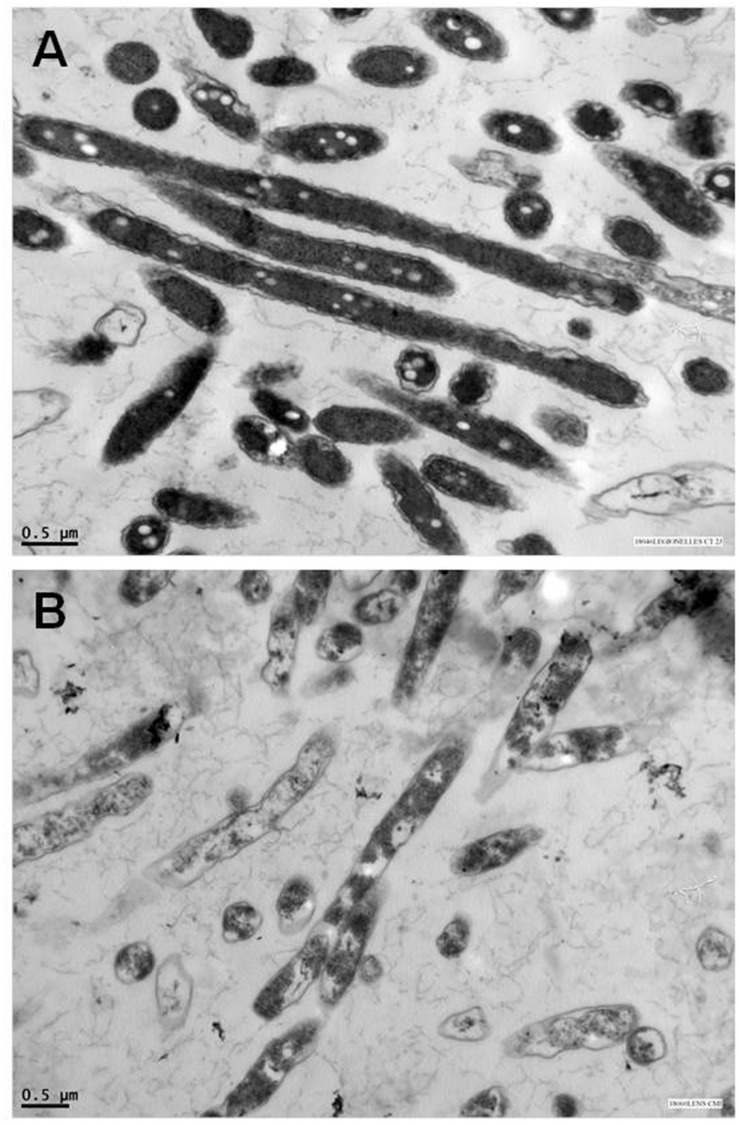
In regard to EOs composition and relative abundance variabilities, their antibacterial activity could not be link to one specific mechanism as cells possess several targets (Carson et al., 2002). Indeed, EOs can act by degrading the cell wall (Helander et al., 1998), inducing damages to the cytoplasmic membrane (Ultee et al., 2002) and to membrane proteins (Ultee et al., 1999), causing the leakage of cell contents (Lambert et al., 2001), coagulating the cytoplasm (Gustafson et al., 1998) and depleting the proton motive force (Ultee and Smid, 2001). To date, only Chaftar et al. (2015b) have investigated EOs action against L. pneumophila. Indeed, scanning electron microscopy analysis highlighted morphological alterations of bacteria when treated with T. vulgaris EO as cells appeared shorter, flattened, and expanded compared to the untreated ones. Transmission electron microscopy experiments indicated that treated L. pneumophila cells were less homogeneous and electron-dense than the untreated control, suggesting a loss of membrane integrity (Figure 4). Authors hypothesized that carvacrol, which constituted 88.5% of the present oil, might destabilize the cytoplasmic membrane and acts as a proton exchanger, as it was shown to alter cell membranes fluidity and permeability, due to its lipophilic properties. However, precise mechanistic data are needed to validate this hypothesis.
FIGURE 4.
Anti-Legionella activity of Thymus vulgaris EO observed by transmission electron microscopy. Micrographs of (A) untreated control cells of L. pneumophila Lens strain and (B) treated L. pneumophila Lens with 70 μg/ml Thymus vulgaris EO (Source: Chaftar et al., 2015b).
Biosurfactants
Biosurfactants are a structurally diverse group of surface-active molecules produced by various microorganisms: bacteria, yeasts or fungi (Pacwa-Plociniczak et al., 2011). Indeed, they are biological amphiphiles composed of a hydrophobic moiety containing saturated, unsaturated, and/or hydroxylated fatty acids or fatty alcohols, and a hydrophilic moiety consisting of mono-, oligo-, or polysaccharides, peptides or proteins (Lang, 2002). Mostly depending on their chemical composition and their molecular weight, biosurfactants are commonly classified as low (including glycolipids, phospholipids, and lipopeptides) and high-molecular weight (polysaccharides, proteins, lipoproteins, and LPS) compounds (Pacwa-Plociniczak et al., 2011). They play critical roles in several biological processes such as the metabolism of hydrophobic substrates, biofilms development and maintenance, biofilms disruption and/or prevention, bacterial motility, host–microbe interactions, stimulation of the induced systemic resistance phenomenon or by acting as natural biocide. Therefore, they are considered of great interest for several biotechnological, biocontrol, and therapeutic applications (Ongena and Jacques, 2008; Sen, 2010; Gudina et al., 2013). While biosurfactants were reported to exhibit lytic and growth-inhibitory activities against a broad range of microorganisms, including viruses, mycoplasmas, bacteria, fungi, and oomycetes, only one study has reported anti-Legionella activity so far (Loiseau et al., 2015). Therefore, several surfactin isoforms were shown to display an antibacterial spectrum almost restricted to the Legionella genus (MICs range 1–4 μg/ml), and also to exhibit a weak activity toward the amoebae Acanthamoebae castellanii, known to be a natural reservoir of L. pneumophila. Surfactin is a major class of cyclic lipopeptides abundantly produced by various Bacillus environmental isolates and remains the best known biosurfactant (Mora et al., 2015).
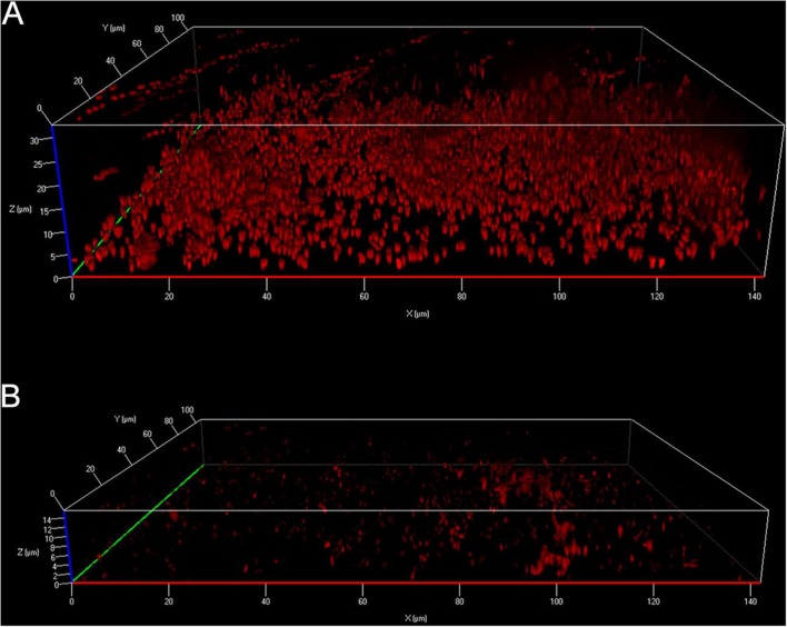
Lipopeptides, which constitute a specific class of microbial secondary metabolites, are well identified as antimicrobial agents (Ines and Dhouha, 2015). However, according to the literature on the antibacterial activities of purified or commercially purchased standard surfactins, few susceptible species have been reported (Table 3). It has to be noted that many studies did not separate surfactins from other produced lipopeptides or only tested the supernatant/crude extracts when performing antibacterial assays. So, the number of sensitive bacterial species is clearly underestimated. Surfactins were also found to breakdown L. pneumophila pre-formed biofilms but did not prevent biofilm attachment (Figure 5) (Loiseau et al., 2015) unlike biofilms produced by Salmonella enterica Serovar Typhimurium (Mireles et al., 2001).
Table 3.
Minimum inhibitory concentrations (MIC) of purified or commercially purchased surfactins against selected bacterial strains.
| Target bacteria | MIC | Producing strain | Antibacterial Assay | Reference |
|---|---|---|---|---|
| E. coli AS1.487 | 15.625 μg/ml | Commercially purchased | Microdilution | Huang et al., 2008 |
| P. syringae pv tomato DC3000 | 25 μg/ml | Commercially purchased | Microdilution | Bais et al., 2004 |
| L. monocytogenes 99/287RB6 strains | 125 μg/ml 250 μg/ml 1 mg/ml | B. subtilis C4 B. subtilis G2III B. subtilis M1 | *WDA | Sabate and Audisio, 2013 |
| S. enteritidis | 6.25 μg/ml | Commercially purchased | Microdilution | Huang et al., 2011 |
| V. anguillarum | 1.5 μg/ml | B. amyloliquefaciens M1 | Not specified | Xu et al., 2014 |
| Legionella sp. | 1–4 μg/ml | Commercially purchased | Microdilution | Loiseau et al., 2015 |
| M. pulmonis MpUR1.1 | 25.9 μg/ml | Commercially purchased | Microdilution | Fassi Fehri et al., 2007 |
*WDA: well diffusion assay.
Minimum inhibitory concentrations were determined against L. pneumophila Lens and for the formylated forms of the peptides except for the peptides indicated by *.
FIGURE 5.
Apotome imaging of surfactin-treated 6-day-old biofilms formed by L. pneumophila. Biofilms were treated 2 h either with (A) ethanol as control or (B) 66 μg/ml surfactin (Source: Loiseau et al., 2015).
Concerning their mechanism of action, surfactins seem to act by direct lysis of negatively charged membranes (Buchoux et al., 2008). This lysis is driven by electrostatic repulsion between negatively charged acidic residues from surfactins and negative charges located on the lipid headgroups after penetration of the lipopeptide into the lipid bilayer, leading to complete destabilization of the planar membrane and small vesicles formation.
Conclusion
Legionella pneumophila appears sensitive to various biomolecules including molecules that are poorly active against others bacteria like surfactin. However, it is important to keep in mind that L. pneumophila is not a routinely used bacterium when determining the antimicrobial potency of a given product in contrast to bacteria such as E. coli, S. aureus, or P. aeruginosa. Therefore, it is easy to understand why there are so little known anti-Legionella molecules available in the literature. On the other hand, the described compounds are very active against L. pneumophila compared to other bacteria. Does L. pneumophila possess some specificity that could explain this sensitivity? As all these compounds are membrane active, maybe a part of the answer is hidden in the composition of L. pneumophila cell envelope. The current knowledge about the structure and molecular composition of its cell envelope was recently reviewed, and authors highlighted several characteristics that deserve more attention (Shevchuk et al., 2011). Starting from the outside and proceeding inward, it appears that L. pneumophila LPS has a unique structure in comparison to the LPS of other Gram-negative bacteria. Due to high levels of long, branched fatty acids and elevated levels of O- and N-acetyl groups, this LPS is highly hydrophobic. The high level of PC is also striking as only 15% of all known bacteria have the ability to synthetize PC (Geiger et al., 2013; Sohlenkamp and Geiger, 2016). Nevertheless, the exact function of this phospholipid in bacterial cell envelopes remains unclear regarding the sensitivity of Legionella to antimicrobial compounds (Geiger et al., 2013). However, it has already been shown that activities of various biomolecules (AMPs, ApoLp-III) are modulated in presence of PC, even if the reason remains poorly understood. Moreover, the fatty acid composition of membranes also influences bactericidal properties. L. pneumophila possesses a high level of branched chain fatty acids, mainly in the stationary phase of growth (Verdon et al., 2011). This level was shown to be involved in the sensitivity of L. pneumophila to warnericin RK. Taken together, all those data highlighted several particularities of the envelope components already shown to be implicated in Legionella virulence (Shevchuk et al., 2011). Another striking feature is the high sensitivity of L. pneumophila to detergent (Verdon et al., 2009a), underlining the key role played by its membrane components. As all described active compounds target this cell compartment, establishing a link between sensitivity and membrane composition/arrangement is tempting. It appears that membrane thickness, fluidity, phospholipid composition and even presence of phospholipids clusters could be key parameters involved in Legionella sensitivity. Anyway, the lack of experimental data about mechanisms of action of active molecules is a bottleneck in the discussion. Further experimental investigations are clearly and rapidly needed to decipher the precise mechanisms of action of such biomolecules. However, the critical analysis of the literature presented here reveals that natural biomolecules could represent potent tools for the biological control of L. pneumophila and/or its natural hosts in water treatment industry, although additional experiments are needed to demonstrate how effective these antagonists would be under field conditions.
Author Contributions
J-MB, SC, and JV conceived and designed the review. J-MB, SC, MS, EP, CL, WA, OL, and JV wrote the paper. JV coordinated the work.
Conflict of Interest Statement
The authors declare that the research was conducted in the absence of any commercial or financial relationships that could be construed as a potential conflict of interest.
Acknowledgments
This work received financial support from the French National Research Program for Environmental and Occupational Health of ANSES, grant EST-2015/1/111.
Footnotes
References
- Abdel-Nour M., Duncan C., Low D. E., Guyard C. (2013). Biofilms: the stronghold of Legionella pneumophila. Int. J. Mol. Sci. 14 21660–21675. 10.3390/ijms141121660 [DOI] [PMC free article] [PubMed] [Google Scholar]
- Abu Kwaik Y., Gao L. Y., Stone B. J., Venkataraman C., Harb O. S. (1998). Invasion of protozoa by Legionella pneumophila and its role in bacterial ecology and pathogenesis. Appl. Environ. Microbiol. 64 3127–3133. [DOI] [PMC free article] [PubMed] [Google Scholar]
- Al-Quadan T., Price C. T., Abu Kwaik Y. (2012). Exploitation of evolutionarily conserved amoeba and mammalian processes by Legionella. Trends Microbiol. 20 299–306. 10.1016/j.tim.2012.03.005 [DOI] [PMC free article] [PubMed] [Google Scholar]
- Andra J., Leippe M. (1999). Candidacidal activity of shortened synthetic analogs of amoebapores and NK-lysin. Med. Microbiol. Immunol. 188 117–124. 10.1007/s004300050113 [DOI] [PubMed] [Google Scholar]
- Bais H. P., Fall R., Vivanco J. M. (2004). Biocontrol of Bacillus subtilis against infection of Arabidopsis roots by Pseudomonas syringae is facilitated by biofilm formation and surfactin production. Plant Physiol. 134 307–319. 10.1104/pp.103.028712 [DOI] [PMC free article] [PubMed] [Google Scholar]
- Bakkali F., Averbeck S., Averbeck D., Idaomar M. (2008). Biological effects of essential oils–a review. Food Chem. Toxicol. 46 446–475. 10.1016/j.fct.2007.09.106 [DOI] [PubMed] [Google Scholar]
- Barker J., Brown M. R., Collier P. J., Farrell I., Gilbert P. (1992). Relationship between Legionella pneumophila and Acanthamoeba polyphaga: physiological status and susceptibility to chemical inactivation. Appl. Environ. Microbiol. 58 2420–2425. [DOI] [PMC free article] [PubMed] [Google Scholar]
- Barker J., Scaife H., Brown M. R. (1995). Intraphagocytic growth induces an antibiotic-resistant phenotype of Legionella pneumophila. Antimicrob. Agents Chemother. 39 2684–2688. 10.1128/AAC.39.12.2684 [DOI] [PMC free article] [PubMed] [Google Scholar]
- Beaute J., Zucs P., de Jong B., European Legionnaires’ Disease Surveillance N. (2013). Legionnaires disease in Europe, 2009-2010. Euro. Surveill. 18:20417. [DOI] [PubMed] [Google Scholar]
- Bechinger B., Lohner K. (2006). Detergent-like actions of linear amphipathic cationic antimicrobial peptides. Biochim. Biophys. Acta 1758 1529–1539. 10.1016/j.bbamem.2006.07.001 [DOI] [PubMed] [Google Scholar]
- Berk S. G., Ting R. S., Turner G. W., Ashburn R. J. (1998). Production of respirable vesicles containing live Legionella pneumophila cells by two Acanthamoeba spp. Appl. Environ. Microbiol. 64 279–286. [DOI] [PMC free article] [PubMed] [Google Scholar]
- Berry D., Xi C., Raskin L. (2006). Microbial ecology of drinking water distribution systems. Curr. Opin. Biotechnol 17 297–302. 10.1016/j.copbio.2006.05.007 [DOI] [PubMed] [Google Scholar]
- Blasco M. D., Esteve C., Alcaide E. (2008). Multiresistant waterborne pathogens isolated from water reservoirs and cooling systems. J. Appl. Microbiol. 105 469–475. 10.1111/j.1365-2672.2008.03765.x [DOI] [PubMed] [Google Scholar]
- Borella P., Guerrieri E., Marchesi I., Bondi M., Messi P. (2005). Water ecology of Legionella and protozoan: environmental and public health perspectives. Biotechnol. Annu. Rev. 11 355–380. 10.1016/S1387-2656(05)11011-4 [DOI] [PubMed] [Google Scholar]
- Bortner C. A., Arnold R. R., Miller R. D. (1989). Bactericidal effect of lactoferrin on Legionella pneumophila: effect of the physiological state of the organism. Can. J. Microbiol. 35 1048–1051. 10.1139/m89-174 [DOI] [PubMed] [Google Scholar]
- Bortner C. A., Miller R. D., Arnold R. R. (1986). Bactericidal effect of lactoferrin on Legionella pneumophila. Infect. Immun. 51 373–377. [DOI] [PMC free article] [PubMed] [Google Scholar]
- Bouyer S., Imbert C., Rodier M. H., Hechard Y. (2007). Long-term survival of Legionella pneumophila associated with Acanthamoeba castellanii vesicles. Environ. Microbiol. 9 1341–1344. 10.1111/j.1462-2920.2006.01229.x [DOI] [PubMed] [Google Scholar]
- Brieland J. K., Fantone J. C., Remick D. G., LeGendre M., McClain M., Engleberg N. C. (1997). The role of Legionella pneumophila-infected Hartmannella vermiformis as an infectious particle in a murine model of Legionnaire’s disease. Infect. Immun. 65 5330–5333. [DOI] [PMC free article] [PubMed] [Google Scholar]
- Buchoux S., Lai-Kee-Him J., Garnier M., Tsan P., Besson F., Brisson A., et al. (2008). Surfactin-triggered small vesicle formation of negatively charged membranes: a novel membrane-lysis mechanism. Biophys. J. 95 3840–3849. 10.1529/biophysj.107.128322 [DOI] [PMC free article] [PubMed] [Google Scholar]
- Burt S. (2004). Essential oils: their antibacterial properties and potential applications in foods–a review. Int. J. Food Microbiol. 94 223–253. 10.1016/j.ijfoodmicro.2004.03.022 [DOI] [PubMed] [Google Scholar]
- Buse H. Y., Lu J., Lu X., Mou X., Ashbolt N. J. (2014). Microbial diversities (16S and 18S rRNA gene pyrosequencing) and environmental pathogens within drinking water biofilms grown on the common premise plumbing materials unplasticized polyvinylchloride and copper. FEMS Microbiol. Ecol. 88 280–295. 10.1111/1574-6941.12294 [DOI] [PubMed] [Google Scholar]
- Campese C., Bitar D., Jarraud S., Maine C., Forey F., Etienne J., et al. (2011). Progress in the surveillance and control of Legionella infection in France, 1998-2008. Int. J. Infect. Dis. 15 e30–e37. 10.1016/j.ijid.2010.09.007 [DOI] [PubMed] [Google Scholar]
- Carson C. F., Mee B. J., Riley T. V. (2002). Mechanism of action of Melaleuca alternifolia (tea tree) oil on Staphylococcus aureus determined by time-kill, lysis, leakage, and salt tolerance assays and electron microscopy. Antimicrob. Agents Chemother. 46 1914–1920. 10.1128/AAC.46.6.1914-1920.2002 [DOI] [PMC free article] [PubMed] [Google Scholar]
- Casteels P., Romagnolo J., Castle M., Casteels-Josson K., Erdjument-Bromage H., Tempst P. (1994). Biodiversity of apidaecin-type peptide antibiotics. Prospects of manipulating the antibacterial spectrum and combating acquired resistance. J. Biol. Chem. 269 26107–26115. [PubMed] [Google Scholar]
- Cervero-Arago S., Rodriguez-Martinez S., Puertas-Bennasar A., Araujo R. M. (2015). Effect of common drinking water disinfectants, chlorine and heat, on free Legionella and amoebae-associated Legionella. PLoS ONE 10:e0134726 10.1371/journal.pone.0134726 [DOI] [PMC free article] [PubMed] [Google Scholar]
- Chaftar N., Girardot M., Labanowski J., Ghrairi T., Hani K., Frere J., et al. (2015a). Comparative evaluation of the antimicrobial activity of 19 essential oils. Adv. Exp. Med. Biol. 10.1007/5584_2015_5011 [Epub ahead of print]. [DOI] [PubMed] [Google Scholar]
- Chaftar N., Girardot M., Quellard N., Labanowski J., Ghrairi T., Hani K., et al. (2015b). Activity of six essential oils extracted from tunisian plants against Legionella pneumophila. Chem Biodivers. 12 1565–1574. 10.1002/cbdv.201400343 [DOI] [PubMed] [Google Scholar]
- Chang C. W., Chang W. L., Chang S. T. (2008a). Influence of pH on bioactivity of cinnamon oil against Legionella pneumophila and its disinfection efficacy in hot springs. Water Res 42 5022–5030. 10.1016/j.watres.2008.09.006 [DOI] [PubMed] [Google Scholar]
- Chang C. W., Chang W. L., Chang S. T., Cheng S. S. (2008b). Antibacterial activities of plant essential oils against Legionella pneumophila. Water Res. 42 278–286. 10.1016/j.watres.2007.07.008 [DOI] [PubMed] [Google Scholar]
- Chang C. W., Kao C. H., Liu Y. F. (2009). Heterogeneity in chlorine susceptibility for Legionella pneumophila released from Acanthamoeba and Hartmannella. J. Appl. Microbiol. 106 97–105. 10.1111/j.1365-2672.2008.03980.x [DOI] [PubMed] [Google Scholar]
- Chmiel E., Palusinska-Szysz M., Zdybicka-Barabas A., Cytrynska M., Mak P. (2014). The effect of Galleria mellonella hemolymph polypeptides on Legionella gormanii. Acta Biochim. Pol. 61 123–127. [PubMed] [Google Scholar]
- Cirillo J. D., Falkow S., Tompkins L. S. (1994). Growth of Legionella pneumophila in Acanthamoeba castellanii enhances invasion. Infect. Immun. 62 3254–3261. [DOI] [PMC free article] [PubMed] [Google Scholar]
- Collier S. A., Stockman L. J., Hicks L. A., Garrison L. E., Zhou F. J., Beach M. J. (2012). Direct healthcare costs of selected diseases primarily or partially transmitted by water. Epidemiol. Infect. 140 2003–2013. 10.1017/S0950268811002858 [DOI] [PMC free article] [PubMed] [Google Scholar]
- Conover G. M., Martinez-Morales F., Heidtman M. I., Luo Z. Q., Tang M., Chen C., et al. (2008). Phosphatidylcholine synthesis is required for optimal function of Legionella pneumophila virulence determinants. Cell Microbiol. 10 514–528. 10.1111/j.1462-5822.2007.01066.x [DOI] [PMC free article] [PubMed] [Google Scholar]
- Cooper I. R., Hanlon G. W. (2010). Resistance of Legionella pneumophila serotype 1 biofilms to chlorine-based disinfection. J. Hosp. Infect. 74 152–159. 10.1016/j.jhin.2009.07.005 [DOI] [PubMed] [Google Scholar]
- Cooper I. R., White J., Mahenthiralingam E., Hanlon G. W. (2008). Long-term persistence of a single Legionella pneumophila strain possessing the mip gene in a municipal shower despite repeated cycles of chlorination. J. Hosp. Infect. 70 154–159. 10.1016/j.jhin.2008.06.015 [DOI] [PubMed] [Google Scholar]
- Cytrynska M., Mak P., Zdybicka-Barabas A., Suder P., Jakubowicz T. (2007). Purification and characterization of eight peptides from Galleria mellonella immune hemolymph. Peptides 28 533–546. 10.1016/j.peptides.2006.11.010 [DOI] [PubMed] [Google Scholar]
- Darveau R. P., Blake J., Seachord C. L., Cosand W. L., Cunningham M. D., Cassiano-Clough L., et al. (1992). Peptides related to the carboxyl terminus of human platelet factor IV with antibacterial activity. J. Clin. Invest. 90 447–455. 10.1172/JCI115880 [DOI] [PMC free article] [PubMed] [Google Scholar]
- Declerck P., Behets J., Delaedt Y., Margineanu A., Lammertyn E., Ollevier F. (2005). Impact of non-Legionella bacteria on the uptake and intracellular replication of Legionella pneumophila in Acanthamoeba castellanii and Naegleria lovaniensis. Microb. Ecol. 50 536–549. 10.1007/s00248-005-0258-0 [DOI] [PubMed] [Google Scholar]
- Declerck P., Behets J., van Hoef V., Ollevier F. (2007). Detection of Legionella spp. and some of their amoeba hosts in floating biofilms from anthropogenic and natural aquatic environments. Water Res. 41 3159–3167. 10.1016/j.watres.2007.04.011 [DOI] [PubMed] [Google Scholar]
- Di Bella M. A., Fedders H., De Leo G., Leippe M. (2011). Localization of antimicrobial peptides in the tunic of Ciona intestinalis (Ascidiacea, Tunicata) and their involvement in local inflammatory-like reactions. Results Immunol. 1 70–75. 10.1016/j.rinim.2011.09.001 [DOI] [PMC free article] [PubMed] [Google Scholar]
- Dominguez A., Alvarez J., Sabria M., Carmona G., Torner N., Oviedo M., et al. (2009). Factors influencing the case-fatality rate of Legionnaires’ disease. Int. J. Tuberc. Lung Dis. 13 407–412. [PubMed] [Google Scholar]
- Donlan R. M., Costerton J. W. (2002). Biofilms: survival mechanisms of clinically relevant microorganisms. Clin. Microbiol. Rev. 15 167–193. 10.1128/CMR.15.2.167-193.2002 [DOI] [PMC free article] [PubMed] [Google Scholar]
- Donlan R. M., Forster T., Murga R., Brown E., Lucas C., Carpenter J., et al. (2005). Legionella pneumophila associated with the protozoan Hartmannella vermiformis in a model multi-species biofilm has reduced susceptibility to disinfectants. Biofouling 21 1–7. 10.1080/08927010500044286 [DOI] [PubMed] [Google Scholar]
- Dorman H. J., Deans S. G. (2000). Antimicrobial agents from plants: antibacterial activity of plant volatile oils. J. Appl. Microbiol. 88 308–316. 10.1046/j.1365-2672.2000.00969.x [DOI] [PubMed] [Google Scholar]
- Dupuy M., Mazoua S., Berne F., Bodet C., Garrec N., Herbelin P., et al. (2011). Efficiency of water disinfectants against Legionella pneumophila and Acanthamoeba. Water Res. 45 1087–1094. 10.1016/j.watres.2010.10.025 [DOI] [PubMed] [Google Scholar]
- Emtiazi F., Schwartz T., Marten S. M., Krolla-Sidenstein P., Obst U. (2004). Investigation of natural biofilms formed during the production of drinking water from surface water embankment filtration. Water Res. 38 1197–1206. 10.1016/j.watres.2003.10.056 [DOI] [PubMed] [Google Scholar]
- Falkinham J. O., Pruden A., Edwards M. (2015). Opportunistic premise plumbing pathogens: increasingly important pathogens in drinking water. Pathogens 4 373–386. 10.3390/pathogens4020373 [DOI] [PMC free article] [PubMed] [Google Scholar]
- Fassi Fehri L., Wróblewski H., Blanchard A. (2007). Activities of antimicrobial peptides and synergy with enrofloxacin against Mycoplasma pulmonis. Antimicrob. Agents Chemother. 51 468–474. 10.1128/AAC.01030-06 [DOI] [PMC free article] [PubMed] [Google Scholar]
- Fedders H., Leippe M. (2008). A reverse search for antimicrobial peptides in Ciona intestinalis: identification of a gene family expressed in hemocytes and evaluation of activity. Dev. Comp. Immunol. 32 286–298. 10.1016/j.dci.2007.06.003 [DOI] [PubMed] [Google Scholar]
- Fedders H., Michalek M., Grotzinger J., Leippe M. (2008). An exceptional salt-tolerant antimicrobial peptide derived from a novel gene family of haemocytes of the marine invertebrate Ciona intestinalis. Biochem. J. 416 65–75. 10.1042/BJ20080398 [DOI] [PubMed] [Google Scholar]
- Fields B. S., Benson R. F., Besser R. E. (2002). Legionella and Legionnaires’ disease: 25 years of investigation. Clin. Microbiol. Rev. 15 506–526. 10.1128/CMR.15.3.506-526.2002 [DOI] [PMC free article] [PubMed] [Google Scholar]
- Fouque E., Yefimova M., Trouilhe M. C., Quellard N., Fernandez B., Rodier M. H., et al. (2015). Morphological study of the encystment and excystment of vermamoeba vermiformis revealed original traits. J. Eukaryot. Microbiol. 62 327–337. 10.1111/jeu.12185 [DOI] [PubMed] [Google Scholar]
- Franco I. S., Shuman H. A., Charpentier X. (2009). The perplexing functions and surprising origins of Legionella pneumophila type IV secretion effectors. Cell Microbiol. 11 1435–1443. 10.1111/j.1462-5822.2009.01351.x [DOI] [PubMed] [Google Scholar]
- Fraser D. W., Tsai T. R., Orenstein W., Parkin W. E., Beecham H. J., Sharrar R. G., et al. (1977). Legionnaires’ disease: description of an epidemic of pneumonia. N. Engl. J. Med. 297 1189–1197. 10.1056/NEJM197712012972201 [DOI] [PubMed] [Google Scholar]
- Garcia M. T., Jones S., Pelaz C., Millar R. D., Abu Kwaik Y. (2007). Acanthamoeba polyphaga resuscitates viable non-culturable Legionella pneumophila after disinfection. Environ. Microbiol. 9 1267–1277. 10.1111/j.1462-2920.2007.01245.x [DOI] [PubMed] [Google Scholar]
- Garduno R. A., Garduno E., Hiltz M., Hoffman P. S. (2002). Intracellular growth of Legionella pneumophila gives rise to a differentiated form dissimilar to stationary-phase forms. Infect. Immun. 70 6273–6283. 10.1128/IAI.70.11.6273-6283.2002 [DOI] [PMC free article] [PubMed] [Google Scholar]
- Geiger O., Lopez-Lara I. M., Sohlenkamp C. (2013). Phosphatidylcholine biosynthesis and function in bacteria. Biochim. Biophys. Acta 1831 503–513. 10.1016/j.bbalip.2012.08.009 [DOI] [PubMed] [Google Scholar]
- Gelhaus C., Jacobs T., Andra J., Leippe M. (2008). The antimicrobial peptide NK-2 the core region of mammalian NK-lysin, kills intraerythrocytic Plasmodium falciparum. Antimicrob. Agents Chemother. 52 1713–1720. 10.1128/AAC.01342-07 [DOI] [PMC free article] [PubMed] [Google Scholar]
- Giao M. S., Wilks S., Azevedo N. F., Vieira M. J., Keevil C. W. (2009). Incorporation of natural uncultivable Legionella pneumophila into potable water biofilms provides a protective niche against chlorination stress. Biofouling 25 335–341. 10.1080/08927010902802232 [DOI] [PubMed] [Google Scholar]
- Gudina E. J., Rangarajan V., Sen R., Rodrigues L. R. (2013). Potential therapeutic applications of biosurfactants. Trends Pharmacol. Sci. 34 667–675. 10.1016/j.tips.2013.10.002 [DOI] [PubMed] [Google Scholar]
- Guo L., Lim K. B., Poduje C. M., Daniel M., Gunn J. S., Hackett M., et al. (1998). Lipid A acylation and bacterial resistance against vertebrate antimicrobial peptides. Cell 95 189–198. 10.1016/S0092-8674(00)81750-X [DOI] [PubMed] [Google Scholar]
- Gustafson J. E., Liew Y. C., Chew S., Markham J., Bell H. C., Wyllie S. G., et al. (1998). Effects of tea tree oil on Escherichia coli. Lett. Appl. Microbiol. 26 194–198. 10.1046/j.1472-765X.1998.00317.x [DOI] [PubMed] [Google Scholar]
- Hamilton E., Seal D. V., Hay J. (1996). Comparison of chlorine and chlorine dioxide disinfection for control of Legionella in a hospital potable water supply. J. Hosp. Infect. 32 156–160. 10.1016/S0195-6701(96)90058-4 [DOI] [PubMed] [Google Scholar]
- Haney E. F., Hunter H. N., Matsuzaki K., Vogel H. J. (2009). Solution NMR studies of amphibian antimicrobial peptides: linking structure to function? Biochim. Biophys. Acta 1788 1639–1655. 10.1016/j.bbamem.2009.01.002 [DOI] [PubMed] [Google Scholar]
- Hechard Y., Ferraz S., Bruneteau E., Steinert M., Berjeaud J. M. (2005). Isolation and characterization of a Staphylococcus warneri strain producing an anti-Legionella peptide. FEMS Microbiol. Lett. 252 19–23. 10.1016/j.femsle.2005.03.046 [DOI] [PubMed] [Google Scholar]
- Helander I. M., Alakomi H. L., Latva-Kala K., Mattila-Sandholm T., Pol I., Smid E. J., et al. (1998). Characterization of the action of selected essential oil components on gram-negative bacteria. J. Agric. Food Chem. 46 3590–3595. 10.1021/jf980154m [DOI] [Google Scholar]
- Hilbi H., Hoffmann C., Harrison C. F. (2011). Legionella spp. outdoors: colonization, communication and persistence. Environ. Microbiol. Rep. 3 286–296. 10.1111/j.1758-2229.2011.00247.x [DOI] [PubMed] [Google Scholar]
- Hindahl M. S., Iglewski B. H. (1984). Isolation and characterization of the Legionella pneumophila outer membrane. J. Bacteriol. 159 107–113. [DOI] [PMC free article] [PubMed] [Google Scholar]
- Huang X., Suo J., Cui Y. (2011). Optimization of antimicrobial activity of surfactin and polylysine against Salmonella enteritidis in milk evaluated by a response surface methodology. Foodborne Pathog. Dis. 8 439–443. 10.1089/fpd.2010.0738 [DOI] [PubMed] [Google Scholar]
- Huang X., Wei Z., Zhao G., Gao X., Yang S., Cui Y. (2008). Optimization of sterilization of Escherichia coli in milk by surfactin and fengycin using a response surface method. Curr. Microbiol. 56 376–381. 10.1007/s00284-007-9066-8 [DOI] [PubMed] [Google Scholar]
- Hubber A., Roy C. R. (2010). Modulation of host cell function by Legionella pneumophila type IV effectors. Annu. Rev. Cell Dev. Biol. 26 261–283. 10.1146/annurev-cellbio-100109-104034 [DOI] [PubMed] [Google Scholar]
- Ines M., Dhouha G. (2015). Lipopeptide surfactants: production, recovery and pore forming capacity. Peptides 71 100–112. 10.1016/j.peptides.2015.07.006 [DOI] [PubMed] [Google Scholar]
- Isberg R. R., O’Connor T. J., Heidtman M. (2009). The Legionella pneumophila replication vacuole: making a cosy niche inside host cells. Nat. Rev. Microbiol. 7 13–24. 10.1038/nrmicro1967 [DOI] [PMC free article] [PubMed] [Google Scholar]
- Jacobs T., Bruhn H., Gaworski I., Fleischer B., Leippe M. (2003). NK-lysin and its shortened analog NK-2 exhibit potent activities against Trypanosoma cruzi. Antimicrob. Agents Chemother. 47 607–613. 10.1128/AAC.47.2.607-613.2003 [DOI] [PMC free article] [PubMed] [Google Scholar]
- Jena P., Mishra B., Leippe M., Hasilik A., Griffiths G., Sonawane A. (2011). Membrane-active antimicrobial peptides and human placental lysosomal extracts are highly active against mycobacteria. Peptides 32 881–887. 10.1016/j.peptides.2011.03.002 [DOI] [PubMed] [Google Scholar]
- Jenssen H., Hancock R. E. (2009). Antimicrobial properties of lactoferrin. Biochimie 91 19–29. 10.1016/j.biochi.2008.05.015 [DOI] [PubMed] [Google Scholar]
- Jjemba P. K., Johnson W., Bukhari Z., LeChevallier M. W. (2015). Occurrence and control of Legionella in recycled water systems. Pathogens 4 470–502. 10.3390/pathogens4030470 [DOI] [PMC free article] [PubMed] [Google Scholar]
- Juglal S., Govinden R., Odhav B. (2002). Spice oils for the control of co-occurring mycotoxin-producing fungi. J. Food Prot. 65 683–687. [DOI] [PubMed] [Google Scholar]
- Karpouhtsis I., Pardali E., Feggou E., Kokkini S., Scouras Z. G., Mavragani-Tsipidou P. (1998). Insecticidal and genotoxic activities of oregano essential oils. J. Agric. Food Chem. 46 1111–1115. 10.1021/jf970822o [DOI] [Google Scholar]
- Kim B. R., Anderson J. E., Mueller S. A., Gaines W. A., Kendall A. M. (2002). Literature review–efficacy of various disinfectants against Legionella in water systems. Water Res. 36 4433–4444. 10.1016/S0043-1354(02)00188-4 [DOI] [PubMed] [Google Scholar]
- Koubar M., Rodier M. H., Garduno R. A., Frere J. (2011). Passage through Tetrahymena tropicalis enhances the resistance to stress and the infectivity of Legionella pneumophila. FEMS Microbiol. Lett. 325 10–15. 10.1111/j.1574-6968.2011.02402.x [DOI] [PubMed] [Google Scholar]
- Laabei M., Jamieson W. D., Yang Y., van den Elsen J., Jenkins A. T. (2014). Investigating the lytic activity and structural properties of Staphylococcus aureus phenol soluble modulin (PSM) peptide toxins. Biochim. Biophys. Acta 1838 3153–3161. 10.1016/j.bbamem.2014.08.026 [DOI] [PubMed] [Google Scholar]
- Laird K., Kurzbach E., Score J., Tejpal J., Chi Tangyie G., Phillips C. (2014). Reduction of Legionella spp. in water and in soil by a citrus plant extract vapor. Appl. Environ. Microbiol. 80 6031–6036. 10.1128/AEM.01275-14 [DOI] [PMC free article] [PubMed] [Google Scholar]
- Lambert R. J., Skandamis P. N., Coote P. J., Nychas G. J. (2001). A study of the minimum inhibitory concentration and mode of action of oregano essential oil, thymol and carvacrol. J. Appl. Microbiol. 91 453–462. 10.1046/j.1365-2672.2001.01428.x [DOI] [PubMed] [Google Scholar]
- Lang S. (2002). Biological amphiphiles (microbial biosurfactants). Curr. Opin. Colloid Interface Sci. 7 12–20. 10.1016/S1359-0294(02)00007-9 [DOI] [Google Scholar]
- Lau H. Y., Ashbolt N. J. (2009). The role of biofilms and protozoa in Legionella pathogenesis: implications for drinking water. J. Appl. Microbiol. 107 368–378. 10.1111/j.1365-2672.2009.04208.x [DOI] [PubMed] [Google Scholar]
- Lee J. Y., Boman A., Sun C. X., Andersson M., Jornvall H., Mutt V., et al. (1989). Antibacterial peptides from pig intestine: isolation of a mammalian cecropin. Proc. Natl. Acad. Sci. U.S.A. 86 9159–9162. 10.1073/pnas.86.23.9159 [DOI] [PMC free article] [PubMed] [Google Scholar]
- Lee K. H., Fitton J. E., Wuthrich K. (1987). Nuclear magnetic resonance investigation of the conformation of delta-haemolysin bound to dodecylphosphocholine micelles. Biochim. Biophys. Acta 911 144–153. 10.1016/0167-4838(87)90003-3 [DOI] [PubMed] [Google Scholar]
- Leippe M. (1995). Ancient weapons: NK-lysin, is a mammalian homolog to pore-forming peptides of a protozoan parasite. Cell 83 17–18. 10.1016/0092-8674(95)90229-5 [DOI] [PubMed] [Google Scholar]
- Liu Z., Stout J. E., Boldin M., Rugh J., Diven W. F., Yu V. L. (1998). Intermittent use of copper-silver ionization for Legionella control in water distribution systems: a potential option in buildings housing individuals at low risk of infection. Clin. Infect. Dis. 26 138–140. 10.1086/516283 [DOI] [PubMed] [Google Scholar]
- Loiseau C., Schlusselhuber M., Bigot R., Bertaux J., Berjeaud J. M., Verdon J. (2015). Surfactin from Bacillus subtilis displays an unexpected anti-Legionella activity. Appl. Microbiol. Biotechnol. 99 5083–5093. 10.1007/s00253-014-6317-z [DOI] [PubMed] [Google Scholar]
- Loret J. F., Greub G. (2010). Free-living amoebae: biological by-passes in water treatment. Int. J. Hyg. Environ. Health 213 167–175. 10.1016/j.ijheh.2010.03.004 [DOI] [PubMed] [Google Scholar]
- Mallegol J., Fernandes P., Melano R. G., Guyard C. (2014). Antimicrobial activity of solithromycin against clinical isolates of Legionella pneumophila serogroup 1. Antimicrob. Agents Chemother. 58 909–915. 10.1128/AAC.01639-13 [DOI] [PMC free article] [PubMed] [Google Scholar]
- Marchand A., Augenstreich J., Loiseau C., Verdon J., Lecomte S., Berjeaud J. M. (2015). Effect of amino acid substitution in the staphylococcal peptides warnericin RK and PSMalpha on their anti-Legionella and hemolytic activities. Mol. Cell. Biochem. 405 159–167. 10.1007/s11010-015-2407-1 [DOI] [PubMed] [Google Scholar]
- Marchand A., Verdon J., Lacombe C., Crapart S., Hechard Y., Berjeaud J. M. (2011). Anti-Legionella activity of staphylococcal hemolytic peptides. Peptides 32 845–851. 10.1016/j.peptides.2011.01.025 [DOI] [PubMed] [Google Scholar]
- Mari M., Bertolini P., Pratella G. C. (2003). Non-conventional methods for the control of post-harvest pear diseases. J. Appl. Microbiol. 94 761–766. 10.1046/j.1365-2672.2003.01920.x [DOI] [PubMed] [Google Scholar]
- Martinez-Morales F., Schobert M., Lopez-Lara I. M., Geiger O. (2003). Pathways for phosphatidylcholine biosynthesis in bacteria. Microbiology 149 3461–3471. 10.1099/mic.0.26522-0 [DOI] [PubMed] [Google Scholar]
- McDade J. E., Shepard C. C., Fraser D. W., Tsai T. R., Redus M. A., Dowdle W. R. (1977). Legionnaires’ disease: isolation of a bacterium and demonstration of its role in other respiratory disease. N. Engl. J. Med. 297 1197–1203. 10.1056/NEJM197712012972202 [DOI] [PubMed] [Google Scholar]
- Mireles J. R., II, Toguchi A., Harshey R. M. (2001). Salmonella enterica serovar typhimurium swarming mutants with altered biofilm-forming abilities: surfactin inhibits biofilm formation. J. Bacteriol. 183 5848–5854. 10.1128/JB.183.20.5848-5854.2001 [DOI] [PMC free article] [PubMed] [Google Scholar]
- Mkaddem M., Bouajila J., Ennajar M., Lebrihi A., Mathieu F., Romdhane M. (2009). Chemical composition and antimicrobial and antioxidant activities of Mentha (longifolia L. and viridis) essential oils. J. Food Sci. 74 M358–M363. 10.1111/j.1750-3841.2009.01272.x [DOI] [PubMed] [Google Scholar]
- Mogoa E., Bodet C., Legube B., Hechard Y. (2010). Acanthamoeba castellanii: cellular changes induced by chlorination. Exp. Parasitol. 126 97–102. 10.1016/j.exppara.2009.12.005 [DOI] [PubMed] [Google Scholar]
- Mogoa E., Bodet C., Morel F., Rodier M. H., Legube B., Hechard Y. (2011). Cellular response of the amoeba Acanthamoeba castellanii to chlorine, chlorine dioxide, and monochloramine treatments. Appl. Environ. Microbiol. 77 4974–4980. 10.1128/AEM.00234-11 [DOI] [PMC free article] [PubMed] [Google Scholar]
- Mondello F., Girolamo A., Scaturro M., Ricci M. L. (2009). Determination of Legionella pneumophila susceptibility to Melaleuca alternifolia Cheel (tea tree) oil by an improved broth micro-dilution method under vapour controlled conditions. J. Microbiol. Methods 77 243–248. 10.1016/j.mimet.2009.02.012 [DOI] [PubMed] [Google Scholar]
- Mora I., Cabrefiga J., Montesinos E. (2015). Cyclic lipopeptide biosynthetic genes and products, and inhibitory activity of plant-associated Bacillus against phytopathogenic bacteria. PLoS ONE 10:e0127738 10.1371/journal.pone.0127738 [DOI] [PMC free article] [PubMed] [Google Scholar]
- Neves I. A., da Camara C. A. (2011). Acaricidal activity against Tetranychus urticae and essential oil composition of four croton species from Caatinga biome in northeastern Brazil. Nat. Prod. Commun. 6 893–899. [PubMed] [Google Scholar]
- Ongena M., Jacques P. (2008). Bacillus lipopeptides: versatile weapons for plant disease biocontrol. Trends Microbiol. 16 115–125. 10.1016/j.tim.2007.12.009 [DOI] [PubMed] [Google Scholar]
- Pacwa-Plociniczak M., Plaza G. A., Piotrowska-Seget Z., Cameotra S. S. (2011). Environmental applications of biosurfactants: recent advances. Int. J. Mol. Sci. 12 633–654. 10.3390/ijms12010633 [DOI] [PMC free article] [PubMed] [Google Scholar]
- Palusinska-Szysz M., Zdybicka-Barabas A., Cytrynska M., Wdowiak-Wrobel S., Chmiel E., Gruszecki W. I. (2015). Analysis of cell surface alterations in Legionella pneumophila cells treated with human apolipoprotein E. Pathog. Dis. 73 1–8. 10.1111/2049-632X.12214 [DOI] [PubMed] [Google Scholar]
- Palusinska-Szysz M., Zdybicka-Barabas A., Pawlikowska-Pawlega B., Mak P., Cytrynska M. (2012). Anti-Legionella dumoffii activity of Galleria mellonella defensin and apolipophorin III. Int. J. Mol. Sci. 13 17048–17064. 10.3390/ijms131217048 [DOI] [PMC free article] [PubMed] [Google Scholar]
- Prashar A., Terebiznik M. R. (2015). Legionella pneumophila: homeward bound away from the phagosome. Curr. Opin. Microbiol. 23 86–93. 10.1016/j.mib.2014.11.008 [DOI] [PubMed] [Google Scholar]
- Reichling J., Koch C., Stahl-Biskup E., Sojka C., Schnitzler P. (2005). Virucidal activity of a beta-triketone-rich essential oil of Leptospermum scoparium (manuka oil) against HSV-1 and HSV-2 in cell culture. Planta Med. 71 1123–1127. 10.1055/s-2005-873175 [DOI] [PubMed] [Google Scholar]
- Richards C. L., Broadaway S. C., Eggers M. J., Doyle J., Pyle B. H., Camper A. K., et al. (2015). Detection of pathogenic and non-pathogenic bacteria in drinking water and associated biofilms on the crow reservation, Montana, USA. Microb. Ecol. 10.1007/s00248-015-0595-6 [Epub ahead of print]. [DOI] [PMC free article] [PubMed] [Google Scholar]
- Robey M., O’Connell W., Cianciotto N. P. (2001). Identification of Legionella pneumophila rcp, a pagP-like gene that confers resistance to cationic antimicrobial peptides and promotes intracellular infection. Infect. Immun. 69 4276–4286. 10.1128/IAI.69.7.4276-4286.2001 [DOI] [PMC free article] [PubMed] [Google Scholar]
- Rodrigues I. A., Azevedo M. M., Chaves F. C., Bizzo H. R., Corte-Real S., Alviano D. S., et al. (2013). In vitro cytocidal effects of the essential oil from Croton cajucara (red sacaca) and its major constituent 7- hydroxycalamenene against Leishmania chagasi. BMC Complement Altern. Med. 13:249 10.1186/1472-6882-13-249 [DOI] [PMC free article] [PubMed] [Google Scholar]
- Rogers J., Dowsett A. B., Dennis P. J., Lee J. V., Keevil C. W. (1994). Influence of temperature and plumbing material selection on biofilm formation and growth of Legionella pneumophila in a model potable water system containing complex microbial flora. Appl. Environ. Microbiol. 60 1585–1592. [DOI] [PMC free article] [PubMed] [Google Scholar]
- Sabate D. C., Audisio M. C. (2013). Inhibitory activity of surfactin, produced by different Bacillus subtilis subsp. subtilis strains, against Listeria monocytogenes sensitive and bacteriocin-resistant strains. Microbiol. Res. 168 125–129. 10.1016/j.micres.2012.11.004 [DOI] [PubMed] [Google Scholar]
- Saby S., Vidal A., Suty H. (2005). Resistance of Legionella to disinfection in hot water distribution systems. Water Sci. Technol. 52 15–28. [PubMed] [Google Scholar]
- Saddi M., Sanna A., Cottiglia F., Chisu L., Casu L., Bonsignore L., et al. (2007). Antiherpevirus activity of Artemisia arborescens essential oil and inhibition of lateral diffusion in Vero cells. Ann. Clin. Microbiol. Antimicrob. 6:10 10.1186/1476-0711-6-10 [DOI] [PMC free article] [PubMed] [Google Scholar]
- Sanli-Yurudu N. O., Kimiran-Erdem A., Cotuk A. (2007). Studies on the efficacy of Chloramine T trihydrate (N-chloro-p-toluene sulfonamide) against planktonic and sessile populations of different Legionella pneumophila strains. Int. J. Hyg. Environ. Health 210 147–153. 10.1016/j.ijheh.2006.08.004 [DOI] [PubMed] [Google Scholar]
- Schlusselhuber M., Humblot V., Casale S., Methivier C., Verdon J., Leippe M., et al. (2015). Potent antimicrobial peptides against Legionella pneumophila and its environmental host, Acanthamoeba castellanii. Appl. Microbiol. Biotechnol. 99 4879–4891. 10.1007/s00253-015-6381-z [DOI] [PubMed] [Google Scholar]
- Schroder-Borm H., Willumeit R., Brandenburg K., Andra J. (2003). Molecular basis for membrane selectivity of NK-2 a potent peptide antibiotic derived from NK-lysin. Biochim. Biophys. Acta 1612 164–171. 10.1016/S0005-2736(03)00115-9 [DOI] [PubMed] [Google Scholar]
- Schwartz T., Hoffmann S., Obst U. (2003). Formation of natural biofilms during chlorine dioxide and u.v. disinfection in a public drinking water distribution system. J. Appl. Microbiol. 95 591–601. 10.1046/j.1365-2672.2003.02019.x [DOI] [PubMed] [Google Scholar]
- Sen R. (2010). Surfactin: biosynthesis, genetics and potential applications. Adv. Exp. Med. Biol. 672 316–323. 10.1007/978-1-4419-5979-9_24 [DOI] [PubMed] [Google Scholar]
- Seow Y. X., Yeo C. R., Chung H. L., Yuk H. G. (2014). Plant essential oils as active antimicrobial agents. Crit. Rev. Food Sci. Nutr. 54 625–644. 10.1080/10408398.2011.599504 [DOI] [PubMed] [Google Scholar]
- Shevchuk O., Jager J., Steinert M. (2011). Virulence properties of the Legionella pneumophila cell envelope. Front. Microbiol. 2:74 10.3389/fmicb.2011.00074 [DOI] [PMC free article] [PubMed] [Google Scholar]
- Silva N., Alves S., Goncalves A., Amaral J. S., Poeta P. (2013). Antimicrobial activity of essential oils from Mediterranean aromatic plants against several foodborne and spoilage bacteria. Food Sci. Technol. Int. 19 503–510. 10.1177/1082013212442198 [DOI] [PubMed] [Google Scholar]
- Simoes L. C., Simoes M., Vieira M. J. (2010). Influence of the diversity of bacterial isolates from drinking water on resistance of biofilms to disinfection. Appl. Environ. Microbiol. 76 6673–6679. 10.1128/AEM.00872-10 [DOI] [PMC free article] [PubMed] [Google Scholar]
- Siqueiros-Cendon T., Arevalo-Gallegos S., Iglesias-Figueroa B. F., Garcia-Montoya I. A., Salazar-Martinez J., Rascon-Cruz Q. (2014). Immunomodulatory effects of lactoferrin. Acta Pharmacol. Sin. 35 557–566. 10.1038/aps.2013.200 [DOI] [PMC free article] [PubMed] [Google Scholar]
- Sohlenkamp C., Geiger O. (2016). Bacterial membrane lipids: diversity in structures and pathways. FEMS Microbiol. Rev. 40 133–159. 10.1093/femsre/fuv008 [DOI] [PubMed] [Google Scholar]
- Solorzano-Santos F., Miranda-Novales M. G. (2012). Essential oils from aromatic herbs as antimicrobial agents. Curr. Opin. Biotechnol. 23 136–141. 10.1016/j.copbio.2011.08.005 [DOI] [PubMed] [Google Scholar]
- Srikanth S., Berk S. G. (1994). Adaptation of amoebae to cooling tower biocides. Microb. Ecol. 27 293–301. 10.1007/BF00182412 [DOI] [PubMed] [Google Scholar]
- Steinert M., Hentschel U., Hacker J. (2002). Legionella pneumophila: an aquatic microbe goes astray. FEMS Microbiol. Rev. 26 149–162. 10.1111/j.1574-6976.2002.tb00607.x [DOI] [PubMed] [Google Scholar]
- Steinert M., Ockert G., Luck C., Hacker J. (1998). Regrowth of Legionella pneumophila in a heat-disinfected plumbing system. Zentralbl. Bakteriol. 288 331–342. 10.1016/S0934-8840(98)80005-4 [DOI] [PubMed] [Google Scholar]
- Tariku Y., Hymete A., Hailu A., Rohloff J. (2011). In vitro evaluation of antileishmanial activity and toxicity of essential oils of Artemisia absinthium and Echinops kebericho. Chem. Biodivers. 8 614–623. 10.1002/cbdv.201000331 [DOI] [PubMed] [Google Scholar]
- Tegmark K., Morfeldt E., Arvidson S. (1998). Regulation of agr-dependent virulence genes in Staphylococcus aureus by RNAIII from coagulase-negative staphylococci. J. Bacteriol. 180 3181–3186. [DOI] [PMC free article] [PubMed] [Google Scholar]
- Thomas V., Bouchez T., Nicolas V., Robert S., Loret J. F., Levi Y. (2004). Amoebae in domestic water systems: resistance to disinfection treatments and implication in Legionella persistence. J. Appl. Microbiol. 97 950–963. 10.1111/j.1365-2672.2004.02391.x [DOI] [PubMed] [Google Scholar]
- Ultee A., Bennik M. H., Moezelaar R. (2002). The phenolic hydroxyl group of carvacrol is essential for action against the food-borne pathogen Bacillus cereus. Appl. Environ. Microbiol. 68 1561–1568. 10.1128/AEM.68.4.1561-1568.2002 [DOI] [PMC free article] [PubMed] [Google Scholar]
- Ultee A., Kets E. P., Smid E. J. (1999). Mechanisms of action of carvacrol on the food-borne pathogen Bacillus cereus. Appl. Environ. Microbiol. 65 4606–4610. [DOI] [PMC free article] [PubMed] [Google Scholar]
- Ultee A., Smid E. J. (2001). Influence of carvacrol on growth and toxin production by Bacillus cereus. Int. J. Food Microbiol. 64 373–378. 10.1016/S0168-1605(00)00480-3 [DOI] [PubMed] [Google Scholar]
- Verdon J., Berjeaud J. M., Lacombe C., Hechard Y. (2008). Characterization of anti-Legionella activity of warnericin RK and delta-lysin I from Staphylococcus warneri. Peptides 29 978–984. 10.1016/j.peptides.2008.01.017 [DOI] [PubMed] [Google Scholar]
- Verdon J., Falge M., Maier E., Bruhn H., Steinert M., Faber C., et al. (2009a). Detergent-like activity and alpha-helical structure of warnericin RK, an anti-Legionella peptide. Biophys. J. 97 1933–1940. 10.1016/j.bpj.2009.06.053 [DOI] [PMC free article] [PubMed] [Google Scholar]
- Verdon J., Girardin N., Lacombe C., Berjeaud J. M., Hechard Y. (2009b). delta-hemolysin, an update on a membrane-interacting peptide. Peptides 30 817–823. 10.1016/j.peptides.2008.12.017 [DOI] [PubMed] [Google Scholar]
- Verdon J., Labanowski J., Sahr T., Ferreira T., Lacombe C., Buchrieser C., et al. (2011). Fatty acid composition modulates sensitivity of Legionella pneumophila to warnericin RK, an antimicrobial peptide. Biochim. Biophys. Acta 1808 1146–1153. 10.1016/j.bbamem.2010.12.011 [DOI] [PubMed] [Google Scholar]
- Walker J. T., Mackerness C. W., Mallon D., Makin T., Williets T., Keevil C. W. (1995). Control of Legionella pneumophila in a hospital water system by chlorine dioxide. J. Ind. Microbiol. 15 384–390. 10.1007/BF01569995 [DOI] [PubMed] [Google Scholar]
- Wang H., Edwards M., Falkinham J. O., III, Pruden A. (2012). Molecular survey of the occurrence of Legionella spp., Mycobacterium spp., Pseudomonas aeruginosa, and amoeba hosts in two chloraminated drinking water distribution systems. Appl. Environ. Microbiol. 78 6285–6294. 10.1128/AEM.01492-12 [DOI] [PMC free article] [PubMed] [Google Scholar]
- Weers P. M., Ryan R. O. (2006). Apolipophorin III: role model apolipoprotein. Insect. Biochem. Mol. Biol. 36 231–240. 10.1016/j.ibmb.2006.01.001 [DOI] [PubMed] [Google Scholar]
- Willumeit R., Kumpugdee M., Funari S. S., Lohner K., Navas B. P., Brandenburg K., et al. (2005). Structural rearrangement of model membranes by the peptide antibiotic NK-2. Biochim. Biophys. Acta 1669 125–134. 10.1016/j.bbamem.2005.01.011 [DOI] [PubMed] [Google Scholar]
- Wingender J., Flemming H. C. (2011). Biofilms in drinking water and their role as reservoir for pathogens. Int. J. Hyg. Environ. Health 214 417–423. 10.1016/j.ijheh.2011.05.009 [DOI] [PubMed] [Google Scholar]
- Xu H. M., Rong Y. J., Zhao M. X., Song B., Chi Z. M. (2014). Antibacterial activity of the lipopetides produced by Bacillus amyloliquefaciens M1 against multidrug-resistant Vibrio spp. isolated from diseased marine animals. Appl. Microbiol. Biotechnol. 98 127–136. 10.1007/s00253-013-5291-1 [DOI] [PubMed] [Google Scholar]
- Xu L., Luo Z. Q. (2013). Cell biology of infection by Legionella pneumophila. Microbes Infect. 15 157–167. 10.1016/j.micinf.2012.11.001 [DOI] [PMC free article] [PubMed] [Google Scholar]
- Yu V. L., Plouffe J. F., Pastoris M. C., Stout J. E., Schousboe M., Widmer A., et al. (2002). Distribution of Legionella species and serogroups isolated by culture in patients with sporadic community-acquired legionellosis: an international collaborative survey. J. Infect. Dis. 186 127–128. 10.1086/341087 [DOI] [PubMed] [Google Scholar]
- Zdybicka-Barabas A., Palusinska-Szysz M., Gruszecki W. I., Mak P., Cytrynska M. (2014). Galleria mellonella apolipophorin III - an apolipoprotein with anti-Legionella pneumophila activity. Biochim. Biophys. Acta 1838 2689–2697. 10.1016/j.bbamem.2014.07.003 [DOI] [PubMed] [Google Scholar]


