Preface 2009
We deeply appreciate the great contributions of many physicians in the registry of esophageal cancer cases. The Comprehensive Registry of Esophageal Cancer in Japan, 2009 was published here, despite some delay. The registry complies with the Act for the Protection of Personal Information. The encryption with a HASH function is used for ‘‘anonymity in an unlinkable fashion’’.
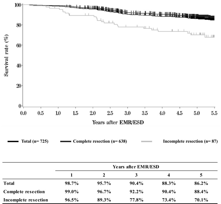
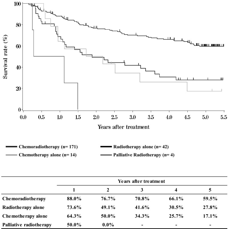
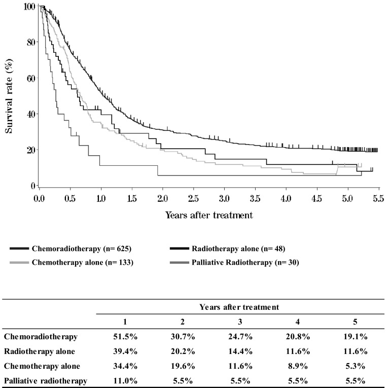
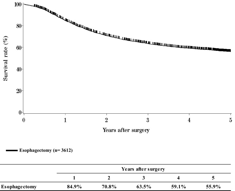
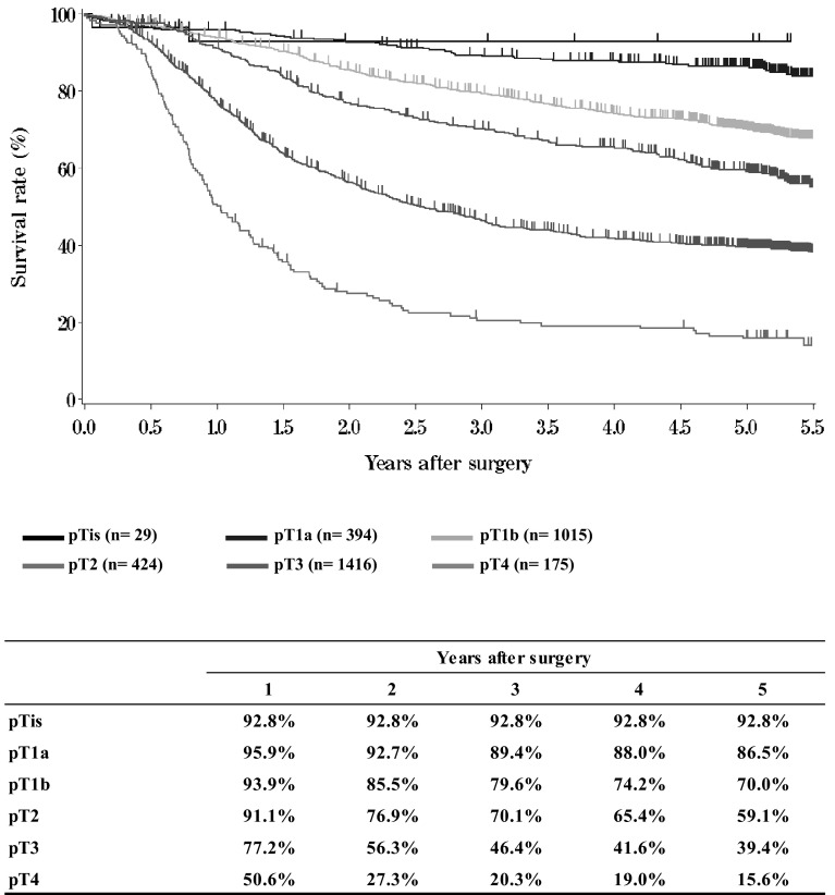
We briefly summarized the Comprehensive Registry of Esophageal Cancer in Japan, 2009. Japanese Classification of Esophageal Cancer 10th and UICC TNM Classification 6th were used for cancer staging according to the subjected year. A total of 6260 cases were registered from 276 institutions in Japan. Tumor locations were cervical: 4.4 %, upper thoracic: 11.9 %, middle thoracic: 48.0 %, lower thoracic: 27.7 % and EG junction: 6.6 %. Superficial carcinomas (Tis, T1a, T1b) were 36.7 %. As for the histologic type of biopsy specimens, squamous cell carcinoma and adenocarcinoma accounted for 90.5 and 3.8 %, respectively. Regarding clinical results, the 5-year survival rates of patients treated using endoscopic mucosal resection, concurrent chemoradiotherapy, radiotherapy alone, chemotherapy alone, or esophagectomy were 86.2, 27.9, 20.2, 5.8, and 55.9 %, respectively. Esophagectomy was performed in 3844 cases. Concerning the approach used for esophagectomy, 24.9 % of the cases were treated thoracoscopically. The operative mortality (within 30 days after surgery) was 1.01 % and the hospital mortality was 4.76 %.
We hope that this Comprehensive Registry of Esophageal Cancer in Japan, 2009 will help to improve all aspects of the diagnosis and treatment of esophageal cancer in Japan.
Contents
-
I.Clinical factors of esophageal cancer patients treated in 2009
- Institution-registered cases in 2009
- Patient Background
-
II.Results of endoscopically treated patients in 2009
- Table9Details of endoscopic treatment
- Table10Complications of EMR/ESD
- Table11Pathological depth of tumor invasion of EMR/ESD specimens
- Figure1Survival of patients treated with EMR/ESD
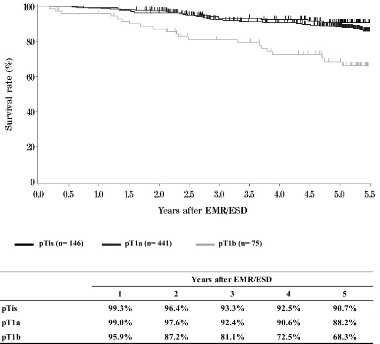
- Figure2Survival of patients treated with EMR/ESD according to the pathological depth of tumor invasion (pT)
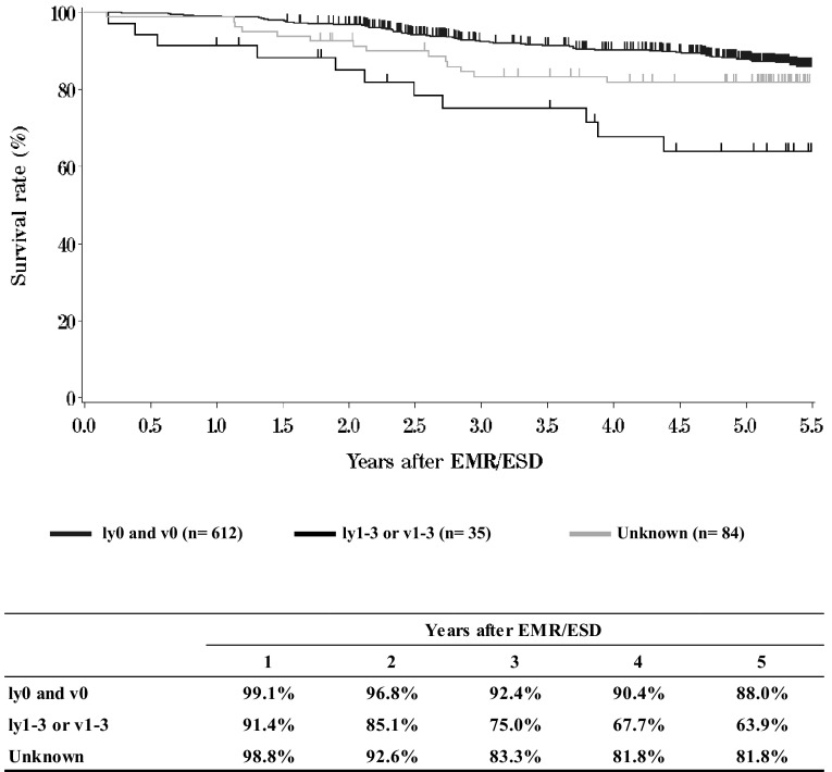
- Figure3Survival of patients treated with EMR/ESD according to the lymphatic and venous invasion
-
III. Results in patients treated with chemotherapy and/or radiotherapy in 2009
-
IV.Results in patients underwent esophagectomy in 2009
- Table14Treatment modalities of esophagectomy
- Table15Tumor location
- Table16Approaches to tumor resection
- Table17Video-assisted surgery
- Table18Fields of lymph node dissection according to the location of the tumor
- Table19Reconstruction route
- Table20Organs used for reconstruction
- Table21Histological classification
- Table22Depth of tumor invasion, pT (JES 10th)
- Table23Pathological grading of lymph node metastasis, pN (JES 10th)
- Table24Numbers of the metastatic nodes
- Table25Pathological findings of distant organ metastasis, pM (JES 10th)
- Table26Residual tumor, R
- Table27Causes of death
- Figure6Survival of patients who underwent esophagectomy
- Figure7Survival of patients who underwent esophagectomy according to clinical stage (JES TNM 10th)
- Figure8Survival of patients who underwent esophagectomy according to clinical stage (UICC TNM 6th)
- Figure9Survival of patients who underwent esophagectomy according to the depth of tumor invasion (JES 10th: pT)
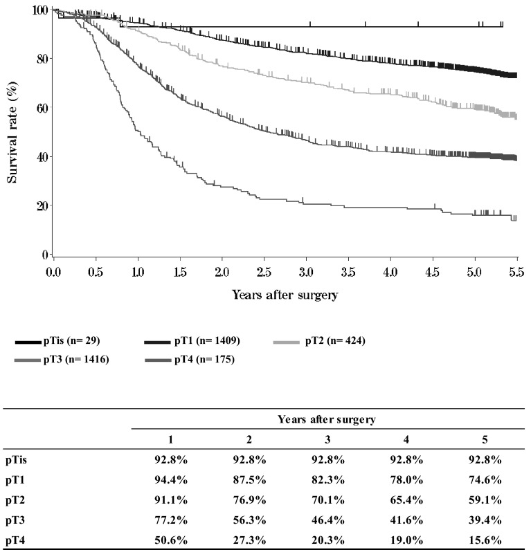
- Figure10Survival of patients who underwent esophagectomy according to the depth of tumor invasion (UICC TNM 6th: pT)
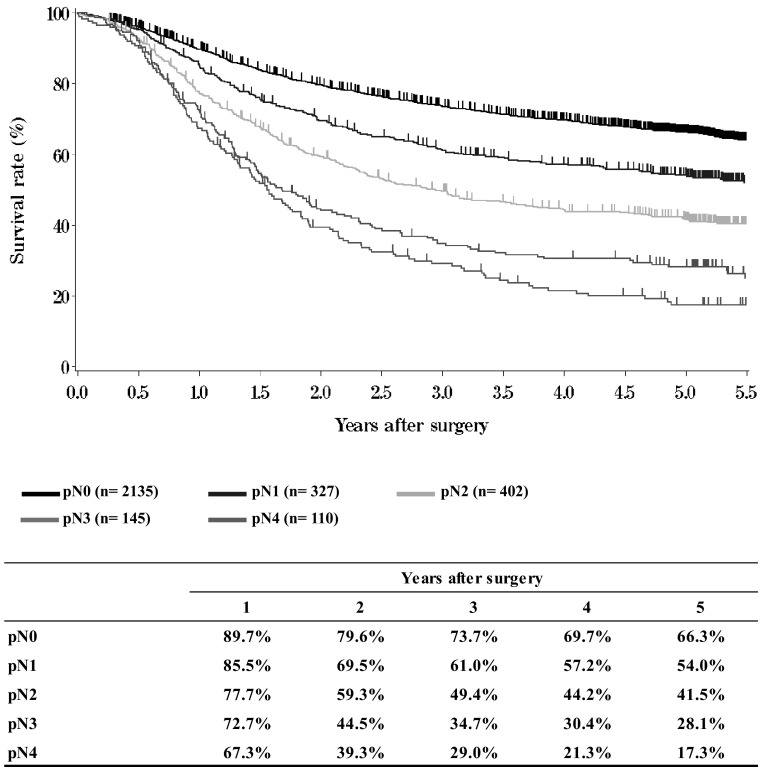
- Figure11Survival of patients who underwent esophagectomy according to lymph node metastasis (JES 10th: pN)
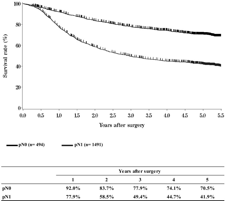
- Figure12Survival of patients who underwent esophagectomy according to lymph node metastasis (UICC TNM 6th: pN)
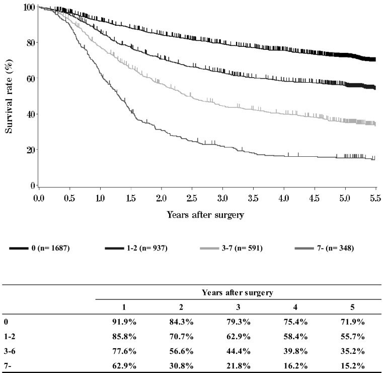
- Figure13Survival of patients who underwent esophagectomy according to number of metastatic nodes
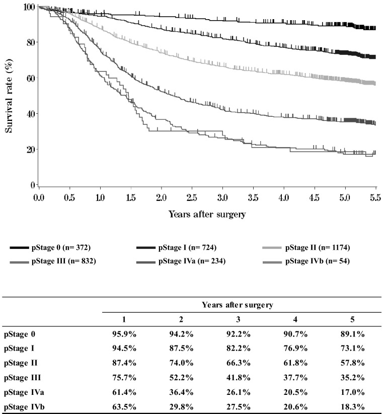
- Figure14Survival of patients who underwent esophagectomy according to pathological stage (JES 10th)
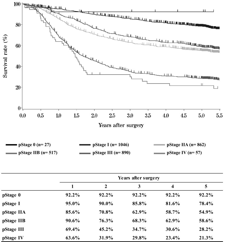
- Figure15Survival of patients who underwent esophagectomy according to pathological stage (UICC TNM 6th)
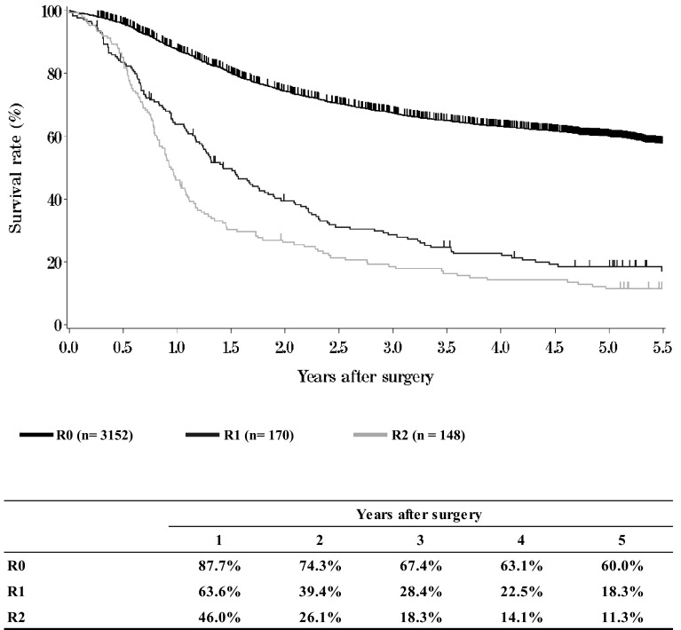
- Figure16Survival of patients who underwent esophagectomy according to residual tumor (R)
Table 1.
Age and gender
| Age | Male | Female | Unknown | Cases (%) |
|---|---|---|---|---|
| ~29 | 6 | 1 | 0 | 7 (0.1 %) |
| 30–39 | 12 | 6 | 0 | 18 (0.3 %) |
| 40–49 | 121 | 34 | 0 | 155 (2.5 %) |
| 50–59 | 946 | 173 | 1 | 1120 (17.9 %) |
| 60–69 | 2332 | 354 | 0 | 2686 (42.9 %) |
| 70–79 | 1575 | 270 | 1 | 1846 (29.5 %) |
| 80–89 | 303 | 75 | 0 | 378 (6.0 %) |
| 90~ | 16 | 3 | 0 | 19 (0.3 %) |
| Unknown | 27 | 4 | 0 | 31 (0.5 %) |
| Total | 5338 | 920 | 2 | 6260 (100 %) |
Table 2.
Primary treatment
| Treatments | Cases (%) |
|---|---|
| Surgery | 3943 (63.0 %) |
| Esophagectomy | 3844 (61.8 %) |
| Palliative | 99 (1.2 %) |
| Chemotherapy/radiotherapy | 1383 (22.1 %) |
| Endoscopic treatment | 932 (14.9 %) |
| Others | 2 (0.0 %) |
| Total | 6260 (100 %) |
Table 3.
Tumor location
| Location of tumor | Endoscopic treatment (%) | Chemotherapy and/or radiotherapy (%) | Palliative surgery (%) | Esophagectomy (%) | Other (%) | Total (%) |
|---|---|---|---|---|---|---|
| Cervical | 18 (1.9 %) | 112 (8.1 %) | 9 (9.1 %) | 137 (3.6 %) | 0 | 276 (4.4 %) |
| Upper thoracic | 105 (11.3 %) | 184 (13.3 %) | 19 (19.2 %) | 437 (11.4 %) | 0 | 745 (11.9 %) |
| Middle thoracic | 511 (54.8 %) | 665 (48.1 %) | 50 (50.5 %) | 1778 (46.3 %) | 1 (50.0 %) | 3005 (48.0 %) |
| Lower thoracic | 243 (26.1 %) | 325 (23.5 %) | 18 (18.2 %) | 1147 (29.8 %) | 0 | 1733 (27.7 %) |
| E > G | 40 (4.3 %) | 38 (2.7 %) | 2 (2.0 %) | 245 (6.4 %) | 0 | 325 (5.2 %) |
| E = G | 5 (0.5 %) | 6 (0.4 %) | 0 | 41 (1.1 %) | 0 | 52 (0.8 %) |
| G > E | 0 | 2 (0.1 %) | 0 | 34 (0.9 %) | 0 | 36 (0.6 %) |
| Unknown | 10 (1.1 %) | 51 (3.7 %) | 1 (1.0 %) | 25 (0.7 %) | 1 (50.0 %) | 88 (1.4 %) |
| Total | 932 (100 %) | 1383 (100 %) | 99 (100 %) | 3844 (100 %) | 2 (100 %) | 6260 (100 %) |
E esophageal, G gastric
Table 4.
Histologic types of biopsy specimens
| Histologic types | Cases (%) |
|---|---|
| Squamous cell carcinoma | 5665 (90.5 %) |
| Squamous cell carcinoma | 3827 (61.1 %) |
| Well differentiated | 354 (5.7 %) |
| Moderately differentiated | 1140 (18.2 %) |
| Poorly differentiated | 344 (5.5 %) |
| Adenocarcinoma | 296 (4.7 %) |
| Adenosquamous carcinoma | 13 (0.2 %) |
| Mucoepidermoid carcinoma | 1 (0.0 %) |
| Basaloid carcinoma | 22 (0.4 %) |
| Neuroendocrine cell carcinoma | 14 (0.2 %) |
| Undifferentiated carcinoma | 10 (0.2 %) |
| Malignant melanoma | 7 (0.1 %) |
| Carcinosarcoma | 17 (0.3 %) |
| Other tumors | 28 (0.4 %) |
| Unknown | 187 (3.0 %) |
| Total | 6260 (100 %) |
Table 5.
Depth of tumor invasion, cT (UICC TNM 6th)
| cT | Cases (%) |
|---|---|
| cTX | 29 (0.5 %) |
| cT0 | 11 (0.2 %) |
| cTis | 157 (2.5 %) |
| cT1 | 359 (5.7 %) |
| cT1a | 650 (10.4 %) |
| cT1b | 1134 (18.1 %) |
| cT2 | 868 (13.9 %) |
| cT3 | 2252 (36.0 %) |
| cT4 | 701 (11.2 %) |
| Unknown | 99 (1.6 %) |
| Total | 6260 (100 %) |
Table 6.
Lymph node metastasis, cN (UICC TNM 6th)
| cN | Cases (%) |
|---|---|
| cNX | 72 (1.2 %) |
| cN0 | 2920 (46.6 %) |
| cN1 | 3157 (50.4 %) |
| Unknown | 111 (1.8 %) |
| Total | 6260 (100 %) |
Table 7.
Distant metastasis, cM (UICC TNM 6th)
| cM | Cases (%) |
|---|---|
| cMX | 57 (0.9 %) |
| cM0 | 5295 (84.6 %) |
| cM1 | 223 (3.6 %) |
| cM1a | 141 (2.3 %) |
| cM1b | 466 (7.4 %) |
| Total | 6260 (100 %) |
Table 8.
Clinical Stage (UICC TNM 6th)
| Location of tumor | Endoscopic treatment (%) | Chemotherapy and/or radiotherapy (%) | Palliative surgery (%) | Esophagectomy (%) | Other (%) | Total (%) |
|---|---|---|---|---|---|---|
| 0 | 131 (14.1 %) | 6 (0.4 %) | 1 (1.0 %) | 13 (0.3 %) | 0 | 151 (2.4 %) |
| I | 658 (70.6 %) | 152 (11.0 %) | 2 (2.0 %) | 964 (25.1 %) | 0 | 1776 (28.4 %) |
| IIA | 6 (0.6 %) | 125 (9.0 %) | 7 (7.1 %) | 717 (18.7 %) | 0 | 855 (13.7 %) |
| IIB | 7 (0.8 %) | 98 (7.1 %) | 2 (2.0 %) | 555 (14.4 %) | 0 | 662 (10.6 %) |
| III | 29 (3.1 %) | 452 (32.7 %) | 62 (62.6 %) | 1243 (32.3 %) | 1 (50.0 %) | 1787 (28.5 %) |
| IV | 10 (1.1 %) | 139 (10.1 %) | 7 (7.1 %) | 44 (1.1 %) | 0 | 200 (3.2 %) |
| IVA | 5 (0.5 %) | 53 (3.8 %) | 1 (1.0 %) | 81 (2.1 %) | 0 | 140 (2.2 %) |
| IVB | 18 (1.9 %) | 265 (19.2 %) | 12 (12.1 %) | 156 (4.1 %) | 0 | 451 (7.2 %) |
| Unknown | 68 (7.3 %) | 93 (6.7 %) | 5 (5.1 %) | 71 (1.8 %) | 1 (50.0 %) | 238 (3.8 %) |
| Total | 932 (100 %) | 1383 (100 %) | 99 (100 %) | 3844 (100 %) | 2 (100 %) | 6260 (100 %) |
Table 9.
Details of endoscopic treatment
| Treatment details | Cases (%) |
|---|---|
| EMR | 201 (21.6 %) |
| EMR + ESD | 11 (1.2 %) |
| EMR + YAG laser | 7 (0.8 %) |
| ESD | 607 (65.1 %) |
| ESD + other treatment | 7 (0.8 %) |
| PDT | 2 (0.2 %) |
| PDT + YAG laser | 2 (0.2 %) |
| YAG laser | 10 (1.1 %) |
| Esophageal stenting | 70 (7.5 %) |
| Esophageal stenting + tracheal stenting | 2 (0.2 %) |
| Tracheal stenting | 4 (0.4 %) |
| Others | 5 (0.5 %) |
| Unknown | 4 (0.4 %) |
| Total | 753 (100 %) |
EMR endoscopic mucosal resection, ESD endoscopic submucosal dissection, YAG: yttrium aluminum garnet, PDT photodynamic therapy
Table 10.
Complications of EMR/ESD
| Complications of EMR/ESD | Cases (%) |
|---|---|
| None | 766 (91.8 %) |
| Perforation | 16 (1.9 %) |
| Bleeding | 2 (0.2 %) |
| Mediastinitis | 0 |
| Stenosis | 42 (5.0 %) |
| Others | 7 (0.8 %) |
| Unknown | 1 (0.1 %) |
| Total | 834 (100 %) |
Table 11.
Pathological depth of tumor invasion of EMR/ESD specimens
| Pathological depth of tumor invasion | Cases (%) |
|---|---|
| pTX | 1 (0.1 %) |
| pT0 | 5 (0.6 %) |
| pTis | 166 (19.9 %) |
| pT1a | 507 (60.9 %) |
| pT1b | 86 (10.3 %) |
| pT2 | 0 |
| Unknown | 68 (8.2 %) |
| Total | 833 (100 %) |
Fig. 1.
Survival of patients treated with EMR/ESD
Fig. 2.
Survival of patients treated with EMR/ESD according to the pathological depth of tumor invasion (pT)
Fig. 3.
Survival of patients treated with EMR/ESD according to the lymphatic and venous invasion
Table 12.
Dose of radiation (non-surgically treated cases)
| Dose of radiation (Gy) | Definitive | Palliative (%) | Recurrence (%) | Others (%) | Unknown (%) | Total (%) | |
|---|---|---|---|---|---|---|---|
| Radiation alone (%) | With chemotherapy (%) | ||||||
| –29 | 5 (4.1 %) | 18 (2.3 %) | 23 (11.0 %) | 2 (6.1 %) | 0 | 1 (5.6 %) | 49 (4.1 %) |
| 30–39 | 1 (0.8 %) | 15 (1.9 %) | 25 (11.9 %) | 3 (9.1 %) | 1 (7.1 %) | 0 | 45 (3.8 %) |
| 40–49 | 11 (8.9 %) | 40 (5.1 %) | 31 (14.8 %) | 9 (27.3 %) | 8 (57.1 %) | 1 (5.6 %) | 100 (8.4 %) |
| 50–59 | 24 (19.5 %) | 199 (25.3 %) | 47 (22.4 %) | 8 (24.2 %) | 2 (14.3 %) | 1 (5.6 %) | 281 (23.7 %) |
| 60–69 | 74 (60.2 %) | 493 (62.6 %) | 81 (38.6 %) | 9 (27.3 %) | 2 (14.3 %) | 15 (83.3 %) | 674 (56.8 %) |
| 70- | 6 (7.2 %) | 8 (2.1 %) | 2 (0.0 %) | 0 | 0 | 0 | 16 (2.2 %) |
| Unknown | 2 (1.6 %) | 15 (1.9 %) | 1 (0.5 %) | 2 (6.1 %) | 1 (7.1 %) | 0 | 21 (1.8 %) |
| Total | 123 (100 %) | 788 (100 %) | 210 (100 %) | 33 (100 %) | 14 (100 %) | 18 (100 %) | 1186 (100 %) |
| Median (min–max) | 60.0 (6.0–120.0) | 60.0 (2.0–124.0) | 54.0 (2.0–95.4) | 50.0 (20.0–66.0) | 40.0 (36.0–60.0) | 60.0 (2.0–61.2) | 60.0 (2.0–124.0) |
Table 13.
Dose of radiation (surgically treated cases)
| Dose of radiation (Gy) | Preoperative radiation (%) | Postoperative radiation (%) |
|---|---|---|
| –29 | 3 (1.4 %) | 1 (1.4 %) |
| 30–39 | 54 (24.4 %) | 2 (2.7 %) |
| 40–49 | 132 (59.7 %) | 21 (28.4 %) |
| 50–59 | 9 (4.1 %) | 18 (24.3 %) |
| 60–69 | 15 (6.8 %) | 27 (36.5 %) |
| 70- | 0 | 0 (1.1 %) |
| Unknown | 8 (3.6 %) | 5 (6.8 %) |
| Total | 221 (100 %) | 74 (100 %) |
| Median (min–max) | 40.0 (15.0–66.0) | 50.4 (4.0–64.0) |
Fig. 4.
Survival of patients treated with chemotherapy and/or radiotherapy (cStage I-IIA)
Fig. 5.
Survival of patients treated with chemotherapy and/or radiotherapy (cStage IIB-IVB)
Table 14.
Treatment modalities of esophagectomy
| Treatments | Cases (%) |
|---|---|
| Esophagectomy | 1630 (42.4 %) |
| Esophagectomy + radiotherapy | 65 (1.7 %) |
| Esophagectomy + chemoradiotherapy | 655 (17.0 %) |
| Esophagectomy + chemoradiotherapy + endoscopic treatment | 16 (0.4 %) |
| Esophagectomy + chemoradiotherapy + other treatment | 2 (0.1 %) |
| Esophagectomy + radiotherapy + endoscopic treatment | 3 (0.1 %) |
| Esophagectomy + radiotherapy + other treatment | 1 (0.0 %) |
| Esophagectomy + chemotherapy | 1385 (36.0 %) |
| Esophagectomy + chemotherapy + endoscopic treatment | 8 (0.2 %) |
| Esophagectomy + chemotherapy + other treatment | 2 (0.1 %) |
| Esophagectomy + endoscopic treatment | 77 (2.0 %) |
| Total | 3844 (100 %) |
Table 15.
Tumor location
| Locations | Cases (%) |
|---|---|
| Cervical | 137 (3.6 %) |
| Upper thoracic | 437 (11.4 %) |
| Middle thoracic | 1778 (46.3 %) |
| Lower thoracic | 1147 (29.8 %) |
| E > G | 245 (6.4 %) |
| E = G | 41 (1.1 %) |
| G > E | 34 (0.9 %) |
| Unknown | 25 (0.7 %) |
| Total lesions | 3844 (100 %) |
Table 16.
Approaches to tumor resection
| Approaches | Cases (%) |
|---|---|
| Cervical approach | 132 (3.4 %) |
| Right thoracotomy | 3239 (84.3 %) |
| Left thoracotomy | 66 (1.7 %) |
| Left thoracoabdominal approach | 49 (1.3 %) |
| Laparotomy | 148 (3.9 %) |
| Transhiatal thoracic esophagectomy | 52 (1.4 %) |
| Transhiatal lower esophagectomy | 92 (2.4 %) |
| Sternotomy | 2 (0.1 %) |
| Others | 32 (0.8 %) |
| Unknown | 32 (0.8 %) |
| Total | 3844 (100 %) |
Table 17.
Video-assisted surgery
| Video-assisted surgery | Cases (%) |
|---|---|
| None | 2549 (66.3 %) |
| Thoracoscopy | 554 (14.4 %) |
| Laparoscopy | 124 (3.2 %) |
| Thoracoscopy + laparoscopy | 388 (10.1 %) |
| Mediastinoscopy | 26 (0.7 %) |
| Thoracoscopy + laparoscopy + mediastinoscopy | 4 (0.1 %) |
| Thoracoscopy + other | 11 (0.3 %) |
| Laparoscopy + mediastinoscopy | 5 (0.1 %) |
| Others | 17 (0.4 %) |
| Unknown | 166 (4.3 %) |
| Total | 3844 (100 %) |
Table 18.
Fields of lymph node dissection according to the location of the tumor
| Field of lymphadenectomy | Cervical | Upper thoracic | Middle thoracic | Lower thoracic | E > G | E = G | G > E | Unknown | Total |
|---|---|---|---|---|---|---|---|---|---|
| None | 9 (6.6 %) | 21 (4.8 %) | 76 (4.3 %) | 39 (3.4 %) | 5 (2.0 %) | 4 (9.8 %) | 1 (2.9 %) | 6 (75.0 %) | 161 (4.2 %) |
| C | 51 (37.2 %) | 5 (1.1 %) | 13 (0.7 %) | 3 (0.3 %) | 0 (0.0 %) | 0 | 0 | 1 (12.5 %) | 73 (1.9 %) |
| C + UM | 20 (14.6 %) | 5 (1.1 %) | 0 | 0 | 0 | 0 | 0 | 0 | 25 (0.7 %) |
| C + UM + MLM | 7 (5.1 %) | 9 (2.1 %) | 45 (2.5 %) | 18 (1.6 %) | 2 (0.8 %) | 0 | 0 | 0 (0.0 %) | 81 (2.1 %) |
| C + UM + MLM + A | 35 (25.5 %) | 286 (65.4 %) | 935 (52.6 %) | 435 (37.9 %) | 29 (11.8 %) | 3 (7.3 %) | 1 (2.9 %) | 8 (100.0 %) | 1732 (45.1 %) |
| C + UM + MLM + A + other | 2 (1.5 %) | 4 (0.9 %) | 0 | 1 (0.1 %) | 0 | 0 | 0 | 0 | 7 (0.2 %) |
| C + UM + A | 0 (0.0 %) | 1 (0.2 %) | 1 (0.1 %) | 0 | 0 | 0 | 0 | 0 | 2 (0.1 %) |
| C + MLM + A | 0 | 2 (0.5 %) | 4 (0.2 %) | 4 (0.3 %) | 0 | 0 | 0 | 0 | 10 (0.3 %) |
| C + A | 5 (3.6 %) | 1 (0.2 %) | 3 (0.2 %) | 3 (0.3 %) | 0 | 0 | 0 | 1 (12.5 %) | 13 (0.3 %) |
| UM | 0 | 5 (1.1 %) | 4 (0.2 %) | 5 (0.4 %) | 0 | 0 | 0 | 0 | 14 (0.4 %) |
| UM + MLM | 2 (1.5 %) | 10 (2.3 %) | 17 (1.0 %) | 13 (1.1 %) | 2 (0.8 %) | 0 | 0 | 1 (12.5 %) | 45 (1.2 %) |
| UM + MLM + A | 0 | 64 (14.6 %) | 584 (32.8 %) | 485 (42.3 %) | 81 (33.1 %) | 9 (22.0 %) | 5 (14.7 %) | 2 (25.0 %) | 1230 (32.0 %) |
| UM + MLM + A + other | 0 | 0 (0.0 %) | 1 (0.1 %) | 0 | 0 | 0 | 0 | 0 | 1 (0.0 %) |
| UM + A | 0 | 1 (0.2 %) | 3 (0.2 %) | 3 (0.3 %) | 0 | 0 | 0 | 0 | 7 (0.2 %) |
| MLM | 0 | 3 (0.7 %) | 13 (0.7 %) | 5 (0.4 %) | 3 (1.2 %) | 0 | 0 | 0 (0.0 %) | 24 (0.6 %) |
| MLM + A | 1 (0.7 %) | 12 (2.7 %) | 50 (2.8 %) | 104 (9.1 %) | 91 (37.1 %) | 16 (39.0 %) | 9 (26.5 %) | 3 (37.5 %) | 286 (7.4 %) |
| A | 1 (0.7 %) | 5 (1.1 %) | 20 (1.1 %) | 24 (2.1 %) | 30 (12.2 %) | 9 (22.0 %) | 18 (52.9 %) | 0 | 107 (2.8 %) |
| Unknown | 4 (2.9 %) | 3 (0.7 %) | 9 (0.5 %) | 5 (0.4 %) | 2 (0.8 %) | 0 | 0 | 3 (37.5 %) | 26 (0.7 %) |
| Total | 137 (100 %) | 437 (100 %) | 1778 (100 %) | 1147 (100 %) | 245 (100 %) | 41 (100 %) | 34 (100 %) | 25 (100 %) | 3844 (100 %) |
C bilateral cervical nodes, UM upper mediastinal nodes, MLM middle-lower mediastinal nodes, A abdominal nodes
Table 19.
Reconstruction route
| Reconstruction route | Cases (%) |
|---|---|
| None | 48 (1.2 %) |
| Subcutaneous | 323 (8.4 %) |
| Retrosternal | 1422 (37.0 %) |
| Intrathoracic | 446 (11.6 %) |
| Posterior mediastinal | 1491 (38.8 %) |
| Cervical | 49 (1.3 %) |
| Others | 36 (0.9 %) |
| Unknown | 29 (0.8 %) |
| Total | 3844 (100 %) |
Table 20.
Organs used for reconstruction
| Organs used for reconstruction | Cases (%) |
|---|---|
| None | 51 (1.3 %) |
| Whole stomach | 102 (2.6 %) |
| Gastric tube | 3234 (81.6 %) |
| Jejunum | 213 (5.4 %) |
| Free jejunum | 88 (2.2 %) |
| Colon | 153 (3.9 %) |
| Free colon | 12 (0.3 %) |
| Skin graft | 0 (0.0 %) |
| Others | 93 (2.3 %) |
| Unknown | 18 (0.5 %) |
| Total organs | 3964 (100 %) |
| Total cases | 3844 |
Table 21.
Histological classification
| Histological classification | Cases (%) |
|---|---|
| Squamous cell carcinoma | 3300 (86.7 %) |
| Squamous cell carcinoma | 685 (18.0 %) |
| Well differentiated | 653 (17.2 %) |
| Moderately differentiated | 1521 (40.0 %) |
| Poorly differentiated | 441 (11.6 %) |
| Adenocarcinoma | 222 (5.8 %) |
| Adenosquamous cell carcinoma | 35 (0.9 %) |
| Adenoid cystic carcinoma | 1 (0.0 %) |
| Basaloid carcinoma | 56 (1.5 %) |
| Neuroendocrine cell carcinoma | 17 (0.4 %) |
| Undifferentiated carcinoma | 10 (0.3 %) |
| Other carcinoma | 9 (0.2 %) |
| Carcinosarcoma | 21 (0.6 %) |
| Malignant melanoma | 11 (0.3 %) |
| GIST | 1 (0.0 %) |
| Other | 46 (1.2 %) |
| Unknown | 78 (2.0 %) |
| Total | 3807 (100 %) |
Table 22.
Depth of tumor invasion, pT (JES 10th)
| pT category | Cases (%) |
|---|---|
| pTX | 24 (0.6 %) |
| pT0 | 94 (2.4 %) |
| pTis | 29 (0.8 %) |
| pT1a | 422 (11.0 %) |
| pT1b | 1065 (27.7 %) |
| pT2 | 454 (11.8 %) |
| pT3 | 1518 (39.5 %) |
| pT4 | 127 (3.3 %) |
| pT4a | 27 (0.7 %) |
| pT4b | 30 (0.8 %) |
| Unknown | 54 (1.4 %) |
| Total | 3844 (100 %) |
Table 23.
Pathological grading of lymph node metastasis, pN (JES 10th)
| Lymph node metastasis | Cases (%) |
|---|---|
| pN0 | 2270 (59.1 %) |
| pN1 | 492 (12.8 %) |
| pN2 | 584 (15.2 %) |
| pN3 | 225 (5.9 %) |
| pN4 | 185 (4.8 %) |
| Unknown | 88 (2.3 %) |
| Total | 3844 (100 %) |
Table 24.
Numbers of the metastatic nodes
| Numbers of lymph node metastasis | Cases (%) |
|---|---|
| 0 | 1779 (46.3 %) |
| 1-2 | 985 (25.6 %) |
| 3-6 | 640 (16.6 %) |
| 7- | 376 (9.8 %) |
| Unknown | 64 (1.7 %) |
| Total | 3844 (100 %) |
Table 25.
Pathological findings of distant organ metastasis, pM (JES 10th)
| Distant metastasis | Cases (%) |
|---|---|
| pMX | 53 (1.4 %) |
| pM0 | 3733 (97.1 %) |
| pM1 | 58 (1.5 %) |
| Total | 3844 (100 %) |
Table 26.
Residual tumor, R
| Residual tumor | Cases (%) |
|---|---|
| RX | 156 (4.1 %) |
| R0 | 3345 (87.0 %) |
| R1 | 187 (4.9 %) |
| R2 | 156 (4.1 %) |
| Total | 3844 (100 %) |
Table 27.
Causes of death
| Cause of death | Cases (%) |
|---|---|
| Death due to recurrence | 1139 (72.8 %) |
| Death due to other cancer | 65 (4.2 %) |
| Death due to other disease (rec +) | 44 (2.8 %) |
| Death due to other disease (rec-) | 179 (11.4 %) |
| Death due to other disease (rec?) | 7 (0.4 %) |
| Operative death* | 39 (2.5 %) |
| Postoperative hospital death** | 40 (2.6 %) |
| Unknown | 52 (3.3 %) |
| Total of death cases | 1565 (100 %) |
rec recurrence
* Operative death means death within 30 days after operation in or out of hospital
** Hospital death is defined as death during the same hospitalization, regardless of department at time of death
Operative mortality after esophagectomy: 1.01 %
Hospital mortality after esophagectomy: 4.76 % 
Fig. 6.
Survival of patients who underwent esophagectomy
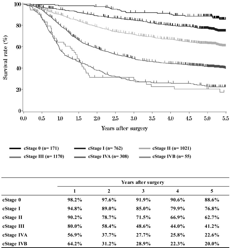
Fig. 7.
Survival of patients who underwent esophagectomy according to clinical stage (JES TNM 10th)
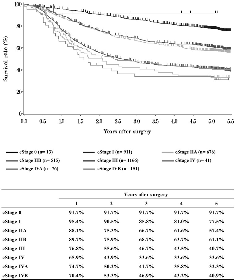
Fig. 8.
Survival of patients who underwent esophagectomy according to clinical stage (UICC TNM 6th)
Fig. 9.
Survival of patients who underwent esophagectomy according to the depth of tumor invasion (JES 10th: pT)
Fig. 10.
Survival of patients who underwent esophagectomy according to the depth of tumor invasion (UICC TNM 6th: pT)
Fig. 11.
Survival of patients who underwent esophagectomy according to lymph node metastasis (JES 10th: pN)
Fig. 12.
Survival of patients who underwent esophagectomy according to lymph node metastasis (UICC TNM 6th: pN)
Fig. 13.
Survival of patients who underwent esophagectomy according to number of metastatic nodes
Fig. 14.
Survival of patients who underwent esophagectomy according to pathological stage (JES 10th)
Fig. 15.
Survival of patients who underwent esophagectomy according to pathological stage (UICC TNM 6th)
Fig. 16.
Survival of patients who underwent esophagectomy according to residual tumor (R)
I. Clinical factors of esophageal cancer patients treated in 2009
Institution-registered cases in 2009
| Institutions |
|---|
| Aichi Cancer Center |
| Aizawa Hospital |
| Akita University Hospital |
| Aomori Municipal Hospital |
| Aomori Prefectural Central Hospital |
| Arao Municipal Hospital |
| Asahikawa Medical College Hospital |
| Chiba Cancer Center |
| Chiba Medical Center |
| Chiba University Hospital |
| Chibaken Saiseikai Narashino Hospital |
| Dokkyo Medical University Hospital |
| Ehime University Hospital |
| Foundation for Detection of Early Gastric Carcinoma |
| Fuchu Hospital |
| Fujioka General Hospital |
| Fujisawa Shounandai Hospital |
| Fujita Health University |
| Fukui Prefectural Hospital |
| Fukui University Hospital |
| Fukuoka Dental College and Dental Hospital |
| Fukuoka Saiseikai General Hospital? |
| Fukuoka University Hospital |
| Fukuoka Wajiro Hospital |
| Fukushima Medical University Hospital |
| Fukuyama City Hopital |
| Gifu Prefectural General Medical Center |
| Gifu University Hospital |
| Gunma Central General Hospital |
| Gunma Prefectural Cancer Center |
| Gunma University Hospital |
| Gunmaken Saiseikai Maebashi Hospital |
| Hachinohe City Hospital |
| Hakodate Goryokaku Hospital |
| Hakodate National Hospital |
| Hamamatsu University School of Medicine, University Hospital |
| Handa City Hospital |
| Hannan Chuo Hospital |
| Heartlife Hospital |
| Higashiosaka City General Hospital |
| Hino Memorial Hospital |
| Hiratsuka City Hospital |
| Hiratsuka Kyosai Hospital |
| Hirosaki University Hospital |
| Hiroshima City Asa Hospital |
| Hiroshima University Research Institute for Radiation Biology Medicine |
| Hitachi General Hospital |
| Hofu Institute of Gastroenterology |
| Hokkaido Kin-Ikyo Chuo Hospital |
| Hokkaido University Hospital |
| Hyogo Cancer Center |
| Hyogo College of Medicine |
| Hyogo Prefectural Nishinomiya Hospital |
| Ibaraki Prefectural Central Hospital |
| Iizuka Hospital |
| Imazu Surgical Clinic |
| Inazawa City Hospital |
| Internatinal University of Health and Welfare Hospital |
| Isehara Kyodo Hospital |
| Ishikawa Prefectural Central Hospital |
| Iwakuni Medical Center |
| Iwate Medical University Hospital |
| Iwate Prefectural Chubu Hospital |
| Iwate Prefectural Isawa Hospital |
| Japanese Red Cross Fukui Hospital |
| Japanese Red Cross Ishinomaki Hospital |
| Japanese Red Cross Kyoto Daini Hospital? |
| Japanese Red Cross Maebashi Hospital |
| Japanese Red Cross Nagaoka Hospital |
| Japanese Red Cross Narita Hospital |
| Japanese Red Cross Nasu Hospital |
| Jichi Medical University Hospital |
| Juntendo University Hospital |
| Juntendo University Shizuoka Hospital |
| Junwakai Memorial Hospital |
| Kagawa Rosai Hospital |
| Kagawa University Hospital |
| Kagoshima Kenritsu Satsunan Hospital |
| Kagoshima University Hospital |
| Kameda General Hospital |
| Kanagawa Cancer Center |
| Kanazawa Medical University Hospital |
| Kanazawa University Hospital |
| Kansai Medical University Hirakata Hospital |
| Kansai Rosai Hospital |
| Kashiwa Kousei General Hospital |
| Kawakita General Hospital |
| Kawasaki Medical School Hospital |
| Kawasaki Medical School Kawasaki Hospital |
| Kawasaki Municipal Hospital |
| Kawasaki Municipal Ida Hospital |
| Keio University Hospital |
| Keiyukai Sapporo Hospital |
| Kikuna Memorial Hospital |
| Kinki Central Hospital |
| Kinki University Hospital |
| Kiryu Kosei General Hospital |
| Kishiwada City Hospital |
| Kitaakita Municipal Hospital |
| Kitakyushu Municipal Medical Center |
| Kitano Hospital |
| Kitasato University Hospital |
| Kobe City Medical Center General Hospital |
| Kobe University Hospital |
| Kochi University Hospital |
| Kokura Memorial Hospital |
| Kumamoto City Hospital |
| Kumamoto University Hospital |
| Kurashiki Central Hospital |
| Kurume General Hospital |
| Kurume University Hospital |
| Kuwana West Medical Center |
| Kyoto University Hospital |
| Kyushu Central Hospital of the Mutual Aid Association of Public School Teachers |
| Kyushu University Beppu Hospital |
| Kyushu University Hospital |
| Kyushu Medical Center |
| Machida Municipal Hospital |
| Matsuda Hospital |
| Matsushita Memorial Hospital |
| Matsuyama Red Cross Hospital |
| Mie University Hospital |
| Mino City Hospital |
| Mito Red Cross Hospital |
| Mitsui Memorial Hospital |
| Miyazaki Konan Hospital |
| Murakami General Hospital |
| Musashimurayama Hospital |
| Musashino Red Cross Hospital |
| Nagahama City Hospital |
| Nagano Red Cross Hospital |
| Nagaoka Chuo General Hospital |
| Nagasaki University Hospital |
| Nagayoshi General Hospital |
| Nagoya City University Hospital |
| Nagoya City West Medical Center |
| Nagoya Daiichi Red Cross Hospital |
| Nagoya University Hospital |
| Nara Hospital Kinki University Faculty of Medicine |
| Nara Medical University Hospital |
| National Cancer Center Hospital |
| National Cancer Center Hospital East |
| National Center for Global Health and Medicine |
| National Defense Medical College Hospital |
| National Hospital Organization Beppu Medical Center |
| National Hospital Organization Chiba-East-Hospital |
| National Hospital Organization Fukuoka-higashi Medical Center |
| National Hospital Organization Kure Medical Center |
| National Hospital Organization Kyoto Medical Center |
| National Hospital Organization Kyushu Cancer Center |
| National Hospital Organization Matsumoto National Hospital |
| National Hospital Organization Nagasaki Medical Center. |
| National Hospital Organization Nagoya Medical Center |
| National Hospital Organization Osaka National Hospital |
| National Hospital Organization Tokyo Medical Center |
| Niigata Cancer Center Hospital |
| Niigata City General Hospital |
| Niigata Prefectural Shibata Hospital |
| Niigata University Medical and Dental Hospital |
| Nikko Memorial Hospital |
| Nippon Medical School Chiba Hokusoh Hospital |
| Nippon Medical School Hospital |
| Nippon Medical School Musashi Kosugi Hospital |
| Nippon Medical School Tama Nagayama Hospital |
| Nishi-Kobe Medical Center |
| Nishinomiya Municipal Central Hospital |
| Numazu City Hospital |
| Obihiro Kousei General Hospital |
| Ohta General Hospital Foundation Ohta Nishinouchi Hospital |
| Oita Red Cross Hospital |
| Oita University Hospital |
| Okayama Saiseikai General Hospital |
| Okayama University Hospital |
| Onomichi Municipal Hospital |
| Osaka City General Hospital |
| Osaka City University Hospital |
| Osaka Hospital of Japan Seafarers relief Association |
| Osaka Medical Center for Cancer and Cardiovascular Diseases |
| Osaka Medical College Hospital |
| Osaka Police Hospital |
| Osaka Prefectural Hospital Organization Osaka General Medical Center |
| Osaka Red Cross Hospital |
| Osaka University Hospital |
| Otsu Municipal Hospital |
| Otsu Red Cross Hospital |
| Rakusei Hospital |
| Ryukyu University Hospital |
| Saga University Hospital |
| Saga-ken Medical Center Koseikan |
| Saiseikai Fukushima General Hospital |
| Saiseikai Hiroshima Hospital |
| Saiseikai Kyoto Hospital |
| Saiseikai Utsunomiya Hospital |
| Saitama City Hospital |
| Saitama Medical Center Jichi Medical University |
| Saitama Medical University Hospital |
| Saitama Medical University Saitama International Medical Center |
| Saitama Medical University Saitama Medical Center |
| Saitama Medical Center |
| Sakai City Medical Center |
| Saku Central Hospital |
| Sanin Rosai Hospital |
| Sano Kousei General Hospital |
| Sendai City Hospital |
| Shiga Medical Center for Adults |
| Shiga University of Medical Science Hospital |
| Shikoku Cancer Center |
| Shimada Hospital |
| Shimane University Hospital |
| Shimizu Welfare Hospital |
| Shinshu University Hospital |
| Shizuoka Cancer Center |
| Shizuoka City Shizuoka Hospital |
| Shizuoka General Hospital |
| Showa University Hospital |
| Showa University Northern Yokohama Hospital |
| Showa University Koto-Toyosu Hospital |
| Social Insurance Omuta Tenryo Hospital |
| Social Insurance Tagawa Hospital |
| Yokohama Chuo Hospital |
| Sonoda Daiichi Hospital |
| St. Marianna University School of Medical Hospital |
| St. Luke’s International Hospital |
| Sugita Genpaku Memorial Obama Municipal Hospital |
| Suita Municipal Hospital |
| Takasago Municipal Hospital |
| Takatsuki Red Cross Hospital |
| Teikyo University Hospital |
| Tenri Hospital |
| The Cancer Institute Hospital of JFCR |
| The Jikei University Hospital |
| The Research Center Hospital for Charged Particle Therapy of NIRS |
| Toho University Omori Medical Center |
| Toho University Sakura Medical Center |
| Tohoku Kosai Hospital |
| Tohoku University Hospital |
| Tokai University Hachioji Hospital |
| Tokai University Hospital |
| Tokai University Tokyo Hospital |
| Tokushima Municipal Hospital |
| Tokushima Red Cross Hospital |
| Tokushima University Hospital |
| Tokyo Dental College Ichikawa General Hospital |
| Tokyo Medical and Dental University Hospital |
| Tokyo Medical University Hospital |
| Tokyo Medical University Ibaraki Medical Center |
| Tokyo Metropolitan Cancer and Infectious Center Komagome Hospital |
| Tokyo Metropolitan Health and Medical Corporation Toshima Hospital |
| Tokyo University Hospital |
| Tokyo Women’s Medical University Hospital |
| Tokyo Women’s Medical University Medical Center East |
| Tonan Hospital |
| Tone Chuou Hospital |
| Tottori Prefectural Central Hospital |
| Tottori University Hospital |
| Toyama Prefectural Central Hospital |
| Toyama University Hospital |
| Tsuchiura Kyodo Hospital |
| Tsukuba University Hospital |
| Tsuruoka Municipal Shonai Hospital |
| “University Hospital, Kyoto Prefectural University of Medicine” |
| University of Miyazaki Hospital |
| Wakayama Medical University Hospital |
| Yamagata Prefectural and Sakata Municipal Hospital Organization |
| Yamagata Prefectural Central Hospital |
| Yamagata Prefectural Shinjo Hospital |
| Yamagata University Hospital |
| Yamaguchi University Hospital |
| Yamaguchi-ken Saiseikai Shimonoseki General Hospital |
| Yamanashi Prefectural Central Hospital |
| Yamanashi University Hospital |
| Yao Municipal Hospital |
| Yokohama City Municipal Hospital |
| Yokohama City University Hospital |
| Yokohama City University Medical Center |
| Yuri General Hospital |
(Total 276 institutions)
Patient Background
II. Results of endoscopically treated patients in 2009
III. Results in patients treated with chemotherapy and/or radiotherapy in 2009
IV. Results in patients who underwent esophagectomy in 2009
Acknowledgments
This study was supported by Health and Labour Sciences Research Grants for Promotion of Cancer Control Programs (H26-Cancer Policy-General-014) from the Ministry of Health, Labour and Welfare of Japan.
Compliance with ethical standards
Ethical Statement
All procedures were in accordance with the ethical standards of the responsible committee on human experimentation (institutional and national) and with the Helsinki Declaration of 1964 and later versions.
Conflict of interest
All authors have nothing to disclose with regard to commercial support.
Footnotes
These data were first made available on January 2016, as the Comprehensive Registry of Esophageal Cancer in Japan, 2009. Not all the tables and figures are reprinted here.
The authors were members of the Registration Committee for Esophageal Cancer, the Japan Esophageal Society, and made great contributions to the preparation of this material.


