

Abstract
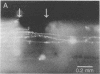
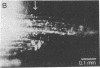
The ability of neurons in the central nervous system to grow through a lesion and restore conduction has been analyzed in a developing spinal cord. The preparation consists of the entire central nervous system of the newly born opossum (Monodelphis domestica), isolated and maintained in culture. Cell division, cell migration, and reflexes are maintained in such preparations for up to 8 days in culture. In the present experiments, massive lesions were produced by crushing the spinal cord, which abolished all conduction for a day. By 2-3 days after injury, electrical conduction across the crush could be observed. After 4-5 days, clear recovery had occurred: the amplitude of the conducted volley was comparable to that in acute preparations. In such preparations, the spinal cord had largely regained its normal appearance at the crush site. Axons stained by carbocyanine dyes or horseradish peroxidase had, by 4 days, grown in profusion through the lesion and several millimeters beyond it. These experiments demonstrate that neurons in the central nervous system of newly born mammals, unlike those in adults, can respond to injury by rapid and extensive outgrowth in the absence of peripheral nerve bridges or antibodies that neutralize inhibitory factors of myelin. With rapid and reliable regeneration occurring in vitro, it becomes practicable to assay the effects of molecules that promote or inhibit the restoration of functional connections.
Full text
PDF


Images in this article


Selected References
These references are in PubMed. This may not be the complete list of references from this article.
- Aguayo A. J., Rasminsky M., Bray G. M., Carbonetto S., McKerracher L., Villegas-Pérez M. P., Vidal-Sanz M., Carter D. A. Degenerative and regenerative responses of injured neurons in the central nervous system of adult mammals. Philos Trans R Soc Lond B Biol Sci. 1991 Mar 29;331(1261):337–343. doi: 10.1098/rstb.1991.0025. [DOI] [PubMed] [Google Scholar]
- David S., Aguayo A. J. Axonal elongation into peripheral nervous system "bridges" after central nervous system injury in adult rats. Science. 1981 Nov 20;214(4523):931–933. doi: 10.1126/science.6171034. [DOI] [PubMed] [Google Scholar]
- Honig M. G., Hume R. I. Dil and diO: versatile fluorescent dyes for neuronal labelling and pathway tracing. Trends Neurosci. 1989 Sep;12(9):333-5, 340-1. [PubMed] [Google Scholar]
- Kalil K., Reh T. A light and electron microscopic study of regrowing pyramidal tract fibers. J Comp Neurol. 1982 Nov 1;211(3):265–275. doi: 10.1002/cne.902110305. [DOI] [PubMed] [Google Scholar]
- Nicholls J. G., Stewart R. R., Erulkar S. D., Saunders N. R. Reflexes, fictive respiration and cell division in the brain and spinal cord of the newborn opossum, Monodelphis domestica, isolated and maintained in vitro. J Exp Biol. 1990 Sep;152:1–15. doi: 10.1242/jeb.152.1.1. [DOI] [PubMed] [Google Scholar]
- Saunders N. R., Adam E., Reader M., Møllgård K. Monodelphis domestica (grey short-tailed opossum): an accessible model for studies of early neocortical development. Anat Embryol (Berl) 1989;180(3):227–236. doi: 10.1007/BF00315881. [DOI] [PubMed] [Google Scholar]
- Savio T., Schwab M. E. Lesioned corticospinal tract axons regenerate in myelin-free rat spinal cord. Proc Natl Acad Sci U S A. 1990 Jun;87(11):4130–4133. doi: 10.1073/pnas.87.11.4130. [DOI] [PMC free article] [PubMed] [Google Scholar]
- Schnell L., Schwab M. E. Axonal regeneration in the rat spinal cord produced by an antibody against myelin-associated neurite growth inhibitors. Nature. 1990 Jan 18;343(6255):269–272. doi: 10.1038/343269a0. [DOI] [PubMed] [Google Scholar]
- Schwab M. E. Nerve fibre regeneration after traumatic lesions of the CNS; progress and problems. Philos Trans R Soc Lond B Biol Sci. 1991 Mar 29;331(1261):303–306. doi: 10.1098/rstb.1991.0021. [DOI] [PubMed] [Google Scholar]
- Stewart R. R., Zou D. J., Treherne J. M., Møllgård K., Saunders N. R., Nicholls J. G. The intact central nervous system of the newborn opossum in long-term culture: fine structure and GABA-mediated inhibition of electrical activity. J Exp Biol. 1991 Nov;161:25–41. doi: 10.1242/jeb.161.1.25. [DOI] [PubMed] [Google Scholar]
- Vidal-Sanz M., Bray G. M., Villegas-Pérez M. P., Thanos S., Aguayo A. J. Axonal regeneration and synapse formation in the superior colliculus by retinal ganglion cells in the adult rat. J Neurosci. 1987 Sep;7(9):2894–2909. doi: 10.1523/JNEUROSCI.07-09-02894.1987. [DOI] [PMC free article] [PubMed] [Google Scholar]
- Zou D. J., Treherne J. M., Stewart R. R., Saunders N. R., Nicholls J. G. Regulation of GABAB receptors by histamine and neuronal activity in the isolated spinal cord of neonatal opossum in culture. Proc Biol Sci. 1991 Oct 22;246(1315):77–82. doi: 10.1098/rspb.1991.0127. [DOI] [PubMed] [Google Scholar]