

Abstract
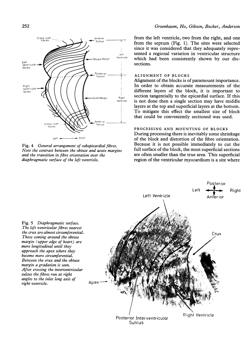
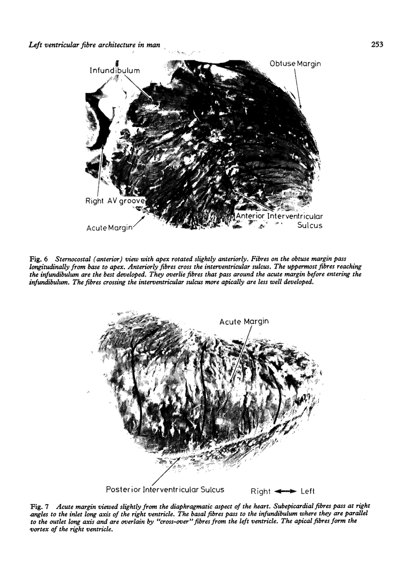
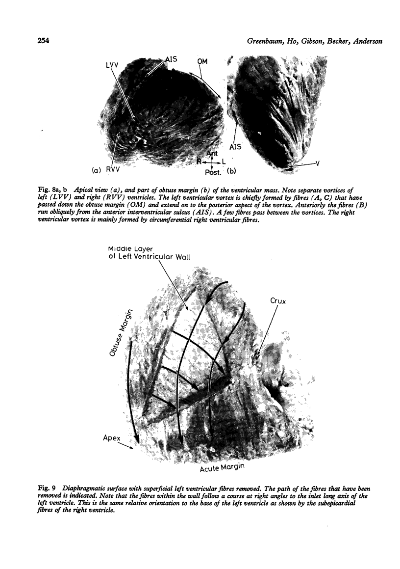
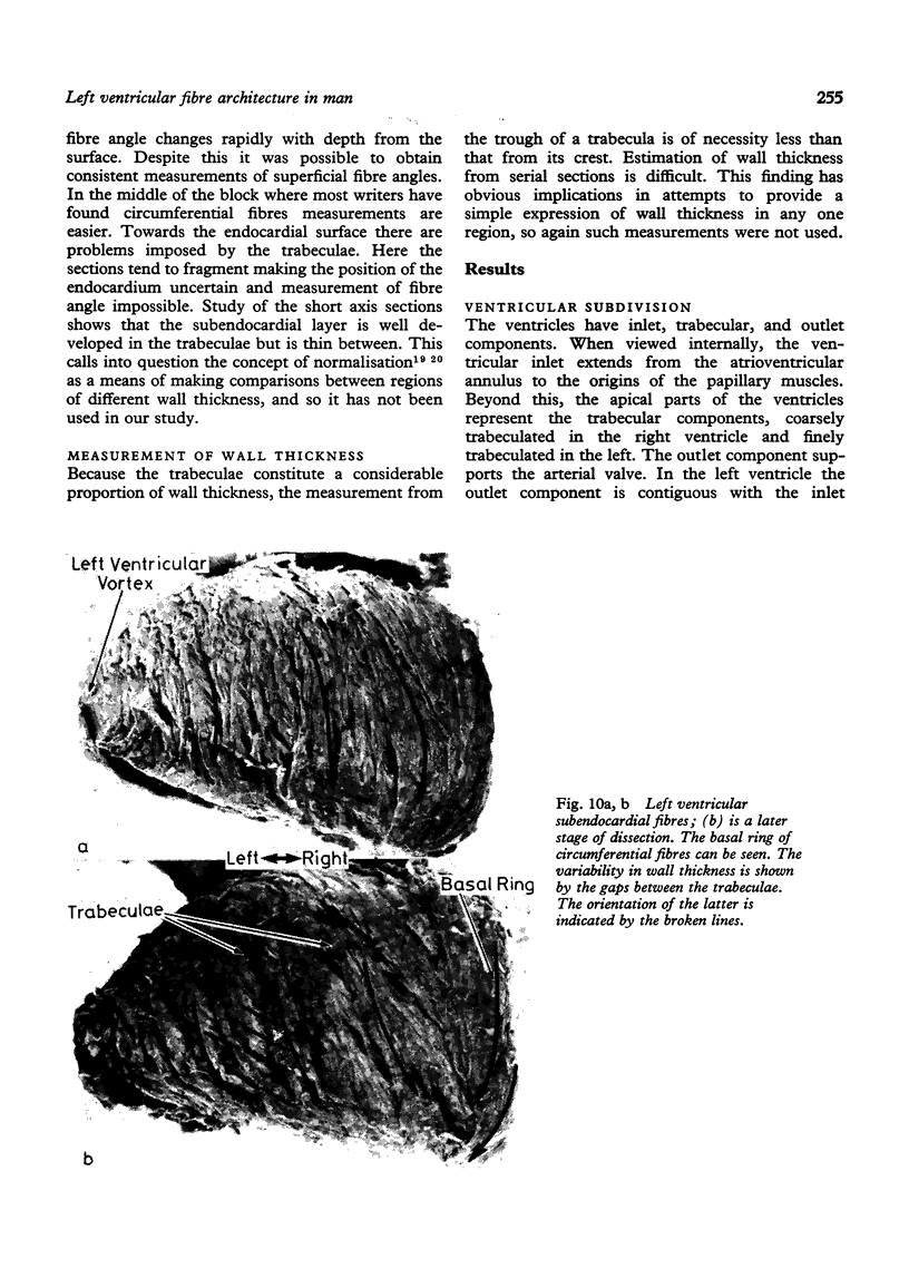
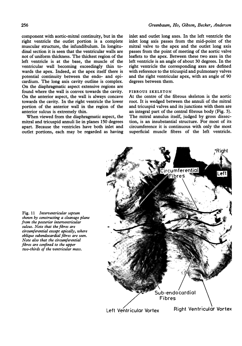
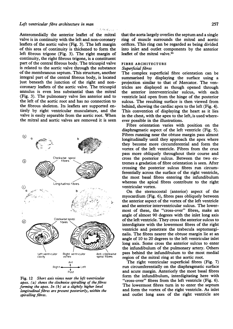
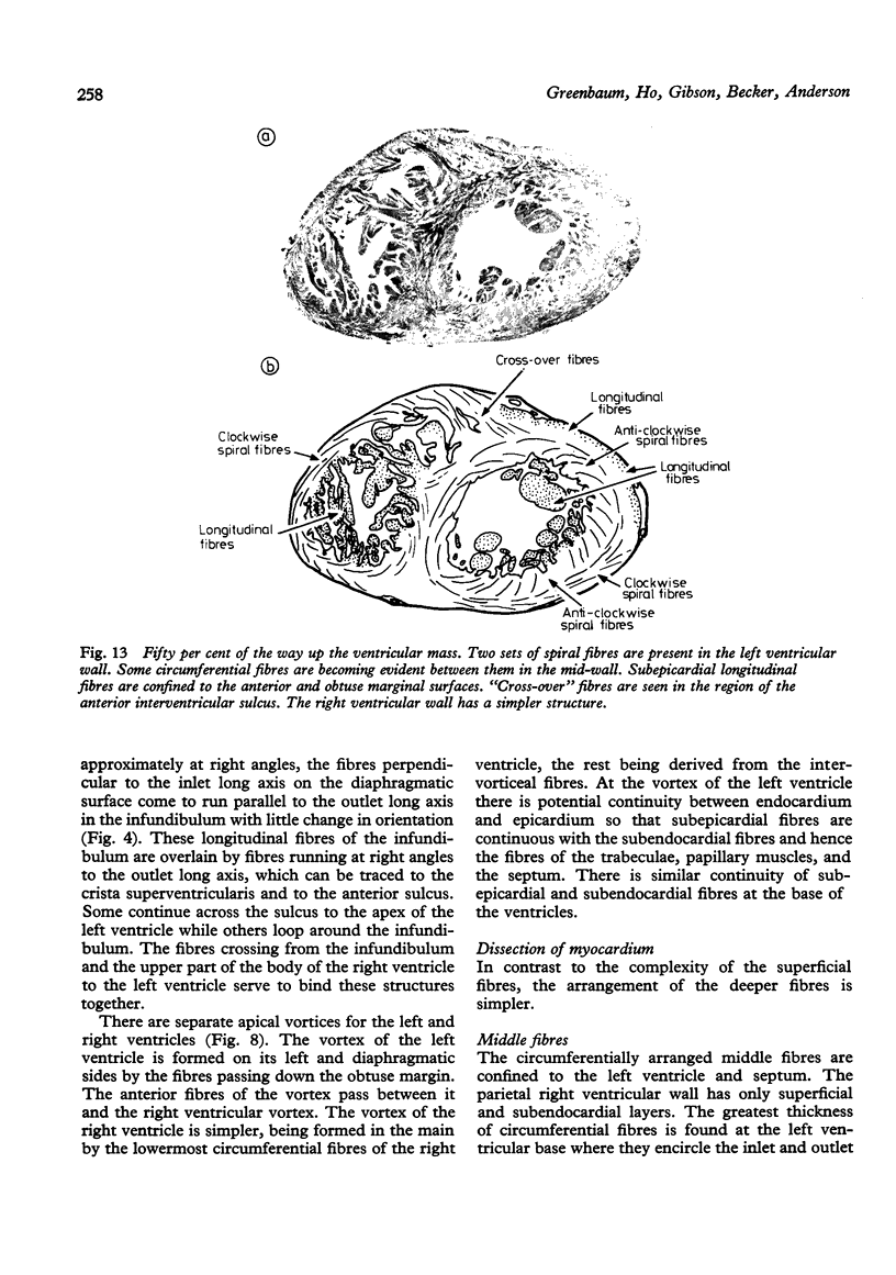
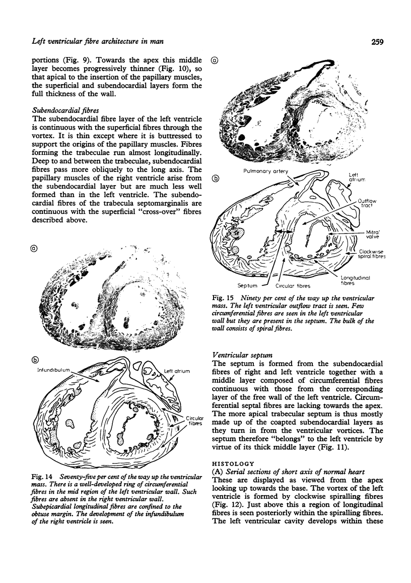
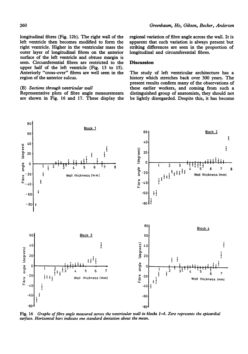
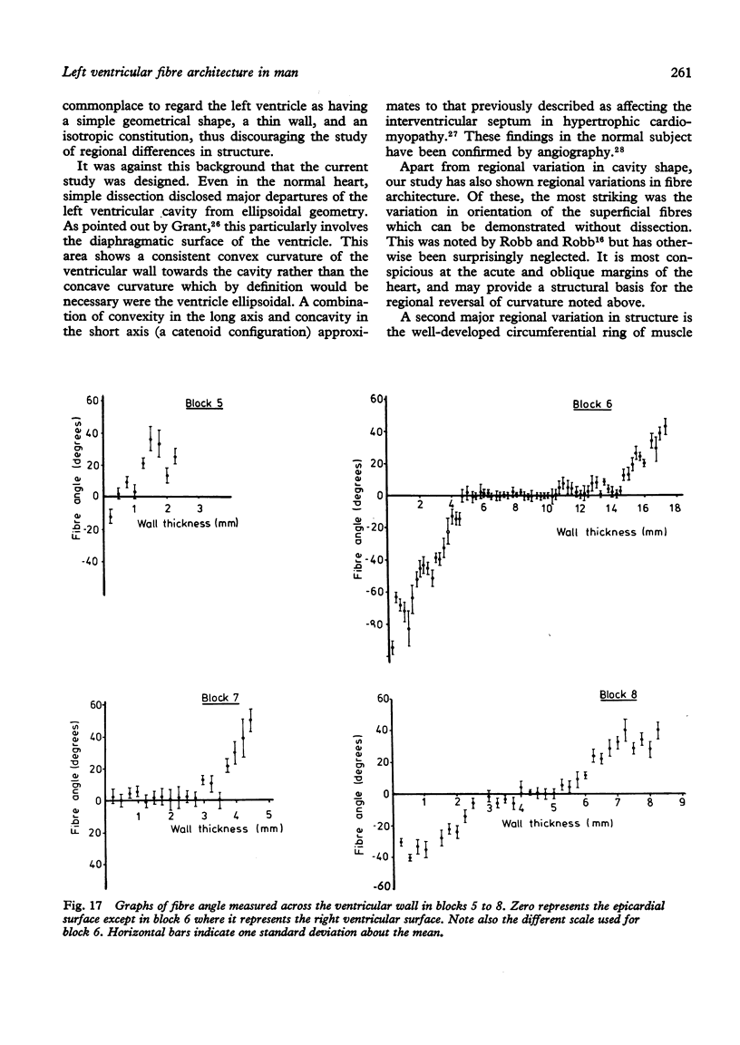
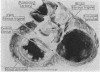
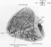
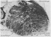
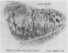
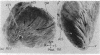
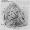
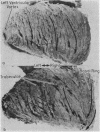
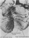
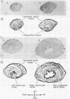
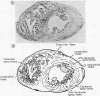
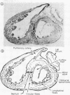
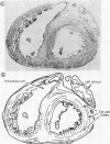
In order to investigate the possibility of regional variation of ventricular structure, 25 normal postmortem human hearts were studied by inspection of cavity shape and subepicardial fibre orientation, by dissection, and by the histology of sections in two orthogonal planes. Ventricular architecture was complex. Inlet and outlet long axes were separated by 30 degrees in the left ventricle. In the right the corresponding figure was 90 degrees. The thickest part of the left ventricular wall was at the base. At the apex there was potential endo- and epicardial continuity. Left ventricular cavity shape departed significantly from any simple geometric figure, there being, consistently, regions of both positive and negative curvature on the diaphragmatic aspect. The presence of trabeculae caused considerable variation in wall thickness. Striking variation was found in the arrangement of subepicardial muscle fibres. Most pronounced was the contrast between the longitudinal arrangement of fibres observed on the oblique margin and the circumferential arrangement of those on the acute. On the diaphragmatic surface of the left ventricle, fibres near the crux and apex ran circumferentially while those between ran obliquely; those on the diaphragmatic surface of the right ventricle also ran circumferentially. Deeper in the myocardium the arrangement was simpler. In the mid-wall of the left ventricle fibres were circumferential, best developed towards the base and in the upper part of the septum. Near the apex of the left ventricle and in the mid-wall of the right ventricle such fibres were sparse. The subendocardial region consisted of longitudinally directed fibres forming the trabeculae and papillary muscles, while fibres deep to and between the trabeculae coursed more obliquely. These findings were confirmed by histology. Models based on uniform myocardial fibre structure cannot explain wall movement in normal subjects, and are likely to have significant limitations if used to investigated left ventricular function in disease.
Full text
PDF
Images in this article
Selected References
These references are in PubMed. This may not be the complete list of references from this article.
- Flett R. L. The Musculature of the Heart, with its Application to Physiology, and a Note on Heart Rupture. J Anat. 1928 Jul;62(Pt 4):439–475. [PMC free article] [PubMed] [Google Scholar]
- GRANT R. P. Architectonics of the heart. Am Heart J. 1953 Sep;46(3):405–431. doi: 10.1016/0002-8703(53)90331-7. [DOI] [PubMed] [Google Scholar]
- GRANT R. P. NOTES ON THE MUSCULAR ARCHITECTURE OF THE LEFT VENTRICLE. Circulation. 1965 Aug;32:301–308. doi: 10.1161/01.cir.32.2.301. [DOI] [PubMed] [Google Scholar]
- Greenbaum R. A., Gibson D. G. Regional non-uniformity of left ventricular wall movement in man. Br Heart J. 1981 Jan;45(1):29–34. doi: 10.1136/hrt.45.1.29. [DOI] [PMC free article] [PubMed] [Google Scholar]
- Hutchins G. M., Bulkley B. H. Catenoid shape of the interventricular septum: possible cause of idiopathic hypertrophic subaortic stenosis. Circulation. 1978 Sep;58(3 Pt 1):392–397. doi: 10.1161/01.cir.58.3.392. [DOI] [PubMed] [Google Scholar]
- LEV M., SIMKINS C. S. Architecture of the human ventricular myocardium; technic for study using a modification of the Mall-MacCallum method. Lab Invest. 1956 Sep-Oct;5(5):396–409. [PubMed] [Google Scholar]
- Ross M. A., Streeter D. D., Jr Nonuniform subendocardial fiber orientation in the normal macaque left ventricle. Eur J Cardiol. 1975 Oct;3(3):229–247. [PubMed] [Google Scholar]
- Streeter D. D., Jr, Hanna W. T. Engineering mechanics for successive states in canine left ventricular myocardium. I. Cavity and wall geometry. Circ Res. 1973 Dec;33(6):639–655. doi: 10.1161/01.res.33.6.639. [DOI] [PubMed] [Google Scholar]
- Streeter D. D., Jr, Hanna W. T. Engineering mechanics for successive states in canine left ventricular myocardium. II. Fiber angle and sarcomere length. Circ Res. 1973 Dec;33(6):656–664. doi: 10.1161/01.res.33.6.656. [DOI] [PubMed] [Google Scholar]
- Streeter D. D., Jr, Spotnitz H. M., Patel D. P., Ross J., Jr, Sonnenblick E. H. Fiber orientation in the canine left ventricle during diastole and systole. Circ Res. 1969 Mar;24(3):339–347. doi: 10.1161/01.res.24.3.339. [DOI] [PubMed] [Google Scholar]
- Streeter D. D., Jr, Vaishnav R. N., Patel D. J., Spotnitz H. M., Ross J., Jr, Sonnenblick E. H. Stress distribution in the canine left ventricle during diastole and systole. Biophys J. 1970 Apr;10(4):345–363. doi: 10.1016/S0006-3495(70)86306-8. [DOI] [PMC free article] [PubMed] [Google Scholar]