

Abstract
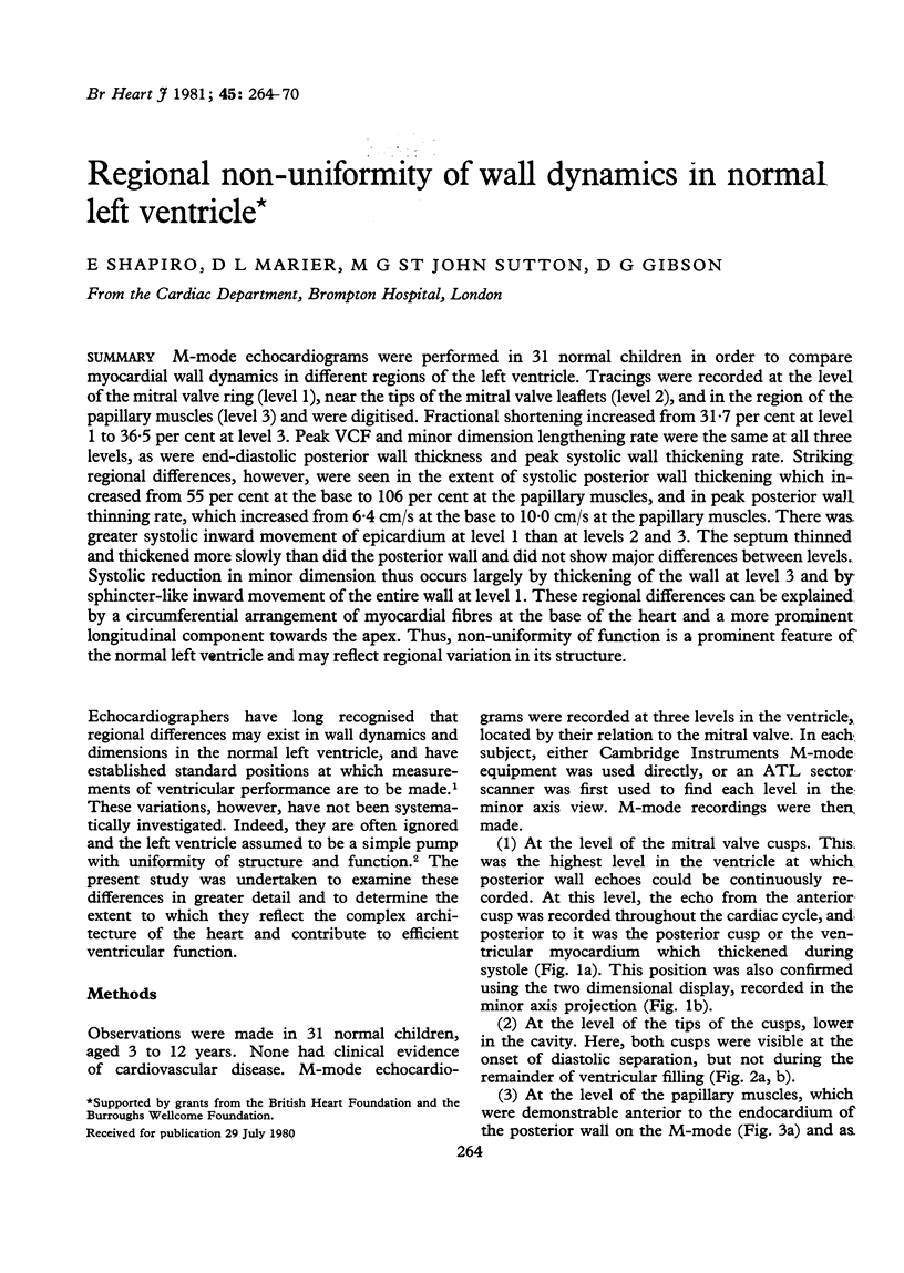
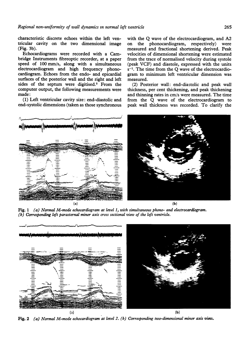
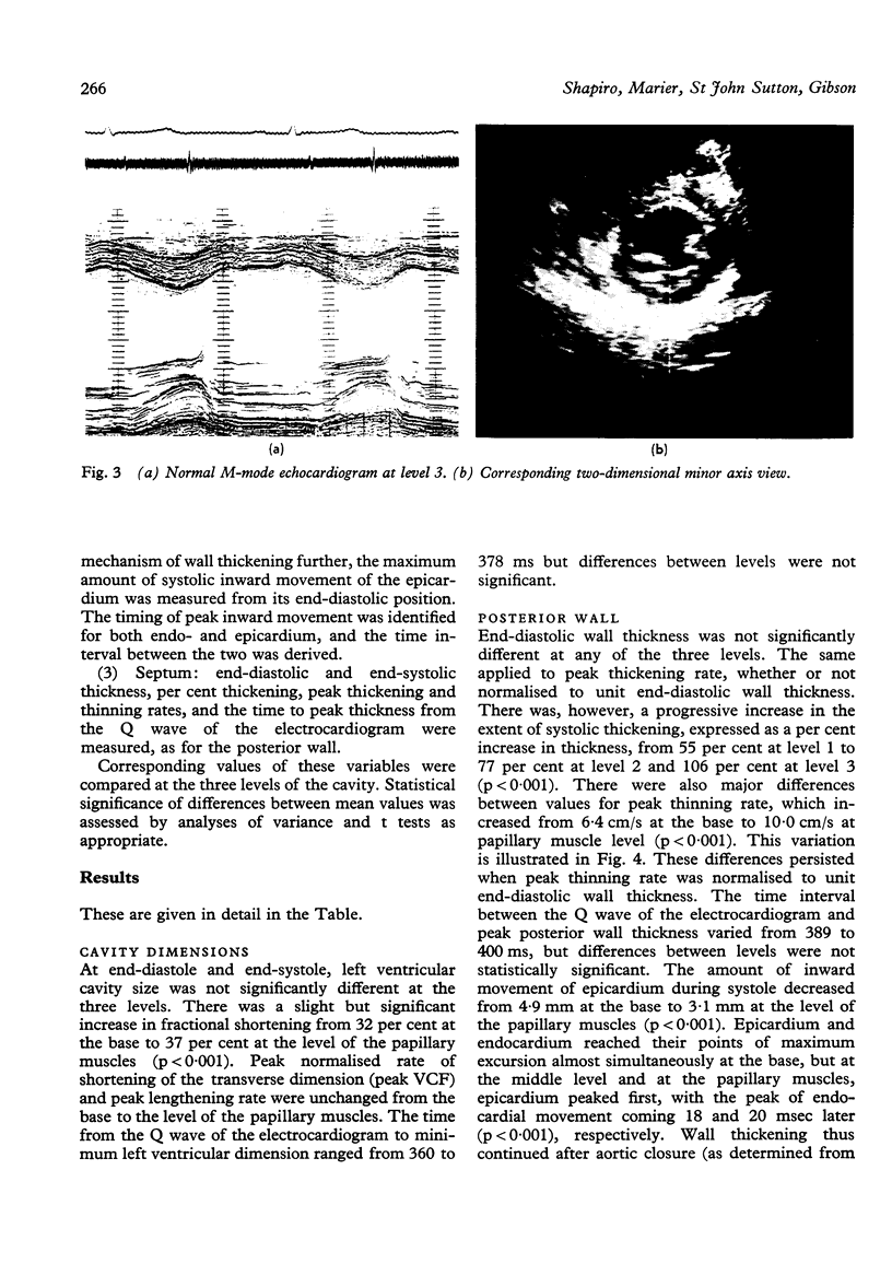
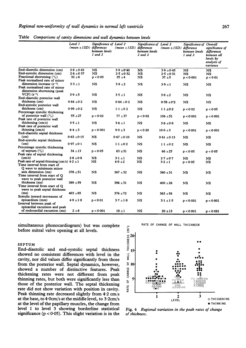
M-mode echocardiograms were performed in 31 normal children in order to compare myocardial wall dynamics in different regions of the left ventricle. Tracings were recorded at the level of the mitral valve ring (level 1), near the tips of the mitral valve leaflets (level 2), and in the region of the papillary muscles (level 3) and were digitised. Fractional shortening increased from 31.7 per cent at level 1 to 36.5 per cent at level 3. Peak VCF and minor dimension lengthening rate were the same at all three levels, as were end-diastolic posterior wall thickness and peak systolic wall thickening rate. Striking regional differences, however, were seen in the extent of systolic posterior wall thickening which increased from 55 per cent at the base to 106 per cent at the papillary muscles, and in peak posterior wall thinning rate, which increased from 6.4 cm/s at the base to 10.0 cm/s at the papillary muscles. There was greater systolic inward movement of epicardium at level 1 than at levels 2 and 3. The septum thinned and thickened more slowly than did the posterior wall and did not show major differences between levels. Systolic reduction in minor dimension thus occurs largely by thickening of the wall at level 3 and by sphincter-like inward movement of the entire wall at level 1. These regional differences can be explained by a circumferential arrangement of myocardial fibres at the base of the heart and a more prominent longitudinal component towards the apex. Thus, non-uniformity of function is a prominent feature of the normal left ventricle and may reflect regional variation in its structure.
Full text
PDF
Images in this article
Selected References
These references are in PubMed. This may not be the complete list of references from this article.
- Armour J. A., Randall W. C. Electrical and mechanical activity of papillary muscle. Am J Physiol. 1970 Jun;218(6):1710–1717. doi: 10.1152/ajplegacy.1970.218.6.1710. [DOI] [PubMed] [Google Scholar]
- Cronin R., Armour J. A., Randall W. C. Function of the in-situ papillary muscle in the canine left ventricle. Circ Res. 1969 Jul;25(1):67–75. doi: 10.1161/01.res.25.1.67. [DOI] [PubMed] [Google Scholar]
- Dumesnil J. G., Ritman E. L., Frye R. L., Gau G. T., Rutherford B. D., Davis G. D. Quantitative determination of regional left ventricular wall dynamics by roentgen videometry. Circulation. 1974 Oct;50(4):700–708. doi: 10.1161/01.cir.50.4.700. [DOI] [PubMed] [Google Scholar]
- Dumesnil J. G., Shoucri R. M., Laurenceau J. L., Turcot J. A mathematical model of the dynamic geometry of the intact left ventricle and its application to clinical data. Circulation. 1979 May;59(5):1024–1034. doi: 10.1161/01.cir.59.5.1024. [DOI] [PubMed] [Google Scholar]
- GRANT R. P. NOTES ON THE MUSCULAR ARCHITECTURE OF THE LEFT VENTRICLE. Circulation. 1965 Aug;32:301–308. doi: 10.1161/01.cir.32.2.301. [DOI] [PubMed] [Google Scholar]
- Gibson D. G., Brown D. Measurement of instantaneous left ventricular dimension and filling rate in man, using echocardiography. Br Heart J. 1973 Nov;35(11):1141–1149. doi: 10.1136/hrt.35.11.1141. [DOI] [PMC free article] [PubMed] [Google Scholar]
- Gibson D. G., Prewitt T. A., Brown D. J. Analysis of left ventricular wall movement during isovolumic relaxation and its relation to coronary artery disease. Br Heart J. 1976 Oct;38(10):1010–1019. doi: 10.1136/hrt.38.10.1010. [DOI] [PMC free article] [PubMed] [Google Scholar]
- Gibson D. G., Sanderson J. E., Traill T. A., Brown D. J., Goodwin J. F. Regional left ventricular wall movement in hypertrophic cardiomyopathy. Br Heart J. 1978 Dec;40(12):1327–1333. doi: 10.1136/hrt.40.12.1327. [DOI] [PMC free article] [PubMed] [Google Scholar]
- Greenbaum R. A., Ho S. Y., Gibson D. G., Becker A. E., Anderson R. H. Left ventricular fibre architecture in man. Br Heart J. 1981 Mar;45(3):248–263. doi: 10.1136/hrt.45.3.248. [DOI] [PMC free article] [PubMed] [Google Scholar]
- Hugenholtz P. G., Kaplan E., Hull E. Determination of left ventricular wall thickness by angiocardiography. Am Heart J. 1969 Oct;78(4):513–522. doi: 10.1016/0002-8703(69)90486-4. [DOI] [PubMed] [Google Scholar]
- LEV M., SIMKINS C. S. Architecture of the human ventricular myocardium; technic for study using a modification of the Mall-MacCallum method. Lab Invest. 1956 Sep-Oct;5(5):396–409. [PubMed] [Google Scholar]
- Popp R. L., Filly K., Brown O. R., Harrison D. C. Effect of transducer placement on echocardiographic measurement of left ventricular dimensions. Am J Cardiol. 1975 Apr;35(4):537–540. doi: 10.1016/0002-9149(75)90837-1. [DOI] [PubMed] [Google Scholar]
- Sallin E. A. Fiber orientation and ejection fraction in the human left ventricle. Biophys J. 1969 Jul;9(7):954–964. doi: 10.1016/S0006-3495(69)86429-5. [DOI] [PMC free article] [PubMed] [Google Scholar]
- Traill T. A., Gibson D. G., Brown D. J. Study of left ventricular wall thickness and dimension changes using echocardiography. Br Heart J. 1978 Feb;40(2):162–169. doi: 10.1136/hrt.40.2.162. [DOI] [PMC free article] [PubMed] [Google Scholar]