Multiple myeloma (MM) is a clonal plasma cell disorder, which remains incurable. The t(4;14) translocation is present in 15% of patients with symptomatic disease and, despite recent therapeutic improvements such as bortezomib treatment, still indicates a poor prognosis.1,2 However, t(4;14) MM is a heterogeneous group, containing both “high risk” and “good risk” patients.3 In addition, the translocation is also detected in some cases of indolent (stage I) MM and even in monoclonal gammopathy of undetermined significance (MGUS).4,5 Prognostic tools capable of predicting the evolution of the different forms of t(4;14) monoclonal gammopathies are currently lacking.
The t(4;14) translocation deregulates two potential oncogenes, FGFR3 and MMSET/WHSC1. Previous studies have shown that FGFR3 expression, which is lost in a subset of t(4;14) MM, does not have a significant impact on patients’ survival.6–9 The MMSET gene, which is over-expressed in all t(4;14) MM, encodes for a histone methyltransferase which is involved in tumor progression and genomic instability.8,10–12 Three major breakpoints within the 5′ coding region of MMSET (MB4-1, MB4-2 and MB4-3) have been observed at 4p16 on chromosome der(4).7,11 Each breakpoint overexpresses a specific IGH/MMSET fusion transcript. While MB4-1 produces a full length MMSET protein, MB4-2 and MB4-3 give different truncated proteins. The aim of this study was to clarify the prognostic significance of each MB4 breakpoint in a large cohort of patients.
We investigated the MB4 breakpoint distribution and prognostic value in a cohort of 294 patients with t(4;14) monoclonal gammopathies including 38 asymptomatic patients (MGUS or stage I MM according to the Durie and Salmon classification) and 256 symptomatic MM patients diagnosed at the Hematology Laboratory of Paris Saint Louis and Nantes. In all cases, quantitative reverse transcriptase polymerase chain reaction (RT-PCR) was performed using cDNA from purified CD138+ bone marrow plasma cells at diagnosis to analyze expression of FGFR3 and the IGH-MMSET fusion transcripts resulting from the three different breakpoints MB4-1, MB4-2 and MB4-3, as described previously.13
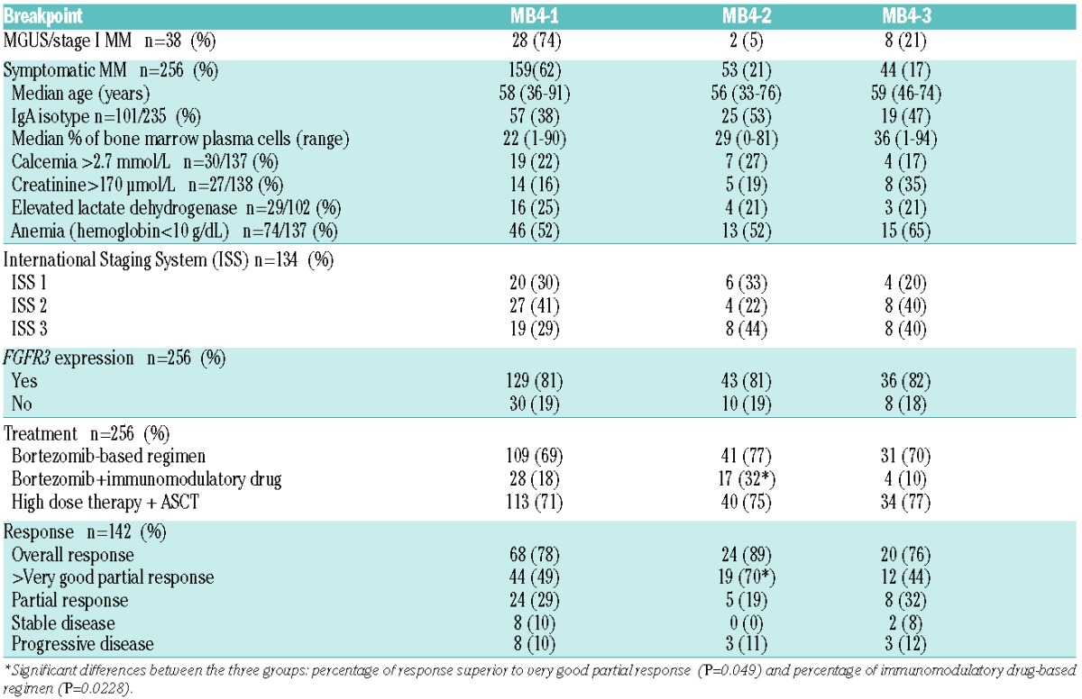
Among the 38 asymptomatic (MGUS/stage I MM) patients [(14 males and 24 females; median age 61 years (range, 35–78) with a median follow-up since diagnosis of 56 months)], RT-PCR analysis of the different IGH/MMSET fusion transcripts revealed a low percentage of the MB4-2 subtype (5%), compared to the MB4-1 and MB4-3 subtypes (74% and 21%, respectively). In contrast, among the patients with symptomatic MM, MB4-2 transcripts were expressed in 21% of cases, as compared to 62% for MB4-1, and 17% for MB4-3. Thus, the MB4-2 breakpoint is rarely observed in t(4;14) indolent monoclonal gammopathy, while it is significantly more frequent in t(4;14) symptomatic MM (P=0.0228) (Table 1). The characteristics of patients with symptomatic MM, including FGFR3 expression, were similar in all MB4 subtypes at diagnosis (Table 1).
Table 1.
Characteristics of MGUS/stage I MM (n=38) and patients with symptomatic MM (n=256), according to the MB4 breakpoint.

In each subgroup, about two-thirds of patients received a bortezomib-containing regimen in frontline therapy and two-thirds of patients had an autologous stem cell transplant. Only four patients received such a transplant after relapse. A combined immunomodulator-bortezomib regimen was used more frequently in MB4-2 patients (32%) than in MB4-1 (18%) (P=0.026) or MB4-3 (10%) patients (P=0.0014).
Overall response rates to the first-line therapy, according to International Myeloma Working Group statements12 were 78%, 89% and 76% in the MB4-1, MB4-2 and MB4-3 sub-groups, respectively. Interestingly, 70% of MB4-2 patients achieved a very good partial response or better, as compared to 49% and 44% of patients with MB4-1 and MB4-3, respectively (P=0.049). Thus, MB4-2 breakpoint was associated with a better quality of response to the front-line treatment, possibly as a result of a more frequent use of a combination of immunomodulatory drugs plus bortezomib.
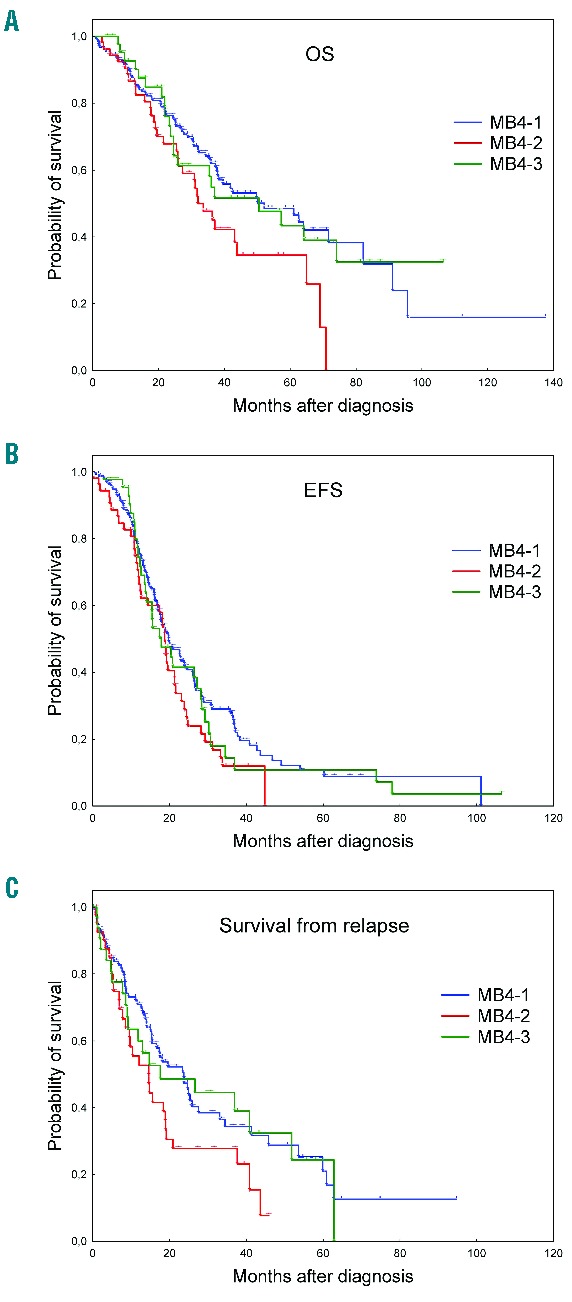
The prognostic impact of each breakpoint (MB4-1, MB4-2 or MB4-3) was analyzed by comparing patients’ outcome (event-free survival, overall survival, survival after the first relapse) using Kaplan-Meier analysis. As shown in Figure 1B, with a median follow-up of 33 months, there was no evidence that event-free survival of the three subgroups was different (P=0.26). In contrast, the overall survival of patients with the MB4-2 breakpoint was shorter than that observed in patients with the other breakpoints (P=0.022) (Figure 1A). Accordingly, survival after first relapse was reduced in the MB4-2 subgroup (median overall survival: 14.6 months versus 23.7 months, P=0.036) (Figure 1C). In multivariate analysis testing MB4 breakpoints, hemoglobin level, calcemia, serum beta-2 microglobulin level and International Staging System score as survival parameters, MB4-2 was an independent prognostic factor for survival [hazard ratio (HR)=1.8; 95% confidence interval (CI): 1.05–3.08; P=0.03)] along with hypercalcemia (HR=3.4; 95%CI: 2.04–5.81; P<0.0001). Thus, symptomatic t(4;14) MM patients with the MB4-2 breakpoint are sensitive to first-line therapy, but develop chemo-resistant relapse and have a poorer outcome.
Figure 1.

(A) Overall survival (B) event-free survival and (C) survival after the first relapse of the 256 patients with symptomatic MM according to the MB4 breakpoint. MB4-1 (n=159): blue curve, MB4-2 (n=54): red curve and MB4-3 (n=43): green curve. In (B) and (C), statistical significance between MB4-2 and the two other subgroups were P=0.022 and P=0.036, respectively (Kaplan-Meier analysis).
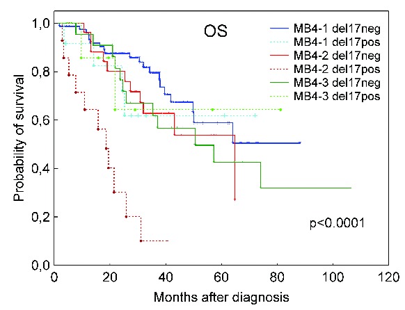
To further investigate the impact of WHSC1 breakpoints on the outcome of patients with t(4;14), deletion of chromosome 17 at p13 [del(17p)], which is associated with high-risk MM, was analyzed by fluorescence in situ hybridization at diagnosis in 162/256 cases for which tumor plasma cells were available. Overall, del(17p) was detected in 34 out of 162 cases (21%). Only 5/34 patients (2 MB4-1, 1 MB4-2 and 2 MB4-3) had a del(17p) in less than 60% of plasma cells (31–45%). The presence of del(17p) was associated with a shortened survival (median survival: 21 versus 40 months, P=0.00766) (data not shown). The del(17p) was statistically more frequently found in tumor cells with MB4-2 (14/41, 34%) as compared to MB4-1 breakpoint (13/90, 14%) (P=0.0216) but was found at a similar rate in plasma cells with MB4-3 breakpoints (7/31, 28%) (P=0.292). The frontline therapy was similar in the del(17p)-positive and -negative MB4 groups. Interestingly, among patients with del(17p), those with the MB4-2 breakpoint had a shorter overall survival than those with the MB4-1 or MB4-3 breakpoint with a median overall survival of 18.6 months (P<0.0001) (Figure 2). In contrast, the MB4 breakpoint had no impact on overall survival in the absence of del(17p) (Figure 2). In multivariate analysis of patients for whom del(17p) status was determined (n=50), MB4-2 was an independent marker of survival (HR: 2.12; 95% CI: 1.20–3.73; P=0.009). These results indicate that patients with del(17p) and the MB4-2 breakpoint constitute a subset of very high risk patients with a very poor prognosis.
Figure 2.

Overall survival of the 162 patients in whom fluorescence in situ hybridization 17p analysis was available, according to the MB4 breakpoint and del(17p). Statistical significance between MB4-2 del(17p) positive (pos.) and the other subgroups was P<0.0001. (Kaplan-Meier analysis) (MB4-1: del17 neg: n=77, del 17 pos n=13; MB4-2: del17 neg n=27, del17 pos n=14; MB4-3: del17 neg n=24, del17 pos n= 7).
Our results contrast with those of a previous study by Keats et al., who found a similar overall survival in the different MB4 subgroups11. However, in that study, survival analysis was performed on a smaller number of patients (n=43). In addition, the survival analysis compared patients with MB4-1 (n=30) to the pooled MB4-2 and MB4-3 subgroup (n=13), according to their ability to encode a full length or a truncated MMSET protein. Pooling MB4-2 and MB4-3 cases may have masked the specific prognostic value of the MB4-2 breakpoint.
At present, the molecular basis for the particularly poor prognosis associated with the MB4-2 breakpoint is not clear. Recent studies have shown that over-expression of MMSET triggers a genome-wide increase of H3K36 dimethylation and drives oncogenic properties in vitro.9–13 In addition, increased H3K36 dimethylation was reported in a series of children with pediatric acute lymphoblastic leukemia expressing mutated or MB4-2-like truncated MMSET.14,15 Thus, it is possible that the higher genome-wide level of H3K36me2 resulting from over-expression of a truncated MB4-2 MMSET protein could render tumor plasma cells more sensitive to first-line therapy, but could eventually drive the emergence of chemoresistant clones that could shorten patients’ survival.
MMSET has also been implicated in the cellular response to DNA damage through its H4K20 histone methyltransferase activity.10 This function requires the phosphorylation of serine 102 by the ATM protein, which facilitates the binding of MMSET at DNA double-strand breaks and the recruitment of 53BP1.10 The removal of Ser102 in truncated MMSET isoforms might therefore enhance genomic instability and promote the emergence of resistant clones in patients. However, Ser102 is deleted in the forms derived from both MB4-2 and MB4-3, and so differential effects on DNA damage are unlikely to account for the different clinical outcomes observed for these two breakpoints. One potentially important difference between MB4-2 and MB4-3 could relate to the position of these breakpoints relative to the 5′ PWWP domain involved in protein-protein interactions. While the MB4-3 breakpoint leads to a truncated MMSET protein lacking the entire PWWP domain, the product expressed from MB4-2 product retains part of this domain.11 Future studies will need to address whether these truncated MMSET proteins could interact with different partner proteins and exert different activities on histone H3 and H4, with specific impact on patients’ outcome.
In our study, a high frequency of del(17p) was observed in plasma cells overexpressing the MB4-2-derived truncated form of MMSET. The combination of both genetic abnormalities severely impairs patients’ outcome. In this retrospective study, del(17p) was specifically included as it was the only poor prognostic marker which had been determined in a large proportion of patients. It is now important to investigate the clinical interactions between the different MMSET breakpoints and other genetic markers associated with poor prognosis, including gain of 1q, del(12p) and del(17p), in prospective studies.
Together, our results indicate that the breakpoint within the MMSET locus may explain, in part, the prognostic heterogeneity of t(4;14) gammopathies. Thus, a systematic identification of the MB4 breakpoint, along with screening for a del(17p), may be useful in the management of patients with t(4;14) MM and may pave the way for specific therapies targeting MMSET activity.
Footnotes
Information on authorship, contributions, and financial & other disclosures was provided by the authors and is available with the online version of this article at www.haematologica.org.
References
- 1.Avet-Loiseau H, Leleu X, Roussel M, et al. Bortezomib plus dexamethasone induction improves outcome of patients with t(4;14) myeloma but not outcome of patients with del(17p). J Clin Oncol 2010;28(30):4630–4634. [DOI] [PubMed] [Google Scholar]
- 2.Moreau P, Facon T, Leleu X, et al. Recurrent 14q32 translocations determine the prognosis of multiple myeloma, especially in patients receiving intensive chemotherapy. Blood 2002;100(5):1579–1583. [DOI] [PubMed] [Google Scholar]
- 3.Moreau P, Attal M, Garban F, et al. Heterogeneity of t(4;14) in multiple myeloma. Long-term follow-up of 100 cases treated with tandem transplantation in IFM99 trials. Leukemia 2007;21(9):2020–2024. [DOI] [PubMed] [Google Scholar]
- 4.López-Corral L, Gutiérrez NC, Vidriales MB, et al. The progression from MGUS to smoldering myeloma and eventually to multiple myeloma involves a clonal expansion of genetically abnormal plasma cells. Clin Cancer Res 2011;17(7):1692–1700. [DOI] [PubMed] [Google Scholar]
- 5.Karlin L, Soulier J, Chandesris O, et al. Clinical and biological features of t(4;14) multiple myeloma: a prospective study. Leuk Lymphoma 2011;52(2):238–246. [DOI] [PubMed] [Google Scholar]
- 6.Keats JJ, Reiman T, Maxwell CA, et al. In multiple myeloma, t(4;14)(p16;q32) is an adverse prognostic factor irrespective of FGFR3 expression. Blood 2003;101(4):1520–1529. [DOI] [PubMed] [Google Scholar]
- 7.Chesi M, Nardini E, Lim RS, et al. The t(4;14) translocation in myeloma dysregulates both FGFR3 and a novel gene, MMSET, resulting in IgH/MMSET hybrid transcripts. Blood 1998;92(9):3025–3034. [PubMed] [Google Scholar]
- 8.Marango J, Shimoyama M, Nishio H, et al. The MMSET protein is a histone methyltransferase with characteristics of a transcriptional corepressor. Blood 2008;111(6):3145–3154. [DOI] [PMC free article] [PubMed] [Google Scholar]
- 9.Kuo AJ, Cheung P, Chen K, et al. NSD2 links dimethylation of histone H3 at lysine 36 to oncogenic programming. Mol Cell 2011;44(4):609–620. [DOI] [PMC free article] [PubMed] [Google Scholar]
- 10.Pei H, Zhang L, Luo K, et al. MMSET regulates histone H4K20 methylation and 53BP1 accumulation at DNA damage sites. Nature 2011;470(7332):124–128. [DOI] [PMC free article] [PubMed] [Google Scholar]
- 11.Keats JJ, Maxwell CA, Taylor BJ, et al. Overexpression of transcripts originating from the MMSET locus characterizes all t(4;14)(p16;q32)-positive multiple myeloma patients. Blood 2005;105(10):4060–4069. [DOI] [PMC free article] [PubMed] [Google Scholar]
- 12.Kyle RA, Rajkumar SV. Criteria for diagnosis, staging, risk stratification and response assessment of multiple myeloma. Leukemia 2009;23(1):3–9. [DOI] [PMC free article] [PubMed] [Google Scholar]
- 13.Chandesris MO, Soulier J, Labaume S, et al. Detection and follow-up of fibroblast growth factor receptor 3 expression on bone marrow and circulating plasma cells by flow cytometry in patients with t(4;14) multiple myeloma. Br J Haematol 2007;136(4):609–614. [DOI] [PubMed] [Google Scholar]
- 14.Jaffe JD, Wang Y, Chan HM, et al. Global chromatin profiling reveals NSD2 mutations in pediatric acute lymphoblastic leukemia. Nat Genet 2013;45(11):1386–1391. [DOI] [PMC free article] [PubMed] [Google Scholar]
- 15.Oyer JA, Huang X, Zheng Y, et al. Point mutation E1099K in MMSET/NSD2 enhances its methyltranferase activity and leads to altered global chromatin methylation in lymphoid malignancies. Leukemia 2014;28(1):198–201. [DOI] [PMC free article] [PubMed] [Google Scholar]


