Abstract
Erythema multiforme (EM) is an acute, self-limited, Type IV hypersensitivity reactions associated with infections and drugs. In this case of acute promyelocytic leukemia, EM diagnosed during the induction phase was mistakenly attributed to vancomycin used to treat febrile neutropenia during that period. However, the occurrence of the lesions of EM again during the consolidation phase with arsenic trioxide (ATO) lead to a re-evaluation of the patient and both the Naranjo and World Health Organization-Uppsala Monitoring Centre scale showed the causality association as “probable.” The rash responded to topical corticosteroids and antihistamines. This rare event of EM being caused by ATO may be attributed to the genetic variation of methyl conjugation in the individual which had triggered the response, and the altered metabolic byproducts acted as a hapten in the subsequent keratinocyte necrosis.
KEY WORDS: Acute promyelocytic leukemia, arsenic trioxide, erythema multiforme, genetic variation
Introduction
Erythema multiforme (EM) is an acute, self-limited, Type IV hypersensitivity reaction associated with certain infections (herpes simplex and mycoplasma),[1] medications, and other triggers such as cold, sunlight, collagen vascular diseases, non-Hodgkin's lymphoma, and multiple myeloma.[2] Common medications causing EM are reportedly sulfa drugs, anticonvulsants, antibiotics (penicillin, ampicillin, tetracyclines, amoxicillin, cefotaxime, cefaclor, cephalexin, ciprofloxacin, erythromycin, minocycline, sulfonamides, trimethoprim-sulfamethoxazole, and vancomycin), anti-tuberculosis drugs (rifampicin, isoniazid, thiacetazone, and pyrazinamide), analgesics (aspirin, phenylbutazone, oxyphenbutazone, and phenazone).[3] However, EM due to arsenic trioxide (ATO) is yet to be reported. A PubMed search with keywords “erythema multiforme, arsenic trioxide” showed no results. Here, we are reporting a case of EM caused due to ATO in a patient of acute promyelocytic leukemia (APL).
Case Report
A 41-year-old female presented with a history of fatigue and intermittent fever of 2 weeks duration, together with easy bruising and menorrhagia. She had no previous history of bleeding manifestation. On examination, she was febrile and had pallor and cutaneous ecchymoses. There was no icterus, sternal tenderness, lymphadenopathy, or organomegaly. Her hemogram showed pancytopenia (hemoglobin 8.6 g/dl, white blood cell count 1.8 × 10⁹/L, and platelet count 30 × 109/L). Examination of peripheral blood smear revealed the presence of abnormal promyelocytes, comprising 28% of total leukocytes. Bone marrow aspiration showed hypercellular marrow with trilineage suppression, with abnormal promyelocytes constituting more than 90% of bone marrow nucleated cells. The bone marrow morphology was suggestive of APL. Immunophenotyping showed these cells to be positive for myeloperoxidase, CD13 and CD33, weakly positive for CD15 and CD64, and negative for human leukocyte antigen-diabetic retinopathy, CD34, and CD117. Bone marrow karyotyping revealed 46 XX, t(15;17)(q22;q21). Real-time polymerase chain reaction for Promyelocytic Leukemia - Retinoic acid receptor alfa (PML-RARA) was positive for the variant form of the fusion protein. She was diagnosed to have APL, Sanz intermediate risk group.
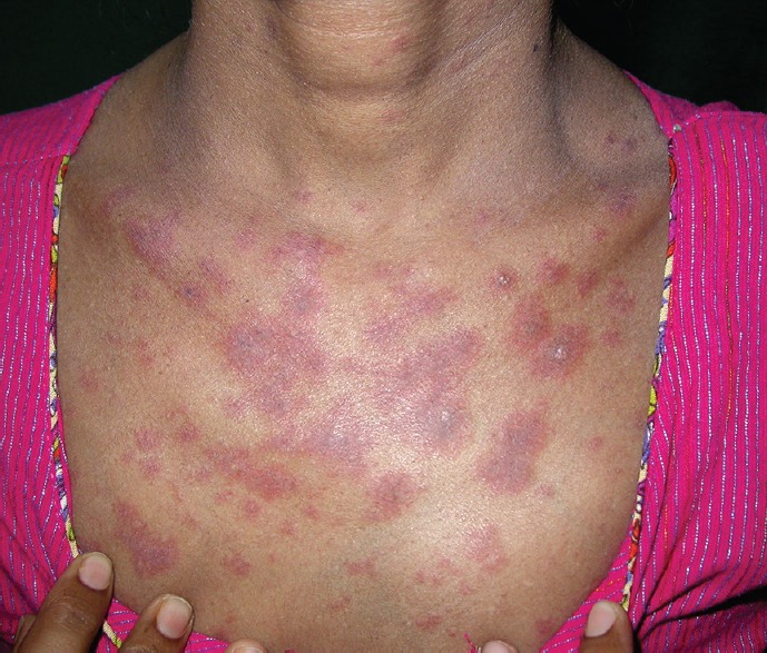
The patient received induction therapy with the combination of all-trans-retinoic acid (ATRA) at 45 mg/m2/day p.o. in two divided doses and ATO 0.15 mg/kg/day intravenous infusion over 2 h. The patient also received vancomycin to tide over the crisis of febrile neutropenia as an empiric antibiotic therapy as per hospital protocol. The induction therapy of ATRA and ATO was started on day 1 and was planned to be continued until bone marrow remission. On day 27 of induction therapy, she developed a few erythematous macular lesions on her upper chest [Figure 1]. The erythematous macules were diagnosed as EM. Naranjo's causality assessment was done, and the scale showed a score of 5 for vancomycin (probable adverse drug reaction [ADR]) and three for the ATRA-ATO combination (possible ADR). World Health Organization-Uppsala Monitoring Centre (WHO-UMC) causality categories fared similarly for either of the two drugs. Thus, the EM lesions were attributed to vancomycin used for febrile neutropenia during induction phase. Vancomycin was stopped and the lesions responded to antihistamine levocetirizine 10 mg/day. Morphological complete remission was documented on day 38 of induction phase.
Figure 1.

Erythematous macular lesions on upper chest
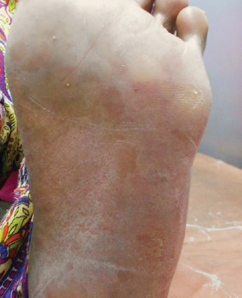
Subsequently, she was planned to receive two cycles of ATO consolidation (0.15 mg/kg/day for 5 days a week for 5 consecutive weeks).[4] On day 15 of the first cycle ATO consolidation, she again developed a painless, mildly pruritic, erythematous plaque, and a few was having necrotic center or dusky erythema on the trunk and face. Palms and soles developed nonblanchable erythematous macule [Figure 2] though oral and genital mucosa was spared. The skin lesions were diagnosed as EM and body surface area involvement approximated 8%. Systemic symptoms were absent. The rash responded to levocetirizine 10 mg and topical betamethasone dipropionate 0.05% w/v twice daily over affected part and subsided within 7 days. ATO was continued due to the favorable benefit: Risk ratio and since only two cycles were planned.
Figure 2.

Nonblanchable erythematous macule on soles
The adverse event report was submitted to pharmacovigilance program of India and bears the worldwide unique number of 2015-11618. The AMC report number is STM-AMC-2528.
Discussion
The recurrence of the rash in spite of nonusage of vancomycin was a cue that some other drug used in the course of therapy might be responsible. The causality assessment of the incidence of EM with use of ATO was done using the Naranjo ADR probability scale and WHO-UMC criteria. The Naranjo scale showed a score of 7, which places the reaction in the “probable” category. According to the WHO-UMC criteria, the case qualifies as “probable” causality category. Thus, the occurrence of EM was attributed to ATO. Severity assessment by the Hartwig's severity assessment scale[5] showed that the severity was “moderate” (Level 3) in nature.
Prophylactic steroids (prednisolone 0.5 mg/kg/day) were used with the subsequent cycle of ATO consolidation. The patient had mild EM skin lesions briefly in second consolidation cycle. At present, she has achieved complete molecular remission after consolidation therapy and was receiving maintenance therapy with ATRA, 6-mercaptopurine, and methotrexate.
EM-induced by drug hypersensitivity involves abnormal or altered metabolism of the responsible drug. Usually, the drugs associated with EM occur in patients who are slow acetylators. Thus, the major metabolism of the drug is carried out by an alternative route, i.e., oxidation by P450 enzymes. This results in the generation of reactive and potentially toxic metabolites. Affected individuals have a defect in detoxifying these reactive metabolites, which then act as hapten by binding to the proteins on epithelial cells covalently. This mounts an immune response leading to a severe skin reaction.[2]
ATO is metabolized by methylation to methylarsonic acid (MAA) and di-MAA.[6] There are pharmacogenetic variations in the activities of enzymes that catalyze S-methylation, O-methylation, N-methylation. These inherited variations are responsible for interindividual differences in the metabolism, effect, and toxicity of drugs that undergo methylation.[7]
EM caused by ATO is rarely reported. There might be a possibility that the genetic variation of methyl conjugation in the individual had triggered the response, and the altered metabolic byproducts acted as a hapten in the subsequent keratinocyte necrosis.
We should bear in mind that early institution of treatment is necessary to prevent the feared Steven–Johnson syndrome in such patients of drug hypersensitivity. Furthermore, one should be cautious in patients who have suffered such an episode earlier as EM tends to recur.
Financial Support and Sponsorship
Nil.
Conflicts of Interest
There are no conflicts of interest.
References
- 1.Ladizinski B, Carter JB, Lee KC, Aaron DM. Diagnosis of herpes simplex virus-induced erythema multiforme confounded by previous infection with Mycoplasma pneumonia. J Drugs Dermatol. 2013;12:707–9. [PubMed] [Google Scholar]
- 2.Plaza JA. Erythema Multiforme. Medscape. [Last accessed on 2016 Feb 01]. Available from: http://www.emedicine.medscape.com/article/1122915-overview .
- 3.Mockenhaupt M, Viboud C, Dunant A, Naldi L, Halevy S, Bouwes Bavinck JN, et al. Stevens-Johnson syndrome and toxic epidermal necrolysis: Assessment of medication risks with emphasis on recently marketed drugs. The EuroSCAR-study. J Invest Dermatol. 2008;128:35–44. doi: 10.1038/sj.jid.5701033. [DOI] [PubMed] [Google Scholar]
- 4.Mathews V, George B, Lakshmi KM, Viswabandya A, Bajel A, Balasubramanian P, et al. Single-agent arsenic trioxide in the treatment of newly diagnosed acute promyelocytic leukemia: Durable remissions with minimal toxicity. Blood. 2006;107:2627–32. doi: 10.1182/blood-2005-08-3532. [DOI] [PubMed] [Google Scholar]
- 5.Hartwig SC, Siegel J, Schneider PJ. Preventability and severity assessment in reporting adverse drug reactions. Am J Hosp Pharm. 1992;49:2229–32. [PubMed] [Google Scholar]
- 6.Yamauchi H, Yamamura Y. Metabolism and excretion of orally administrated arsenic trioxide in the hamster. Toxicology. 1985;34:113–21. doi: 10.1016/0300-483x(85)90161-1. [DOI] [PubMed] [Google Scholar]
- 7.Weinshilboum R. Pharmacogenetics of methylation: Relationship to drug metabolism. Clin Biochem. 1988;21:201–10. doi: 10.1016/s0009-9120(88)80002-x. [DOI] [PubMed] [Google Scholar]


