Abstract
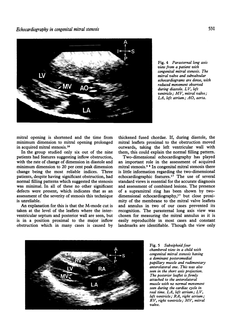
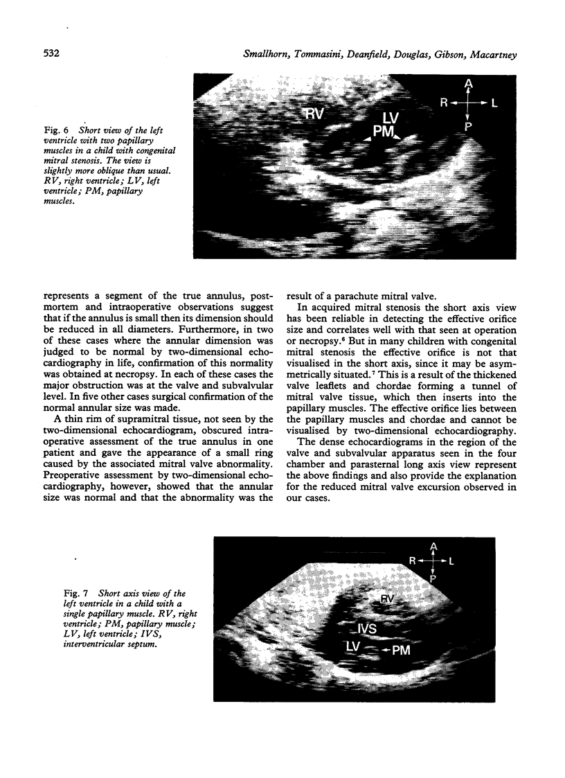
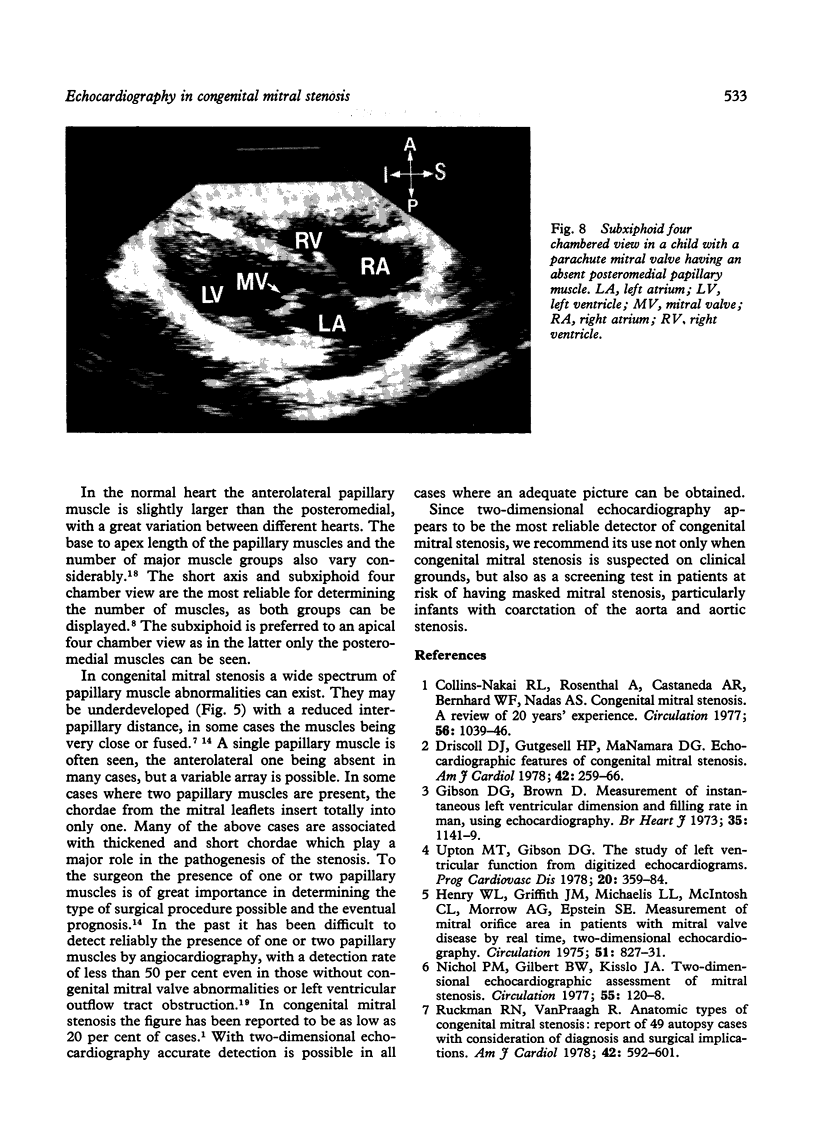
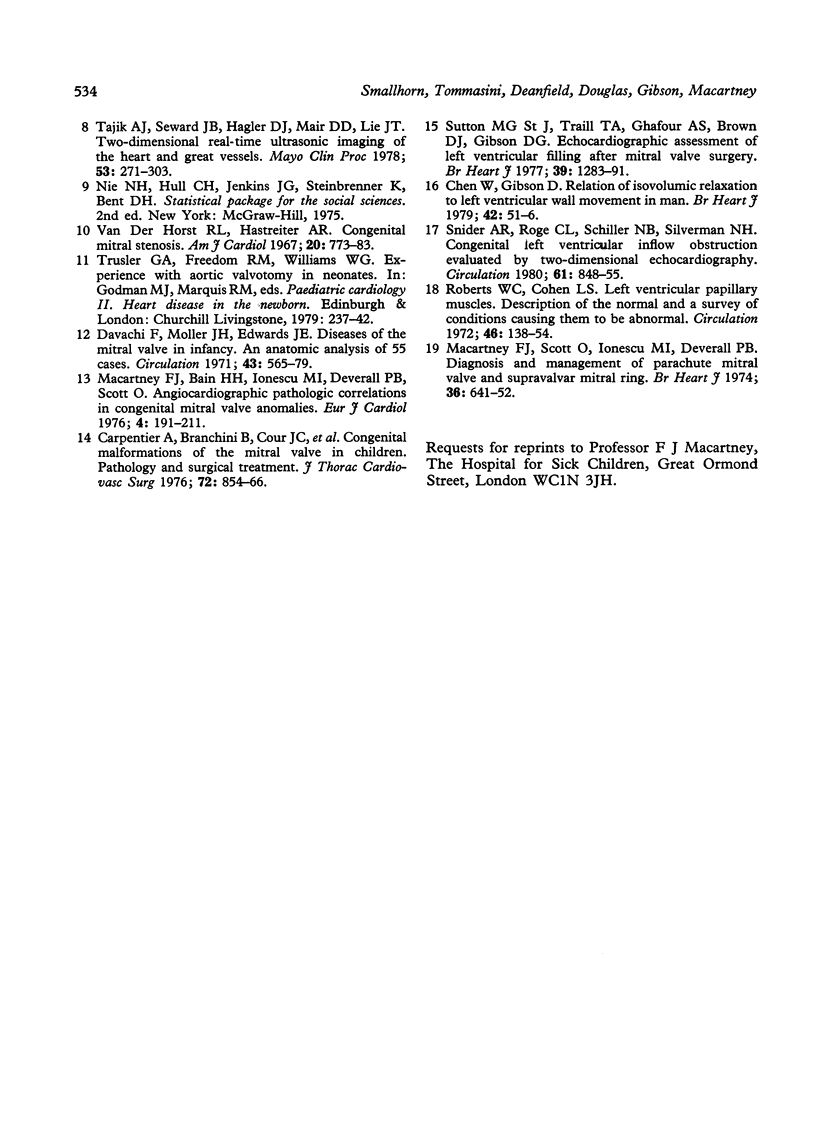
Digitised left ventricular echocardiograms were studied in nine children with congenital mitral stenosis to assess the severity of inflow obstruction. In six children the two prime indices of mitral stenosis were abnormal, with a prolonged time from minimum dimension to 20 per cent dimension change and a reduced peak dimension change during diastole. In three, however, these values did not suggest inflow obstruction, depsite significant gradients at cardiac catheterisation. Two-dimensional echocardiography was performed in 10 children with congenital mitral stenosis to determine the mitral annular size and the morphology of the valve and subvalvular apparatus. The annular size and number of papillary muscles could be assessed along with the detection of combined mitral abnormalities. Two-dimensional studies can reliably delineate the type of mitral abnormality, and should be performed in all cases with congenital heart disease having a high incidence of associated left ventricular inflow obstruction. Digitised M-mode left ventricular echocardiography is in general unreliable in assessing congenital obstruction, though it may be of some value in individual cases.
Full text
PDF
Images in this article
Selected References
These references are in PubMed. This may not be the complete list of references from this article.
- Carpentier A., Branchini B., Cour J. C., Asfaou E., Villani M., Deloche A., Relland J., D'Allaines C., Blondeau P., Piwnica A. Congenital malformations of the mitral valve in children. Pathology and surgical treatment. J Thorac Cardiovasc Surg. 1976 Dec;72(6):854–866. [PubMed] [Google Scholar]
- Chen W., Gibson D. Relation of isovolumic relaxation to left ventricular wall movement in man. Br Heart J. 1979 Jul;42(1):51–56. doi: 10.1136/hrt.42.1.51. [DOI] [PMC free article] [PubMed] [Google Scholar]
- Collins-Nakai R. L., Rosenthal A., Castaneda A. R., Bernhard W. F., Nadas A. S. Congenital mitral stenosis. A review of 20 years' experience. Circulation. 1977 Dec;56(6):1039–1047. doi: 10.1161/01.cir.56.6.1039. [DOI] [PubMed] [Google Scholar]
- Davachi F., Moller J. H., Edwards J. E. Diseases of the mitral valve in infancy. An anatomic analysis of 55 cases. Circulation. 1971 Apr;43(4):565–579. doi: 10.1161/01.cir.43.4.565. [DOI] [PubMed] [Google Scholar]
- Driscoll D. J., Gutgesell H. P., McNamara D. G. Echocardiographic features of congenital mitral stenosis. Am J Cardiol. 1978 Aug;42(2):259–266. doi: 10.1016/0002-9149(78)90908-6. [DOI] [PubMed] [Google Scholar]
- Gibson D. G., Brown D. Measurement of instantaneous left ventricular dimension and filling rate in man, using echocardiography. Br Heart J. 1973 Nov;35(11):1141–1149. doi: 10.1136/hrt.35.11.1141. [DOI] [PMC free article] [PubMed] [Google Scholar]
- Henry W. L., Griffith J. M., Michaelis L. L., McIntosh C. L., Morrow A. G., Epstein S. E. Measurement of mitral orifice area in patients with mitral valve disease by real-time, two-dimensional echocardiography. Circulation. 1975 May;51(5):827–831. doi: 10.1161/01.cir.51.5.827. [DOI] [PubMed] [Google Scholar]
- Macartney F. J., Bain H. H., Ionescu M. I., Deverall P. B., Scott O. Angiocardiographic/pathologic correlations in congenital mitral valve anomalies. Eur J Cardiol. 1976 Jun;4(2):191–211. [PubMed] [Google Scholar]
- Macartney F. J., Scott O., Ionescu M. I., Deverall P. B. Diagnosis and management of parachute mitral valve and supravalvar mitral ring. Br Heart J. 1974 Jul;36(7):641–652. doi: 10.1136/hrt.36.7.641. [DOI] [PMC free article] [PubMed] [Google Scholar]
- Nichol P. M., Gilbert B. W., Kisslo J. A. Two-dimensional echocardiographic assessment of mitral stenosis. Circulation. 1977 Jan;55(1):120–128. doi: 10.1161/01.cir.55.1.120. [DOI] [PubMed] [Google Scholar]
- Roberts W. C., Cohen L. S. Left ventricular papillary muscles. Description of the normal and a survey of conditions causing them to be abnormal. Circulation. 1972 Jul;46(1):138–154. doi: 10.1161/01.cir.46.1.138. [DOI] [PubMed] [Google Scholar]
- Ruckman R. N., Van Praagh R. Anatomic types of congenital mitral stenosis: report of 49 autopsy cases with consideration of diagnosis and surgical implications. Am J Cardiol. 1978 Oct;42(4):592–601. doi: 10.1016/0002-9149(78)90629-x. [DOI] [PubMed] [Google Scholar]
- Snider A. R., Roge C. L., Schiller N. B., Silverman N. H. Congenital left ventricular inflow obstruction evaluated by two-dimensional echocardiography. Circulation. 1980 Apr;61(4):848–855. doi: 10.1161/01.cir.61.4.848. [DOI] [PubMed] [Google Scholar]
- St John Sutton M. G., Traill T. A., Ghafour A. S., Brown D. J., Gibson D. G. Echocardiographic assessment of left ventricular filling after mitral valve surgery. Br Heart J. 1977 Dec;39(12):1283–1291. doi: 10.1136/hrt.39.12.1283. [DOI] [PMC free article] [PubMed] [Google Scholar]
- Tajik A. J., Seward J. B., Hagler D. J., Mair D. D., Lie J. T. Two-dimensional real-time ultrasonic imaging of the heart and great vessels. Technique, image orientation, structure identification, and validation. Mayo Clin Proc. 1978 May;53(5):271–303. [PubMed] [Google Scholar]
- Upton M. T., Gibson D. G. The study of left ventricular function from digitized echocardiograms. Prog Cardiovasc Dis. 1978 Mar-Apr;20(5):359–384. doi: 10.1016/0033-0620(78)90003-8. [DOI] [PubMed] [Google Scholar]
- van der Horst R. L., Hastreiter A. R. Congenital mitral stenosis. Am J Cardiol. 1967 Dec;20(6):773–783. doi: 10.1016/0002-9149(67)90389-x. [DOI] [PubMed] [Google Scholar]


