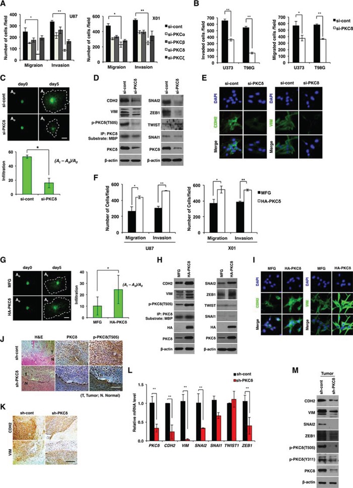
Figure 1. Effect of PKCδ on infiltration of GBM cells through mesenchymal transformation.

A. Migration and invasion assay in GBM cells transfected with control or PKC isoform siRNAs as indicated. B. Migration and invasion assay in GBM cells transfected with control or PKCδ siRNAs. C. Effect of PKCδ depletion on infiltration of U87 GBM cells in collagen-based matrix three-dimensional (3D) culture system. Scale bar, 100 μm. D. Western blot analysis for mesenchymal markers and regulators in U87 GBM cells transfected with control or PKCδ siRNAs. E. Immunocytochemistry for CDH2 and VIM in U87 GBM cells transfected with control or PKCδ siRNAs. F. Migration and invasion assay in GBM cells transduced with MFG or HA-PKCδ. G. Effect of PKCδ on infiltration of U87 GBM cells in collagen-based matrix 3D culture system. Scale bar, 100 μm. H. Western blot analysis for mesenchymal markers and regulators in U87 GBM cells transduced with MFG or HA-PKCδ. I. Immunocytochemistry for CDH2 and VIM in U87 GBM cells transduced with MFG or HA-PKCδ. J, K. Immunohistochemistry for p-PKCδ (J), and CDH2, VIM (K) in orthotopic U87 cell-xenograft tumors. U87 GBM cells were transduced with pSuper or PKCδ shRNA prior to injection to mice. Scale bar, 200 μm. L, M. q-RT PCR (L) and Western blot analysis (M) for mesenchymal markers and regulators in the orthotopic xenograft tumors. β-actin was used for a loading control. *, P < 0.05 versus control; **, p<0.01 versus control.
