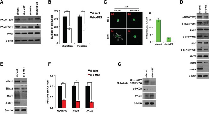
Figure 4. PKCδ is activated by c-MET.

A. Western blot analysis for activation status of PKCδ in U87 GBM cells transfected with control, c-MET, EGFR or EGFRvIII siRNAs. B. Migration and invasion assay in U87 GBM cells transfected with control or c-MET siRNAs. C. Infiltration of X01 GBM cells transfected with control or c-MET siRNAs in collagen-based matrix 3D culture system. Scale bar, 100 μm. D, E. Western blot analysis for activation status of PKCδ, SRC, STAT3 and NOTCH2 (D) or for CDH2, SNAI2 and ZEB1 (E) in U87 GBM cells transfected with control or c-MET siRNAs. F. qRT-PCR for NOTCH-2, JAG1 and -2 in U87 GBM cells transfected by control or c-MET siRNAs. G. Kinase assay of immunoprecipitated c-MET using GSC-PKCδ as a substrate and western blot analysis for p-PKCδ in U87 GBM cells transfected with control or c-MET siRNAs. β-actin was used for a loading control. *, P < 0.05 versus control; **, p<0.01 versus control.
