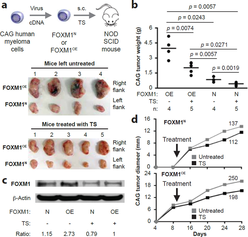
Figure 5. Treatment of NSG mice with thiostrepton inhibits FOXM1OE xenografts more effectively than FOXM1N xenografts.
(a) Scheme of experimental approach (top) and photographic images of myeloma xenografts harvested upon study termination on day 28 (bottom). FOXM1OE and FOXM1N CAG cells were generated using in vitro lentiviral gene transduction, followed by xenografting s.c. into the right and left flank of NSG hosts, respectively. One half of the study group was treated with TS (30 mg/kg IP twice weekly) beginning on day 10 post cell transfer, while the other half was left untreated.
(b) Mean tumor weights (indicated by horizontal lines) in the 4 experimental groups at end of study on day 28 post cell transfer. Tumor weights in TS-treated mice were smaller than in untreated mice (p values of Mann-Whitney tests are indicated) in case of both FOXM1OE and FOXM1N xenografts. The magnitude of TS-dependent tumor reduction was ~8 times higher in OE samples (4.2 g – 2.6 g = 1.6 g) compared to N samples (0.8 g – 0.6 g = 0.2 g).
(c) Representative Western blot of FOXM1 protein levels in FOXM1OE and FOXM1N xenografts collected from TS-treated (“+”) or untreated (“-“) hosts on day 28 post cell transfer. The ratios of FOXM1 to β-actin are indicated below the blot.
(d) Time course of tumor growth in NSG mice treated with TS or left untreated. Mean values (squares) are plotted. Areas under the curve, a metric of tumor growth that ranged from 112 to 250 in 4 experimental groups, are also indicated.

