Abstract
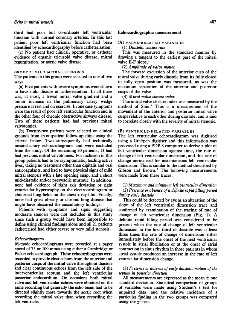
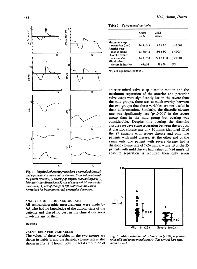
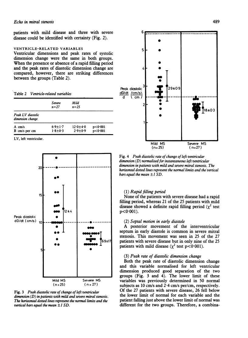
Fifty-two patients with pure mitral stenosis (27 with severe stenosis and 25 with mild stenosis) were studied to assess the ability of different M-mode echocardiographic measurements to separate mild and severe disease. Variables related to valve motion, for example diastolic closure rate, the mitral valve closure index, and the amplitude of valve motion, accurately divided patients with mitral stenosis from normal subjects but did not distinguish usefully between mild and severe disease. In contrast, variables dependent on left ventricular dimension change in diastole, for example the rapid filling period and the peak rate of left ventricular diastolic dimension change, accurately separated mild and severe disease. No patient with severe mitral stenosis had a rapid filling period, whereas 21 of the 25 patients with mild disease did have one. The peak rate of left ventricular diastolic dimension change was less than 10 cm/s or less than 2.4 cm/s per cm when normalised for left ventricular dimension in all patients with severe disease and in only six of the 25 patients with mild disease.
Full text
PDF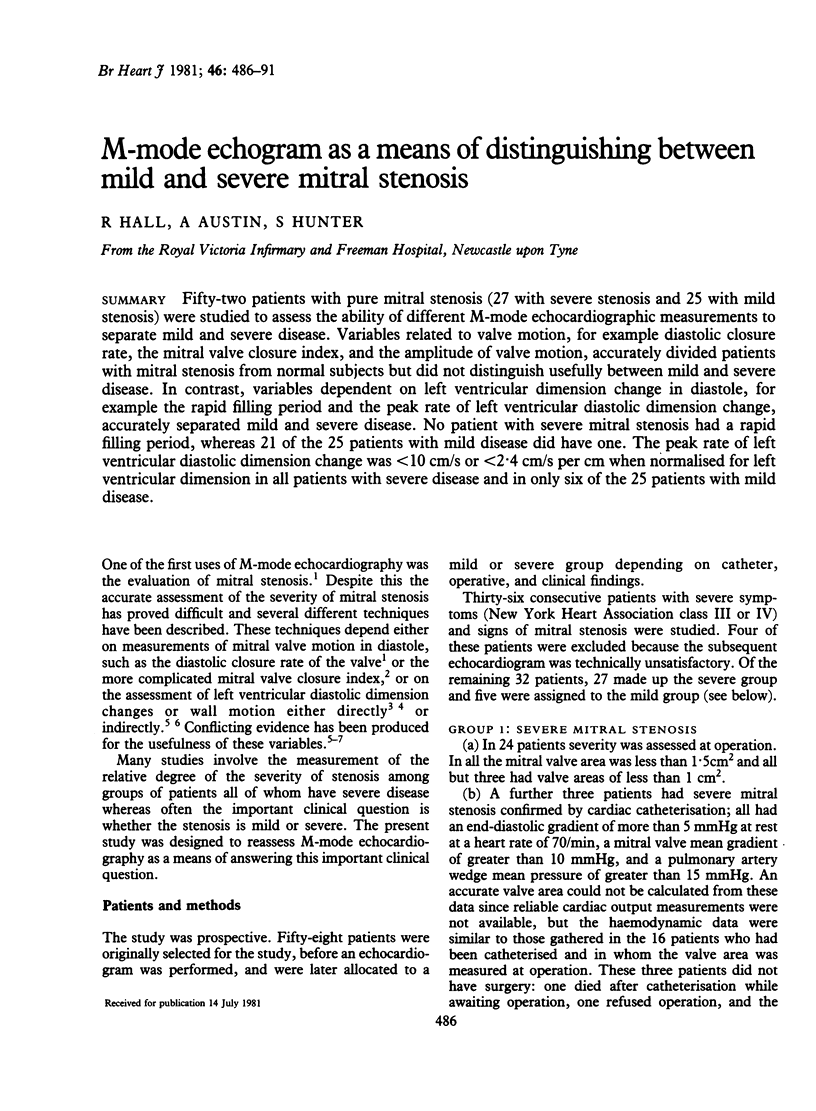
Selected References
These references are in PubMed. This may not be the complete list of references from this article.
- Cope G. D., Kisslo J. A., Johnson M. L., Behar V. S. A reassessment of the echocardiogram in mitral stenosis. Circulation. 1975 Oct;52(4):664–670. doi: 10.1161/01.cir.52.4.664. [DOI] [PubMed] [Google Scholar]
- Edler I. Ultrasoundcardiography in mitral valve stenosis. Am J Cardiol. 1967 Jan;19(1):18–31. doi: 10.1016/0002-9149(67)90258-5. [DOI] [PubMed] [Google Scholar]
- Fukukawa K., Matsuura T., Endo N., Kunishige H., Tohara M., Watanabe T., Matsukubo H., Tsuji Y., Ijichi H. Use of digitised left ventricular echocardiograms in assessment of mitral stenosis. Br Heart J. 1979 Aug;42(2):176–181. doi: 10.1136/hrt.42.2.176. [DOI] [PMC free article] [PubMed] [Google Scholar]
- Gibson D. G., Brown D. Measurement of instantaneous left ventricular dimension and filling rate in man, using echocardiography. Br Heart J. 1973 Nov;35(11):1141–1149. doi: 10.1136/hrt.35.11.1141. [DOI] [PMC free article] [PubMed] [Google Scholar]
- Hall R. J., Clarke S. E., Brown D. Evaluation of posterior aortic wall echogram in diagnosis of mitral valve disease. Br Heart J. 1979 May;41(5):522–528. doi: 10.1136/hrt.41.5.522. [DOI] [PMC free article] [PubMed] [Google Scholar]
- Shiu M. F. Mitral valve closure index. Echocardiographic index of severity of mitral stenosis. Br Heart J. 1977 Aug;39(8):839–843. doi: 10.1136/hrt.39.8.839. [DOI] [PMC free article] [PubMed] [Google Scholar]
- Strunk B. L., London E. J., Fitzgerald J., Popp R. L., Barry W. H. The assessment of mitral stenosis and prosthetic mitral valve obstruction, using the posterior aortic wall echocardiogram. Circulation. 1977 Jun;55(6):885–891. doi: 10.1161/01.cir.55.6.885. [DOI] [PubMed] [Google Scholar]
- Traill T. A., St John Sutton M. G., Gibson D. G. Mitral stenosis with high left ventricular diastolic pressure. Br Heart J. 1979 Apr;41(4):405–411. doi: 10.1136/hrt.41.4.405. [DOI] [PMC free article] [PubMed] [Google Scholar]
- Wise J. R., Jr Echocardiographic evaluation of mitral stenosis using diastolic posterior left ventricular wall motion. Circulation. 1980 May;61(5):1037–1042. doi: 10.1161/01.cir.61.5.1037. [DOI] [PubMed] [Google Scholar]


