Abstract
Background
Stationary equipment devices are often used to improve fitness. The ElliptiGO® was recently developed that blends the elements of an elliptical trainer and bicycle, allowing reciprocal lower limb pedaling in an upright position. However, it is unknown whether the muscle activity used for the ElliptiGO® is similar to walking or cycling. To date, there is no information comparing muscle activity for exercise on the treadmill, stationary upright and recumbent bikes, and the ElliptiGO®.
Purpose/Hypothesis
The purpose of this study was to assess trunk and lower extremity muscle activity among treadmill walking, cycling (recumbent and upright) and the ElliptiGO® cycling. It was hypothesized that the ElliptiGO® and treadmill would elicit similar electromyographic muscle activity responses compared to the stationary bike and recumbent bike during an exercise session.
Study Design
Cohort, repeated measures
Methods
Twelve recreationally active volunteers participated in the study and were assigned a random order of exercise for each of the four devices (ElliptiGO®, stationary upright cycle ergometer, recumbent ergometer, and a treadmill). Two-dimensional video was used to monitor the start and stop of exercise and surface electromyography (SEMG) were used to assess muscle activity during two minutes of cycling or treadmill walking at 40-50% heart rate reserve (HRR). Eight muscles on the dominant limb were used for analysis: gluteus maximus (Gmax), gluteus medius (Gmed), biceps femoris (BF), lateral head of the gastrocnemius (LG), tibialis anterior (TA), rectus femoris (RF). Two trunk muscles were assessed on the same side; lumbar erector spinae at L3-4 level (LES) and rectus abdominus (RA). Maximal voluntary isometric contractions (MVIC) were determined for each muscle and SEMG data were expressed as %MVIC in order to normalize outputs.
Results
The %MVIC for RF during ElliptiGO® cycling was higher than recumbent cycling. The LG muscle activity was highest during upright cycling. The TA was higher during walking compared to recumbent cycling and ElliptiGO® cycling. No differences were found among the the LES and remaining lower limb musculature across devices.
Conclusion
ElliptiGO® cycling was found to elicit sufficient muscle activity to provide a strengthening stimulus for the RF muscle. The LES, RA, Gmax, Gmed, and BF activity were similar across all devices and ranged from low to moderate strength levels of muscle activation. The information gained from this study may assist clinicians in developing low to moderate strengthening exercise protocols when using these four devices.
Level of evidence
3
Keywords: Cycling, electromyography, elliptical, ergometers, lower extremity, muscle activity, treadmill
INTRODUCTION
It is well known that regular physical activity can reduce the risk for cardiovascular disease, Type 2 diabetes, cancer, stroke, obesity, and many other non-communicable diseases.1 In order to improve health and quality of life, it has been recommended that adults should participate in at least 150 minutes per week of moderate-intensity physical activity.2 Running is one of the most popular methods of physical activity which offers many health benefits. However, running is not tolerated by everyone and also has a high incidence of lower extremity injuries.3 There is no one particular cause for running injuries, and are more likely related to several variables such as training intensity, frequency, and distance4 as well as the repetitive impact loading on the joints.5
Health care professionals commonly prescribe stationary cycling or elliptical training as a low-impact alternative to walking or running in order to reduce stress on the hip or knee joints.6,7 These forms of exercise could benefit runners who have lower extremity musculoskeletal injuries since elliptical training has been found to have reduced lower limb loading rates compared to walking.8 Cross training such as a combination of cycling and running has been shown to be an effective way to maintain aerobic capacity for runners.9 White et al10 found that collegiate female distance runners who substituted 50% of their running time for cycling had similar aerobic fitness compared to females who ran 100% of their time during the cross country season.
Cycling or elliptical training has been found to facilitate lower limb coordination or improve reciprocal muscle activity.11-13Researchers have studied the use of cycle ergometry to improve muscle strength among healthy older women,14 people with multiple sclerosis,15 and individuals who were post-stroke.16 These authors found improvements in lower extremity muscle strength, power, and postural control. Macaluso et al reported increased muscle strength and power among older women at 40% of a 2-repetition maximum and at 80% of a 2-repetition maximum using cycling training.14 Elliptical exercise has also been found to result in higher quadriceps and hamstring loading compared to walking as well as lower vertical reaction forces during elliptical cycling compared to walking.8 It appears that cycling and elliptical training are effective exercise modes for muscle strengthening for individuals who prefer these devices instead of running or walking.
Many researchers have analyzed muscle activity during upright cycling,17-22 elliptical cycling,22-24and recumbent cycling,19,21,25,26 however few have compared a combination of these exercise devices18,24and the methodology has varied across studies. Results of research that has assessed muscle activity among various equipment devices have been difficult to compare as methodologies and equipment design vary widely among the literature. For example, the gastrocnemius and gluteus maximus have been found to be less active using a Precor EFX 576i (OH,USA) elliptical trainer24 compared to treadmill walking whereas others who studied five elliptical models (SportsArt Fitness E870, Life Fitness X7, Octane Fitness Pro4500, True Fitness Technology TSXa) reported higher gluteus maximus muscle activity compared to walking beyond over ten meters.22 Burnfield et al22 allowed the participants to self-select a comfortable speed for both treadmill and elliptical training, as compared to the research by Sozen24 who established the exercise intensity at 65% of maximum heart rate which may have resulted in muscle activity differences. An understanding of muscle activity recruitment among exercises devices would enable physical therapists to make better evidence practice decisions for their patients.
Recently, ElliptiGO® (ElliptiGO® Inc., Solana Beach, CA) was developed to resemble a blend of elliptical and running exercise. This device is built on two wheels and can be placed on an indoor trainer or can stand alone and be ridden outdoors. The bike design combines cycling and elliptical pedaling so that the cycling is performed standing up with the lower legs aligned vertically on two platform pedals. The manufacturer contends that the foot platforms are designed to simulate a running-like experience since the platforms are positioned closer together and allow for longer stride length compared to elliptical machines. To the authors' knowledge, no research has been performed comparing muscle activity patterns while using the ElliptiGO® to other modes of exercise such as treadmill walking or cycling. Thus, the purpose of this study was to assess trunk and lower extremity muscle activity among treadmill walking, cycling (recumbent and upright) and the ElliptiGO® cycling. It was hypothesized that the ElliptiGO® and treadmill would be more similar in muscle activity compared to the stationary bike and recumbent bike.
METHODS
Twelve recreationally active, healthy volunteers participated in the study. Recreationally active was defined as someone who participated in recreational activities for at least 30 minutes per day.28 The sample size was determined based on a study with similar independent variables where ten subjects were studied using elliptical training, stationary cycling, treadmill walking and over ground walking.18 Subjects were recruited using a sample of convenience from the university campus and the study involved a single-session research design. Exclusion criteria consisted of anyone diagnosed with musculoskeletal, cardiovascular, vestibular, visual, neurological, or balance disorders, or have a history musculoskeletal injury requiring medical treatment in the past year. The protocol was approved by the Institutional Review Board, project number 607. An informed consent was provided to all subjects explaining the risks and benefits of the study.
Instrumentation
Muscle activity data was collected using telemetry transmitter (8-channel, 12-bit analog-digital converter, Noraxon Myosystem 900 EMG system, Noraxon USA, Inc., Scottsdale, AZ) and a 2-D video recording was obtained during performance of the test movements in order to assist in subsequent analysis. The digital video camera (Canon Optura50, Canon Inc., Lake Success, NY) was placed at the height of the subject's trunk, three meters anterior to the subject for a sagittal plane recording during the exercise testing. A transmitter belt unit powered by a 9V battery was worn to collect surface electromyography (SEMG) signals. The raw SEMG data were sampled at 1000Hz for each of the eight muscles on the dominant limb and converted to a mean amplitude and used for SEMG analyis: gluteus maximus (Gmax), gluteus medius (Gmed), lumbar erector spinae (LES), biceps femoris (BF), lateral head of the gastrocnemius (LG), tibialis anterior (TA), rectus femoris (RF), and rectus abdominus (RA). The amplifier bandwidth frequency ranged from 10Hz highpass to 500 Hz lowpass and common mode rejection=85dB. The raw data was stored in a personal computer and Myoresearch 2.10 software (Noraxon USA, Inc, Scottsdale, AZ) was used to process and analyze the data. The onset of each of the eight muscle contractions during the four exercise modes were marked by when the start of the motion as noted on the video recording and when the muscle SEMG amplitude was 10 µV of baseline. The raw SEMG signals were processed using a full-wave rectification and root-mean-square algorithm at a time constant of 300 milliseconds.
Procedures
Anthropometric measurements were taken, including height and weight using a standard scale, (Detecto, Webb City, MO). Silver-silver chloride snap single surface pre-gelled electrodes (Noraxon USA, Inc., Scottsdale, AZ) were placed in a bipolar configuration on the skin of the dominant leg and torso of the same side of dominant limb. Leg dominance was defined as the preferred limb for kicking a ball.29 The electrodes were positioned parallel to the muscle fiber orientation with an interelectrode distance of approximately 2.0 cm. The skin was prepped by shaving, abrading, and cleaning with isopropyl alcohol prior to electrode placement. The ground lead was placed on the subject's patellar tuberosity contralateral to the subject's dominant limb. Each subsequent lead was positioned on the subject's dominant limb side and parallel to the muscle fibers and described in detail in Table 1.
Table 1.
Electrode placement for the trunk and lower extremity musculature.
| Trunk and Lower Extremity Muscles | Electrode Placement |
|---|---|
| Gluteus Maximus | 3cm inferior and medial to a line drawn between posterior superior iliac spine and posterior greater trochanter 30,31 |
| Anterior portion of Gluteus Medius | 3 cm inferior to the iliac crest30,31 |
| Biceps Femoris | Midway between the ischial tuberosity and the crease of the popliteal fossa30,31 |
| Lateral Gastrocnemius | 2 cm lateral from midline, just distal to knee 30 |
| Tibialis Anterior | ¼ to 1/3 of the distance between knee and ankle, just lateral to shaft of tibia30 |
| Rectus Femoris | Along the longitudinal fibers midway between the anterior superior iliac spine and base of patella30 |
| Upper Rectus Abdominus | 3 cm lateral from midline, and at the midpoint between umbilicus and xiphoid process30 |
| Lumbar Erector Spinae (L3-L4) | 2 cm lateral from spinous process between L3 and L4 in a vertical direction30 |
Table 2.
Means and + standard deviations for electromyographic activity of eight muscles across the four equipment devices.
| ElliptiGO® | Recumbent | Upright | Treadmill | |
|---|---|---|---|---|
| Rectus abdominus | 50.7±26.72 | 41.8±15.33 | 55.0±26.60 | 58.2±20.63 |
| Lumbar ES | 36.3±25.61 | 20.2±17.04 | 36.8±33.13 | 44.7±28.20 |
| Biceps Femoris | 26.8±24.91 | 18.3±13.74 | 32.9±18.34 | 37.4±32.08 |
| Rectus Femoris | 45.8±21.82* | 15.8±6.64* | 30.1±20.00 | 29.9±15.59 |
| Gluteus Maximus | 26.5±17.13 | 15.4±7.66 | 17.9±17.42 | 25.0±18.30 |
| Gluteus Medius | 16.0±7.40 | 17.1±19.81 | 24.4±25.01 | 21.7±13.97 |
| Lateral Gastroc | 47.9±25.72† | 33.3±9.61† 1‡ | 71.2±13.68† | 57.7±23.50‡ |
| Tibialis Anterior | 19.3±10.94§ | 19.3±13.53§ | 30.2±11.12 | 43.3±22.51§ |
Data expressed as percentage of maximum voluntary isometric contraction, (%MVIC).
For rectus femoris muscle, statistically significantly higher %MVIC for ElliptiGO® compared to recumbent cycling, (p=.001).
For lateral gastrocnemius muscle, statistically significantly higher %MVIC for upright cycling compared to recumbent and ElliptiGO® cycling, (p=.03).
For lateral gastrocnemius muscle, statistically significantly higher %MVIC for treadmill walking compared to recumbent cycling, (p=.02).
For tibialis anterior muscle, statistically significantly higher %MVIC for treadmill walking compared to recumbent and ElliptiGO® cycling, (p=.002).
Prior to the start of data collection, subjects participated in a warm-up session consisting of self-selected pace of walking for five minutes. Three maximal voluntary isometric contractions (MVIC) were performed in standard manual muscle test positions for each subject for the eight muscles analyzed. Each test for the MVIC was held for five seconds, followed by a three second rest between contractions, and was performed three times. There was a 30-second rest between muscles tested. The rectus femoris was tested with the subject sitting, and manual resistance applied approximately 40 degrees from full knee extension.32 The subject was positioned in prone for the gluteus maximus, with the knee flexed to 90 degrees and the hip fully extended. Manual resistance was applied on the lower part of the posterior thigh as the hip moved into extension.32 The gluteus medius muscle was assessed in a sidelying position, with the hip in neutral rotation and slightly extended with minimal resistance applied to the distal lower leg as the hip actively moved into abduction. The lumbar erector spinae was tested with the subject in a prone position and trunk off the edge of the table at the level of the anterior superior iliac spine. A second investigator stabilized the lower extremity just above the ankle as the subject extended the lumbar spine to neutral and resistance was applied to the posterior scapulae.33 The biceps femoris was tested in prone position with the knee flexed to 45 degrees and lower leg in external rotation.33 The lateral gastrocnemius was tested in prone position with the foot over the edge of the table and manual resistance was applied to the plantar aspect of the foot.34 The test position for the tibialis anterior was in sitting with knee flexed to 90 degrees and manual resistance applied against the medial and dorsal aspect of the foot.32 The rectus abdominis was tested in supine and hook-lying position. The subject was instructed to place arms across chest and perform a partial curl-up (flexed position) while the investigator applied matched resistance bilaterally shoulders to prevent the motion.35 The average SEMG amplitudes collected during the exercise conditions were later normalized to the highest MIVC value of the three MVIC trials obtained during the manual muscle tests, and expressed as percentage of MIVC, (%MVIC). Following MIVC data collection, participants rested five minutes prior to additional data collection.
The subjects were given verbal instructions and demonstrations for each exercise. Subjects were also given the opportunity to acclimate to the exercise modes by walking and cycling for approximately two minutes prior to data collection. Participants were assigned a random order for each of the four exercise devices; ElliptiGO® Model 3C (ElliptiGO Inc., Solana Beach, CA), stationary upright friction-braked Monark 828E cycle ergometer (Monark Exercise AB, Vansbro, Sweden), recumbent ergometer (Model T4/TRS 4000) (NuStep Inc., Ann Arbor, MI), and a single-belted treadmill (Trackmaster TMX58, Full Vision Inc., Newton, KS).
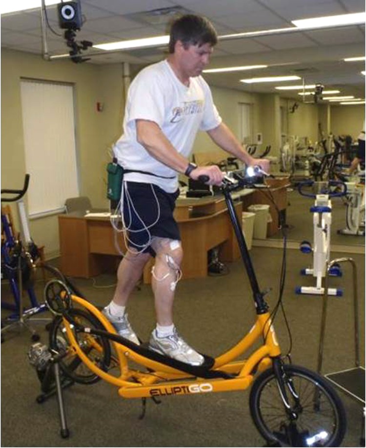
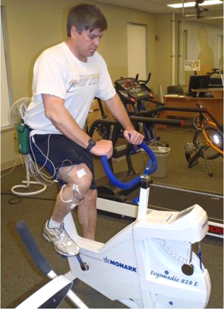
The rear wheel of the ElliptiGO® was placed on a stationary trainer. The subjects were instructed to maintain an upright posture and both feet in contact with the pedals during cycling (Figure 1). The feet were positioned in the center of the pedal with each of the subject's legs perpendicular to the ground. The Monark cycle ergometer seat height was determined by measuring 95% of the distance from the right greater trochanter to the floor with the subject in standing position.36 The handlebars were adjusted for comfort and then remained constant throughout the cycling bout. The participant was instructed to remain seated during the test. The seat and distance from the pedal for each participant was set so that the knees were slightly bent when the legs were maximally extended. Handlebars were adjusted by placing the arm in neutral alignment allowing approximately 60 degrees of elbow flexion. The forefoot was placed on the pedal without straps or clips. Subjects were instructed to remain on their seat and have their hands in contact with handlebars during the cycling bout. (Figure 2)
Figure 1.
ElliptiGO® on stationary trainer at 40% to 50% of heart rate reserve.
Figure 2.
Stationary upright biking at 40% to 50% of heart rate reserve.
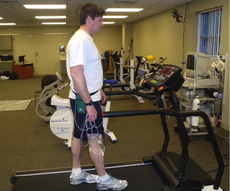
Subjects were asked to walk on the treadmill using a self-selected gait speed. The treadmill was set at 0° incline. They were also instructed to maintain free arm swing without holding onto the rails. (Figure 3)
Figure 3.
Treadmill walking at 40% to 50% heart rate reserve.
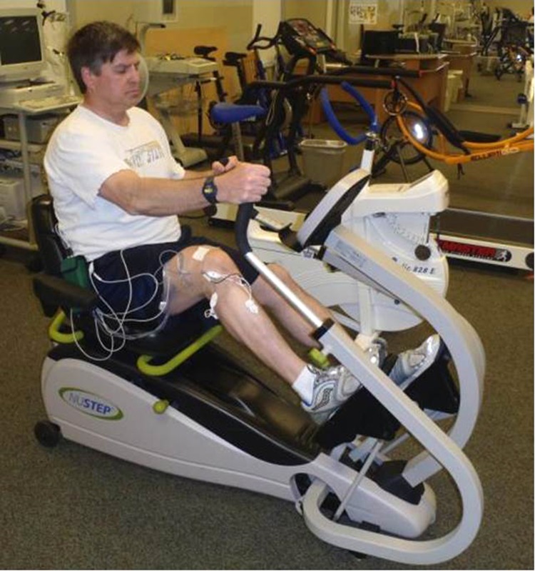
Each subject was fitted to the recumbent bike using a goniometer so that the amount of knee flexion was approximately 15° to 20° of knee flexion during the upstroke and 0° of knee flexion during the down stroke. Subjects were instructed to hold onto handlebars and feet in pedals throughout the cycling duration. (Figure 4)
Figure 4.
Recumbent cycling at 40% to 50% of heart rate reserve.
The exercise duration and intensity were consistent for each of the four exercise devices. Exercise duration was five minutes, (two minutes of acclimation, two minutes of data collection at 40-50% heart rate reserve [HRR], and one minute of cool down). The exercise intensity was determined by calculating maximal heart rate using the Karvonen formula (220-age) and then determining 40-50% of estimated HRR using maximal heart rate.37 Heart rate was recorded using a chest heart rate monitor (Polar heart rate, Polar Electro Inc., Lake Success, NY) during each exercise mode. Subjects were also asked to rate the perceived exertion (RPE) at the completion of each exercise trial. The subjects were given verbal and written instructions on Borg's 10-point RPE scale before each trial.38 A copy of the RPE scale was kept in full view of the subjects during each trial.
The upright cycle ergometer cadence was set at 60 revolutions per minute. The subject was able to monitor the cadence by the digital monitor display on the ergometer. In order to maintain the appropriate exercise intensity for the upright and recumbent bikes, resistance was either added or removed by manipulating the dial tension. ElliptiGO® cycling intensity was changed by either increasing or decreasing the rate of pedal motion. The treadmill speed was adjusted by the investigator from the treadmill control panel throughout the walking bout based on the participant's heart rate response.
Statistical Analysis
Descriptive statistics are reported as means + standard deviations for demographic data and muscle activity. Shapiro-Wilk's W-test was applied to examine normality in the distribution of data. The singular peak maximal voluntary isometric contractions (MVIC) generated from each of the eight manual muscle tests were used to normalize the SEMG amplitudes for each muscle and expressed as %MIVC. Descriptive statistics and ANOVAs were calculated using SPSS version 21.0 (SPSS, Inc, Chicago, IL) software. The normalized SEMG values were analyzed using separate one-way analyses of variance. Post-hoc comparisons of the means of interest were conducted using the Bonferroni procedure and set at p < 0.05.
RESULTS
Twelve participants (six males, six females) with a mean age of 32.42 + 8.3 years, height 168.75 + 7.0cm, and mass 77.65 + 14.4kg were involved in the study. The means and standard deviations of the SEMG results across the eight muscles are presented in Table 1. The mean SEMG data for the lumbar erector spinae (LES) muscles ranged from 20%MVIC for the recumbent cycling to 44%MVIC on the treadmill. There were no main effect differences among the equipment devices for muscle activity for RA (p=.331), lumbar (p=.164), Gmax (p=.255), Gmed (p=.623) or BF (p=.227). Mean SEMG RA muscle activity ranged from 41%MVIC during recumbent cycling to 58%MVIC on the treadmill. Gluteus maximus and medius SEMG mean activity ranged within 15%MVIC to 26%MVIC across the four exercise devices. Biceps femoris average SEMG activity ranged from 18%MVIC during recumbent cycling to 37%MVIC with treadmill walking.
The %MVIC for RF during ElliptiGO® cycling was significantly higher (46% MVIC) compared to recumbent cycling, (16% MVIC), (p=.001). The LG was highest during upright cycling (71% MVIC) and significantly higher than recumbent cycling (p=.0001) and ElliptiGO® cycling (p=.03). Treadmill walking (58% MVIC) also resulted in higher LG activity compared to recumbent cycling, 33% MVIC (p=.02). The greatest tibialis anterior activity was found during treadmill walking (43% MVIC) compared to recumbent cycling (19% MVIC)(p=.002) and ElliptiGO®(19% MVIC)(P=.002).
DISCUSSION
The purpose of the current study was to compare trunk and lower extremity muscle activity during the use of four stationary exercise devices. The results partially supported the hypothesis that the treadmill and ElliptiGO® would elicit similar muscle activity compared to the recumbent and upright stationary cycling. As hypothesized, the lateral gastrocnemius activity was not different between treadmill walking and ElliptiGO® cycling, however, the lateral gastrocnemius muscle activity was also similar during upright stationary cycling. Comparable SEMG activity was found among all four devices for lumbar erector spinae, biceps femoris, rectus abdominus, gluteus medius and gluteus maximus muscles. Contrary to the stated hypothesis, the treadmill elicited higher tibialis anterior muscle activity compared to the ElliptiGO®.
The stationary upright, recumbent, or elliptical devices vary in sitting and pedaling positions, which may explain some of the differences seen in SEMG outputs. For example, these exercise modes differ with the seat-to-crank set frame alignment, which alters trunk alignment and amount of bodyweight supported by limbs. The standard bike seat is positioned above the crank set compared to the recumbent bike which has the seat at the same level and in line with the crank set. The seat also allows the back of recumbent cyclist to be supported and reclined back compared to the standard bike in which the cyclist must lean more forward. Despite these differences in frame and saddle heights, the trunk muscles (rectus abdominus and lumbar erector spinae), hip muscles (biceps femoris, gluteus maximus, gluteus medius) elicited similar muscle activity across the four equipment devices examined in the current study. Other researchers have also found that upper limb and lower limb muscle activity to be highly correlated between the recumbent bike and BWS treadmill walking when walking speed and cycling were matched for similar exercise intensity.25
Trunk Muscles
The rectus abdominus muscle mean SEMG value among the four equipment devices was 51%MVIC. This muscle activity value falls within the activation level needed for strengthening the abdominal muscles.39,40 While the rectus abdominus activity was not assessed in a study by Anderson et al, these authors reported 40% to 60% is an adequate range for muscle strengthening.41 The only exercise that resulted in sufficient muscle activity for lumbar erector spinae strengthening was treadmill walking, (45% MVIC). In contrast, if a low exercise intensity is the focus during the early phase of rehabilitation for lumbar erector spinae muscles, then the ElliptiGO®, upright and recumbent bikes would be appropriate since the muscle activity values fell below the strengthening intensity level.
Hip Muscles
The results of the current study indicate that the ElliptiGO® cycling produced 46% MVIC activity for the rectus femoris muscle which was significantly higher than recumbent biking, (16%MVIC). The higher rectus femoris activity may be because of greater lower extremity weight bearing since the subject is required to perform cycling motion in a standing position on the ElliptiGO® compared to cycling in a recumbent sitting position. In addition, 46%MVIC has been found to be a sufficient level to elicit neuromuscular adaptation needed for strengthening.41 This may be advantageous for runners to maintain the hip flexor and knee extensor muscles using a simulated running gait pattern via the ElliptiGO®. Since elliptical cycling has been found to elicit smaller joint forces compared to walking,8 we can only speculate that limb loading may also be reduced using the ElliptiGO® since it offers a similar pedal motion as the elliptical device.
Treadmill walking and upright cycling both elicited 30%MVIC for the rectus femoris muscle. While treadmill walking requires more weight bearing load compared to upright cycling, there is no foot resistance while waking to advance the limb forward, which may explain the lower muscle activity. In contrast, ElliptiGO® cycling involves overcoming pedal resistance in order to propel the lower limb forward via hip flexion and knee extension motions resulting in higher rectus femoris activity. Lopes et al reported peak mean SEMG values when comparing upright and recumbent cycling at 80rpm and a work rate of 100W.27 These authors found upright and recumbent cycling elicited 23% MVIC and 20% MVIC RF activity, respectively. The current study found a slightly higher value (30%MVIC) for the upright bike and slightly lower EMG activity (16%MVIC) for the recumbent cycle.
The gluteus maximus and gluteus medius muscles elicited values below the strengthening stimulus across all four exercise devices. While exercise devices produced low %MVIC levels, these muscles have been found to serve an important role during walking and cycling. The gluteus medius muscle has been found to be a pelvis stabilizer by limiting the amount of forward rotation during walking.42 During cycling, the gluteus maximus and rectus femoris work together as agonist and antagonist to coordinate muscle activity at the hip.43 Other muscle patterns such as the biceps femoris and plantarflexors (gastrocnemius and soleus) have been found to be synchronized together at the end of the extension phase of cycling and during the extension-to-flexion transition phase.44 Despite these coordinated muscle patterns, the biceps femoris was also found to be below the level for strengthening stimulus for all four exercise devices. Lopes et al also found similar muscle activity values for the semitendinosus muscle during upright and recumbent cycling.27
Lower limb Muscles
The lateral gastrocnemius muscle ranged from 33% MVIC to 71% MVIC. The treadmill elicited 58% MVIC and the upright bike was 71% MVIC which indicates that these exercise modes could be used for sufficient stimulus for calf strengthening. Upright cycling produced the highest lateral gastrocnemius activity, particularly compared to ElliptiGO® and recumbent biking. The likely explanation for the differences between the current study and Sozen24 is that different exercise intensity and devices were used. Sozen24 used the Precor EFX 576i, (OH, USA) elliptical at a 65%maximum heart rate intensity compared to the present study which selected ElliptiGO® cycling at 40-50%maximum heart rate intensity. The high SEMG value may also be explained by the position of the ankle. During this study, the participant's forefoot was placed on the pedal, which allowed for ankle motion. Upright cycling has been found to influence lateral gastrocnemius activity when the ankle is in a dorsiflexed position.45 Cannon et al reported that the lateral gastrocnemius activity increases during dorsiflexion in order to generate knee flexion.45 The gastrocnemius has been found to function as a knee flexor during cycling.19 While the current study did not monitor the amount of ankle motion occurring during the pedal stroke, the participant's ankle was able to move through dorsiflexion and plantarflexion as the pedal moved in the upstroke and down stroke patterns. Lopes found higher medial gastrocnemius activity (34%) for both upright and recumbent cycling compared to rectus femoris, tibialis anterior, and semitendinosus.27 The medial gastrocnemius muscle has been found to be active during pedaling motion.27,46 The current study did not assess medial gastrocnemius activity, however, did find that the lateral gastrocnemius was most active during upright cycling and less active during recumbent cycling. During upright cycling, the plantarflexors have been found to be active during a portion of the upstroke as well as throughout the entire downstroke phase of cycling compared to the dorsiflexor activity only during the initial phase of the upstroke47 which may also explain higher gastrocnemius muscle compared to tibialis anterior activity reported in the present study.
Overall, the tibialis anterior elicited low strengthening values, (19% MVIC and 30% MVIC) during recumbent and upright biking respectively which was similar to values reported during recumbent biking in a separate study.27 The ElliptiGO® involves having the participant stand with the feet placed onto pedal platforms. This type of setup limits the amount of ankle dorsiflexion and plantarflexion motions because the feet do not leave the platform. The cycling motion is also different in that the feet move in an elliptical pattern compared to a circular pattern used by cyclists when on the upright or recumbent ergometers. Treadmill walking involves dorsiflexion and plantarflexion of the ankle whereas the other exercise modes have the feet fixed on platforms, which limit ankle motion. This may explain why the tibialis anterior and lateral gastrocnemius SEMG values were higher during treadmill walking compared to ElliptiGO® and recumbent cycling in which the feet were fixed to platform pedals.
Limitations and Suggestions for Future Research
A significant limitation to the current study was the use of stationary setup for the ElliptiGO® limiting its functional utility as a mobile device. Likewise, upright and recumbent bicycling conditions were stationary and walking was performed on a treadmill. This makes the muscle activity gathered from surface electromyography difficult to compare to the ElliptiGO® while moving on the road, cycling on the road, or over ground running. Future studies should include over ground running and road cycling to assess differences in muscle activity because of decreased stability as compared to stationary trainers or treadmills. While the exercise intensity was maintained between 40% and 50% HRR during use of each of the four exercise devices, the upright bike was the only device that displayed a digital workload and cadence. Thus, while the exercise intensity was based on heart rate, there may have been speed discrepancies during the two minute data collection capture among the exercise devices. Kinematic information was also not assessed to determine actual hip, knee, and ankle joint angles. It is unknown how these exercise devices may affect other populations such as unfit or individuals with musculoskeletal injuries since the subjects recruited were recreationally active, healthy, and free of musculoskeletal injuries in the past year. In addition, the muscles chosen were superficial and limited to eight because of the electromyography system. Cross-talk with the use of SEMG electrodes from adjacent muscles is always a concern in any SEMG study, even with most rigorous methods and electrode placement. It has been suggested that using a standardized method for SEMG placement improves the recordings at each of the muscle sites.48 The current investigation used standardized electrode placement for each of the muscles assessed in order to decrease potential for cross-talk.30,48
CONCLUSIONS
The ElliptiGO® cycling condition elicited sufficient SEMG activity for a stimulus for strengthening of the rectus femoris muscle, which may serve as an alternative means for muscle training without the joint loading incurred with walking or jogging. While joint loading forces were not assessed in this study, others have found that ergometer cycling49 and elliptical training8 result in smaller lower limb joint loads compared to walking. Many of the other muscles such as lumbar erector spinae, rectus abdominus gluteus maximus, gluteus medius, and biceps femoris were similar in muscle activity across the four exercise devices, providing low to moderate strengthening stimuli. Comparable trunk, gluteal, and hamstring muscle SEMG activities suggest that cross training using the equipment devices at 40% to 50% HRR may be a substitute for low-impact exercise. This was the first study to examine muscle activity while using the ElliptiGO®, recumbent and upright bikes, and treadmill walking. The muscle activity information gained from this study may assist clinicians in developing low to moderate strengthening stimuli for various muscles when using the stationary upright and recumbent bikes, treadmill, or the ElliptiGO®.
REFERENCES
- 1.Reiner M Niermann C Jekauc D Woll A. Long-term health benefits of physical activity-a systematic review of longitudinal studies. BMC Public Health 2013;13:813-822. [DOI] [PMC free article] [PubMed] [Google Scholar]
- 2.Garber CE Blissmer B Deschenes MR Franklin BA Lamonte MJ Lee IM Nieman DC Swain DP. American College of Sports Medicine position stand. Quantity and quality of exercise for developing and maintaining cardiorespiratory, musculoskeletal, and neuromotor fitness in apparently healthy adults: guidance for prescribing exercise. Med Sci Sports Exerc. 2011;43:1334-1359. [DOI] [PubMed] [Google Scholar]
- 3.Van Gent RN Siem D van Middelkoop M van Os AG Bierma-Zeinstra SM Koes BW. Incidence and determinants of lower extremity running injuries in long distance runners: a systematic review. Br J Sports Med. 2007;41:469-480. [DOI] [PMC free article] [PubMed] [Google Scholar]
- 4.Yeung EW Yeung SS. A systematic review of interventions to prevent lower limb soft tissue running injuries. Br J Sports Med. 2001;35:383-389. [DOI] [PMC free article] [PubMed] [Google Scholar]
- 5.Hreljac A Marshall R Hume P. Evaluation of lower extremity overuse injury potential in runners. Med Sci Sports Exerc. 2000;32:1635-1641. [DOI] [PubMed] [Google Scholar]
- 6.Johnston TE. Biomechanical considerations for cycling interventions in rehabilitation. Phys Ther. 2007;87:9; 1243-1252. [DOI] [PubMed] [Google Scholar]
- 7.McLeod WD Blackburn TA. Biomechanics of knee rehabilitation with cycling. Am J Sports Med. 1980;8; 175-180. [DOI] [PubMed] [Google Scholar]
- 8.Lu T Chien H Chen H. Joint loading in the lower extremities during Elliptical exercise. Med Sci Sports Exerc. 2007;39:1651-1658. [DOI] [PubMed] [Google Scholar]
- 9.Bonacci J Vleck V Saunders P Blanch P Vicenzini B. Ratings of perceived exertion during cycling is associated with subsequent running economy in triathletes. J Sci and Med in Sport 2012;16:49-53. [DOI] [PubMed] [Google Scholar]
- 10.White LJ Dressendorfer RH Muller SM Ferguson MA. Effectiveness of cycle cross-training between competitive seasons in female distance runners. J Strength and Cond. 2003;2:319-323. [DOI] [PubMed] [Google Scholar]
- 11.Damiano DL Norman T Stanley C Soon Park H. Comparison of elliptical training, stationary cycling, treadmill walking, and overground walking. Gait Posture 2011;34:260-264. [DOI] [PMC free article] [PubMed] [Google Scholar]
- 12.Fujiwara T Liu M Chino N. Effect of pedaling exercise on the hemiplegic lower limb. Am J Phys Med Rehabil. 2003;82:357-363. [DOI] [PubMed] [Google Scholar]
- 13.Hornby TG Kinnaird CR Holleran C Rafferty MR Rodriguez KS Cain JB. Kinematic, Muscular, and Metabolic Responses During Exoskeletal-, Elliptical-, or Therapist-Assisted Stepping in People With Incomplete Spinal Cord. Phys Ther. 2012;62:1278-1291. [DOI] [PMC free article] [PubMed] [Google Scholar]
- 14.Macaluso A Young A Gibb K Rowe D de Vito G. Cycling as a novel approach to resistance training increases muscle strength, power, and selected functional abilities in healthy older women. J Appl Physiol. 2003;95:2544-2553. [DOI] [PubMed] [Google Scholar]
- 15.Cakit BD Nacir B Genc H Saracoglu M Karagoz A Erdern U. Cycling progressive resistance training for people with multiple sclerosis. Am J Phys Med Rehabil. 2010;89:446-457. [DOI] [PubMed] [Google Scholar]
- 16.Brown DA Kautz SA. Increased workload enhances power output during pedaling exercise in persons with poststroke hemiplegia. Stroke 1998;29:598-606. [DOI] [PubMed] [Google Scholar]
- 17.Eisner WD Bode SD Nyland J Caborn DN. Electromyographic timing analysis of forward and backward cycling. Med Sci Sports Exerc. 1999;33:449-455. [DOI] [PubMed] [Google Scholar]
- 18.Prosser LA Stanley CJ Norman TL Park HS Damiano D. Comparison of elliptical training, stationary cycling, treadmill walking, and over ground walking. Gait Posture 2011;33:244-250. [DOI] [PMC free article] [PubMed] [Google Scholar]
- 19.Gregor SM Perell KL Rushatakankovit S Miyamoto E Muffolettto R Gregor RJ. Lower extremity general muscle moment patterns in healthy individuals during recumbent cycling. Clin Biomech (Bristol, Avon). 2002;17:123-129. [DOI] [PubMed] [Google Scholar]
- 20.Ericson MO. Muscular function during ergometer cycling. Scand J Rehab Med 1988;20:35-43. [PubMed] [Google Scholar]
- 21.Hakansson NA Hull ML. Functional roles of the leg muscles when pedaling in the recumbent versus upright position. J Biomech Eng. 2005;127:301-310. [DOI] [PubMed] [Google Scholar]
- 22.Hamzaid NA Smith RM Davis GM. Isokinetic cycling and elliptical stepping: a kinematic and muscle activation analysis. Clin Res Foot Ankle 2013;1:1-6. [Google Scholar]
- 23.Burnfield JM Shu Y Buster T Taylor A. Similarity of joint kinematics and muscle demands between elliptical training and walking: implications for practice. Phys Ther. 2010;90:289-305. [DOI] [PubMed] [Google Scholar]
- 24.Sozen H. Comparison of muscle activation during elliptical trainer, treadmill, and bike exercise. Biol. Sport 2010;27:203-206. [Google Scholar]
- 25.Stoloff R Zehr EP Ferris D. Recumbent stepping has similar but simpler neural control compared to walking. Exp Brain Res. 2007;178(4):427-438. [DOI] [PubMed] [Google Scholar]
- 26.Momeni K Faghri PD Evans M. Lower-extremity joint kinematics and muscle activations during semi-reclined cycling at different workloads in healthy individuals. J Neuro Engineering 2014;11:1-15. [DOI] [PMC free article] [PubMed] [Google Scholar]
- 27.Lopes AD Alouche SR Hakansson N Cohen M. Electromyography during pedaling on upright and recumbent ergometer. Inter J Sports Phys Ther. 2014;9:76-81. [PMC free article] [PubMed] [Google Scholar]
- 28.U.S. Department of Health and Human Services Web Site [Internet]. 2008 Physical Activity Guidelines for Americans. Atlanta (GA): USDHHS;[cited April 29 2015]Available from: http//www.health.gov/paguidelines.
- 29.Scheiders AG Sullivan J O'Malley KJ Clarke SV Knappstein SA Taylor LJ. A Valid and Reliable Clinical Determination of Footedness. PM R. 2010; 2: 835-841. [DOI] [PubMed] [Google Scholar]
- 30.Cram JR Kasman GS Holtz J. Introduction to surface electromyography. Gaithersburg, MD: Aspen, 1998. [Google Scholar]
- 31.Rainoldi A Melchiorri G Caruso I. A method for positioning electrodes during surface EMG recordings in lower limb muscles. J Neuroscience Methods 2004;134:370-43. [DOI] [PubMed] [Google Scholar]
- 32.Kendall PF McCreary EF Provance PG Rodgers MM Romani WA. Muscles: Testing and Function with Posture and Pain. 5th ed. Philadelphia, PA: Lippincott Williams & Williams; 2005:1-480. [Google Scholar]
- 33.Hislop HJ Montgomery J. Daniels and Worthingham's Muscle Testing: Techniques of Manual Examination. 8th ed. Philadelphia, PA: Elsevier/Saunders;2007 [Google Scholar]
- 34.Palmer ML Epler ME. Fundamentals of Muscular Assessment Techniques. 2nd ed. Philadelphia, PA: Lippincott, Williams, Wilkins, 1998. [Google Scholar]
- 35.Juker D McGill S Kropf P Steffen T. Quantitative intramuscular myoelectric activity of lumbar portions of psoas and the abdominal wall during a wide variety of tasks. Med Sci Sports Exerc. 1998;30:301–310. [DOI] [PubMed] [Google Scholar]
- 36.Jorge M Hull ML. Analysis of EMG measurements during bicycle pedaling. J Biomech 1986;19:683-694. [DOI] [PubMed] [Google Scholar]
- 37.Karvonen MJ Kentala E Mustala O. The effects of training on heart rate; a longitudinal study. Ann Med Exp Biol Fenn. 1957;35:307-315. [PubMed] [Google Scholar]
- 38.Borg GAV. An introduction to Borg's RPE-scale. NY Movement Publication, 1985. [Google Scholar]
- 39.Ekstrom RA Donatelli RA Carp KC. Electromyographic analysis of core trunk, hip, and thigh muscles during 9 rehabilitation exercises. J Orthop Sports Phys Ther. 2007;37:754-762. [DOI] [PubMed] [Google Scholar]
- 40.Escamilla RF Lewis C Bell D, et al. Core muscle activation during Swiss ball and traditional abdominal exercises. J Orthop Sports Phys Ther. 2010;40:265-276. [DOI] [PubMed] [Google Scholar]
- 41.Anderson LL Magnusson SP Nielsen M Haleem J Poulsen K Aagaard P. Neuromuscular activation in conventional therapeutic exercises and heavy resistance exercises: implications for rehabilitation. Phys Ther 2006;86:683-697. [PubMed] [Google Scholar]
- 42.Neumann D. Kinesiology of the hip: a focus on muscular activity. J Orthop Sports Phys Ther. 2010;40:82-94. [DOI] [PubMed] [Google Scholar]
- 43.So R Ng J Ng G. Muscle recruitment patterns in cycling: a review. Phys Ther in Sport 2005;6:89-96. [Google Scholar]
- 44.Raasch C ZajacF Ma B Levine S. Muscle coordination of maximum-speed pedaling. J of Biomech. 1997;30:595-602. [DOI] [PubMed] [Google Scholar]
- 45.Cannon DT Kolkhorst FW Cipriani D. Effect of pedaling technique on muscle activity and cycling efficiency. Eur J Appl Physiol. 2007;99:659-664. [DOI] [PubMed] [Google Scholar]
- 46.Ericson MO Nisell R Arborelius UP Ekholm J. Muscular activity during ergometer cycling. Scand J Rehabil Med. 1985;17:53-61. [PubMed] [Google Scholar]
- 47.Barroso F Torricelli D Moreno JC Taylor J Gómez-Soriano J Esteban EB Santos C Pons JL. Similarity of muscle synergies in human walking and cycling: preliminary results. Conf Proc IEEE Eng Med Biol Soc 2013:6933–6936. [DOI] [PubMed] [Google Scholar]
- 48.Criswell E. Cram's Introduction to Surface Electromyography. 2nd ed. Sudberry, MA: Jones and Bartlett, 2011. [Google Scholar]
- 49.Kutzner I Heinlein B Friedmar G Rohlmann A Halder AM Beier A Bergmann G. Loading of the knee joint during ergometer cycling: a telemetric in vivo data. J Orthop Sports Phys Ther. 2012;42:1032-1038. [DOI] [PubMed] [Google Scholar]


