Abstract
Simultaneous imaging systems combining positron emission tomography (PET) and magnetic resonance imaging (MRI) have been actively investigated. A PET/MR imaging system (GE Healthcare) comprised of a time-of-flight (TOF) PET system utilizing silicon photomultipliers (SiPMs) and 3-tesla (3T) MRI was recently installed at our institution. The small-ring (60 cm diameter) TOF PET subsystem of this PET/MRI system can generate images with higher spatial resolution compared with conventional PET systems. We have examined theoretically and experimentally the effect of uniform magnetic fields on the spatial resolution for high-energy positron emitters. Positron emitters including 18F, 124I, and 68Ga were simulated in water using the Geant4 Monte Carlo toolkit in the presence of a uniform magnetic field (0, 3, and 7 Tesla). The positron annihilation position was tracked to determine the 3D spatial distribution of the 511-keV gammy ray emission. The full-width at tenth maximum (FWTM) of the positron point spread function (PSF) was determined. Experimentally, 18F and 68Ga line source phantoms in air and water were imaged with an investigational PET/MRI system and a PET/CT system to investigate the effect of magnetic field on the spatial resolution of PET. The full-width half maximum (FWHM) of the line spread function (LSF) from the line source was determined as the system spatial resolution. Simulations and experimental results show that the in-plane spatial resolution was slightly improved at field strength as low as 3 Tesla, especially when resolving signal from high-energy positron emitters in the air-tissue boundary.
I. Introduction
An integrated system of positron emission tomography (PET) and magnetic resonance imaging (MRI) for simultaneous acquisition has recently gained momentum in its development in research and industry. One manufacturer already has introduced a commercial integrated PET/MRI system for whole-body imaging (Siemens Healthcare) with a 3-T MRI system and avalanche photodiodes (APDs)-based PET system [1]. An integrated time-of-flight (TOF) PET/MR system (GE Healthcare) is a new development, which is currently under investigation for its clinical utility, and has been available at our institution for research. This system is comprised of state-of-the-art PET photodetector technology using silicon photomultipliers (SiPMs) that enable time-of-flight capability within a strong magnetic field. Therefore, it is important to examine the effect of magnetic field on PET imaging performance, particularly its spatial resolution. Previous studies [2–4] reported that the magnetic field reduced the positron annihilation spread in the plane perpendicular to that of the magnetic field, especially for radionuclides with high-energy positron emission. In this study, the effect of magnetic field on positron annihilation spread was investigated using Geant4 Monte Carlo simulations and experiments with the state-of-art TOF-PET/MRI system and a PET/CT system. Although the presence of a magnetic field can improve the in-plane imaging spatial resolution, it is crucial to consider this effect on positron range in all directions when evaluating imaging performance of an integrated PET/MRI system.
II. Methods and Materials
All simulation and image analysis was performed using Python (version 2.7.8).
A. GEANT4 Monte Carlo Simulations
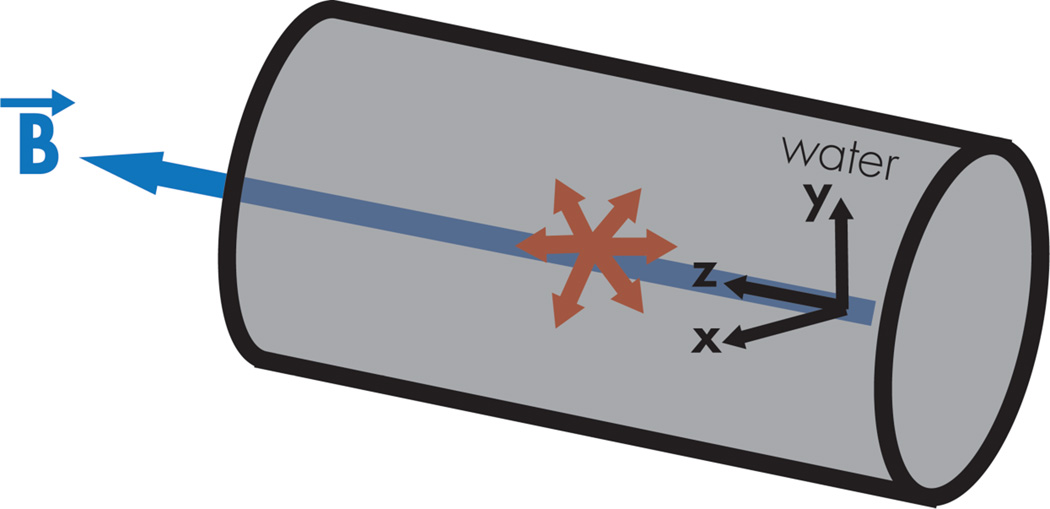
To characterize the effect of magnetic field on positron range, a Monte Carlo simulation application was developed using the Geant4 toolkit (version 4.9.6.p02) [5]. Point sources of positron-emitting radionuclides including 18F (Emax = 633.5 keV), 124I (Emax = 2.14 MeV), and 68Ga (Emax = 1.89 MeV) were simulated in a homogeneous cylinder of water. The Geant4 modular physics lists including G4EmStandardPhysics_opt4, G4RadioactiveDecayPhysics, and G4DecayPhysics were used to model radioactive decay, ionization, multiple scattering, bremsstrahlung, and electron-positron annihilation. A uniform magnetic field (B0) was applied in the z-direction of the cylinder using Geant4 magnetic field constructor. The positron-electron annihilation end-point coordinates were tallied to construct a 3D point spread function (PSF) with an isotropic voxel size of 50-µm. For efficient simulation runtime, any decay product subsequent to the positron annihilation was not tracked. The three-dimensional (3D) PSF was projected into 2D PSF planes along the x and z directions (defined in Fg. 1), and one-dimensional (1D) PSFs were obtained by projecting the 2D PSF plane along vertical and horizontal directions. Half of the 1D PSF was fitted to a bi-exponential function, and the full width tenth maximum (FWTM) was determined numerically as the spatial resolution of the positron range spread. For each batch, five million events were simulated for 18F and 68Ga, and 10 million events were simulated for 124I. Ten batches of simulations were performed for statistical purposes.
Fig. 1.
The GEANT4 Monte Carlo simulation setup is illustrated with an isotropic source at the center of a water-filled cylinder surrounded with a uniform magnetic field along the z-direction. The x, y, and z direction referred in this work is defined.
B. Experimental Imaging
18F and 68Ga line source phantoms were constructed with capillary tubes (0.6-mm inner diameter) placed on top of a 3-mm thick PMMA plate. To determine 3D spatial resolution, the line source phantoms were imaged with the line sources placed close to the center of PET field of view parallel to the x- and z-direction of the system in air. To examine the system spatial resolution close to the clinical setting, the line source was also imaged in water by securing the line source plate on top of a piece of foam in a water-filled cylinder. The line source phantoms were imaged with a simultaneous TOF-PET/MRI system (Investigational PET/MRI from GE Healthcare) and a PET/CT system (GE Discovery VCT) to examine the effect of magnetic field on system spatial resolution. PET images were reconstructed with the 3D Fourier-rebinned filtered back projection algorithm with the GE research toolbox. The line spread function (LSF) of the line source phantoms were obtained from multiple PET image slices, and the full width half maximum (FWHM) of the LSF in x, y, and z directions were determined by fitting the LSF with a Gaussian function. Table I shows the comparison of the PET system and image specification between the TOF-PET/MRI system and the PET/CT system.
Table I.
The PET system and image specification: PET/MRI vs. PET/CT
| Investigational TOF- PET/MRI (3T) |
GE PET/CT Discovery VCT |
|
|---|---|---|
| PET Crystal Size | ||
| In-plane | 3.95 | 4.7 |
| Axial (mm) | 5.3 | 6.3 |
|
PET Detector Ring Diameter (cm) |
60 | 88 |
|
3D PET Acquisition (min per bed / # of bed) |
3.0 / 1 bed | 5.0 / 1 bed |
|
PET Reconstructed Image Voxel Size (mm3) |
1.37 × 1.37 × 2.78 | 1.37 × 1.37 × 3.27 |
III. Results
A. Simulated Positron Point Source
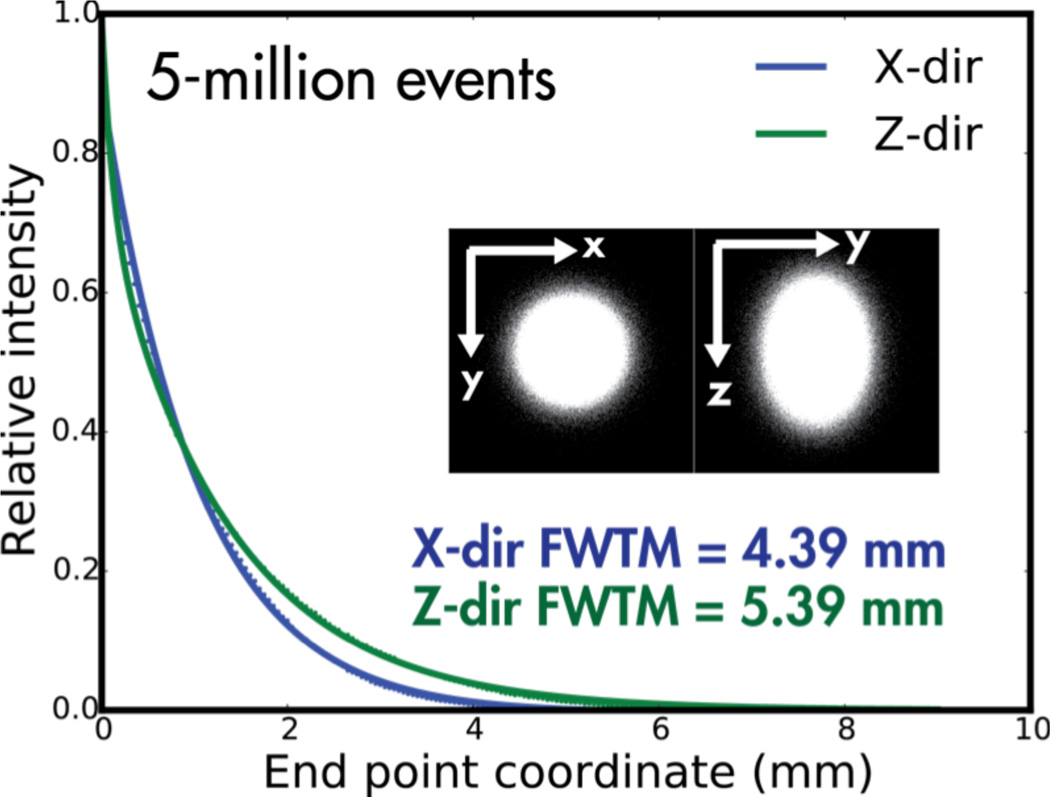
Fig. 2 shows the simulated positron trajectories of 68Ga in water in the plane transverse to the direction of the magnetic field at 0 T, 3 T, and 7 T. The simulated positron PSF of a 68Ga point source in water at 3 T is shown in Fig. 3. Table II lists the FWTM of 18F, 68Ga, and 124I positron spread in water from the Geant4 Monte Carlo simulations. While minimal effect was observed in the FWTM of 18F in all directions, the simulations show that the FWTM of 68Ga and 124I in the x or y-direction was reduced by up to 23% and 93% at 3 T and 7 T, respectively, compared to those at 0 T. The FWTM of 68Ga and 124I in the z-direction was not affected at 3 T and 7 T.
Fig. 2.
The simulated positron trajectories of a 68Ga point source in water is shown with a uniform magnetic field at (a) 0 T, (b) 3 T, and (c) 7 T. The magnetic field was applied in the direction going into the paper plane.
Fig. 3.
The simulated positron PSF of 68Ga in water with a 3 T uniform magnetic field is illustrated in x-direction (blue) and z-direction (green). The dot represents the data, and line is the bi-exponential fitted data. The inset images show the simulated 3D PSF projected in x-y plane and y-z plane.
Table II.
The full-width at tenth maximum (FWTM) of the simulated positron spread for 18F, 124I, and 68Ga in water.
| Positron Spread FWTM (mm) in water | ||||
|---|---|---|---|---|
| 0 T | 3 T | 7 T | ||
| 18F | x | 0.88 ± 0.0006 | 0.86 ± 0.0010 | 0.80 ± 0.0007 |
| y | 0.88 ± 0.0008 | 0.86 ± 0.0006 | 0.80 ± 0.0006 | |
| z | 0.88 ± 0.0010 | 0.88 ± 0.0007 | 0.88 ± 0.0009 | |
| 124I | x | 5.35 ± 0.0078 | 4.41 ± 0.0070 | 3.60 ± 0.7941 |
| y | 5.35 ± 0.0168 | 4.40 ± 0.0059 | 3.60 ± 0.7969 | |
| z | 5.34 ± 0.0142 | 5.35 ± 0.0024 | 5.35 ± 0.0121 | |
| 68Ga | x | 5.39 ± 0.0077 | 4.39 ± 0.0037 | 2.80 ± 0.0032 |
| y | 5.39 ± 0.0084 | 4.39 ± 0.0077 | 2.80 ± 0.0168 | |
| z | 5.38 ± 0.0129 | 5.39 ± 0.0091 | 5.39 ± 0.0142 | |
B. System Spatial Resolution: PET/MRI vs. PET/CT
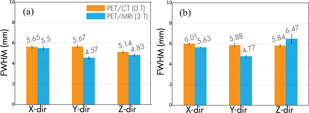
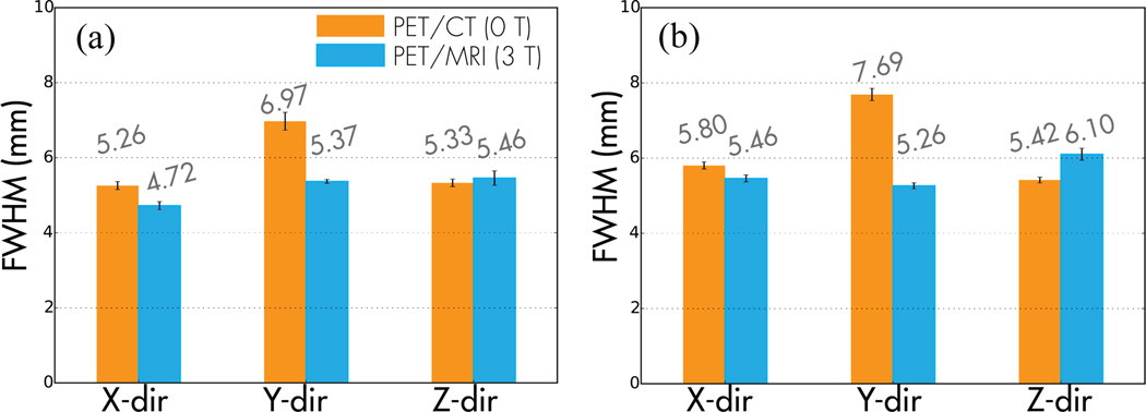
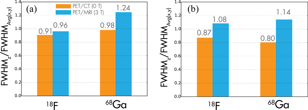
Fig. 4 and Fig. 5 show that the spatial resolution of the PET/MRI system was improved compared to that of the PET/CT system. The spatial resolution of both imaging systems was close to isotropic for 18F when imaged in air and water. For 68Ga, the spatial resolution degraded in all directions compared to 18F for both systems. When the line phantoms were imaged in water, the FWHM of the PET/CT system in y-direction was increased by up to 32% compared to that of x-direction for both 18F and 68Ga. The in-plane spatial resolution (x and y-directions) of the PET/MRI system (3 T) improved by up to 24% compared to the axial spatial resolution (z-direction) when imaged the 68Ga line source phantom with the PET/MRI system. The change in spatial resolution was larger when the line source was imaged in air than in water as shown in Fig. 6. When imaging the phantom in air, the ratio of axial FWHM to in-plane FWHM of the PET/MRI system was larger for 68Ga compared to that in water while the spatial resolution of 18F and 68Ga measured with the PET/CT system was close to isotropic.
Fig. 4.
The system spatial resolution in full-width at half maximum (FWHM) is compared between the PET/CT (orange) and PET/MRI (blue) measured with the (a) 18F and (b) 68Ga line source phantoms imaged in air.
Fig. 5.
The system spatial resolution in full-width at half maximum (FWHM) is compared between the PET/CT (orange) and PET/MRI (blue) measured with the (a) 18F and (b) 68Ga line source phantoms imaged in water.
Fig. 6.
The ratio of FWHM in z-direction to the FWHM averaged in x and y-directions is shown for 18F and 68Ga line source phantoms imaged with the PET/CT system (orange) and the PET/MRI system (blue) in (a) air and (b) water.
IV. Discussion
This study found that there is measurable effect on the PET system spatial resolution from the magnetic field. Simulations show that the improvement in the in-plane spatial resolution (x and y-direction) of 68Ga (Emax = 1.89 MeV) and 124I (Emax = 2.14 MeV) was greater than that of 18F (Emax = 633.5 keV) with the presence of a magnetic field. Fig. 6 shows that the spatial resolution of the PET/CT system (without magnetic field) was close to isotropic even for the high-energy positron emitters such as 68Ga. This finding suggests that the intrinsic positron range without the presence of a magnetic field did not have anisotropic effect on the system spatial resolution. When the same 68Ga phantom was imaged in the PET/MRI system, the in-plane spatial resolution was improved by up to 24% compared to the axial spatial resolution, suggesting that there may be some effect from the presence of magnetic field. The experimental imaging also shows that the effect of magnetic field on the in-plane spatial resolution may be slightly larger when the positrons are emitted in air instead of water. Since the line source was secured on top of a piece of foam during image acquisition, larger positron spread from the water-air boundary attributed to the larger FWHM of the PET/CT system in the y-direction in Fig. 5. Special attention to image quality may be necessary when resolving PET signal in air-tissue boundary such as lung nodules with high-energy positron emitters. Implementing correction using radionuclide-specific PSF models in the PET iterative reconstruction algorithms may be useful.
V. Conclusions
In this study, we have investigated the effect of magnetic field on system spatial resolution with Geant4 Monte Carlo simulations and experimental imaging. The presence of a magnetic field at 3 T was found to have measurable improvement in the system in-plane spatial resolution especially when resolving PET signal in air or air-tissue boundary with high-energy positron emitters.
Acknowledgment
We thank the nuclear medicine and MRI technologist, Vahid Ravanfar, for his tremendous help in data acquisition. We also appreciate for the helpful conversation with the GE Healthcare research team (particularly Floris Jensen and Timothy Deller).
This work was supported in part by GE Healthcare and National Cancer Institute R01 CA154561
References
- 1.Shah NJ, et al. Effects of Magnetic Fields of up to 9.4 T on Resolution and Contrast of PET Images as Measured with an MR-BrainPET. PLoS One. 2014;9:e95250. doi: 10.1371/journal.pone.0095250. [DOI] [PMC free article] [PubMed] [Google Scholar]
- 2.Iida H, Kanno I, Miura S, Murakami M, Takahashi K, Uemura K. A Simulation Study of a Method to Reduce Positron-Annihilation Spread Distributions Using a Strong Magnetic-Field in Positron Emission Tomography. IEEE Transactions on Nuclear Science. 1986 Feb;33:597–600. [Google Scholar]
- 3.Levin CS, Hoffman EJ. Calculation of positron range and its effect on the fundamental limit of positron emission tomography system spatial resolution. Phys Med Biol. 1999 Mar;44:781–799. doi: 10.1088/0031-9155/44/3/019. [DOI] [PubMed] [Google Scholar]
- 4.Wirrwar A, Vosberg H, Herzog H, Halling H, Weber S, MullerGartner HW. 4.5 Tesla magnetic field reduces range of high-energy positrons - Potential implications for positron emission tomography. IEEE Transactions on Nuclear Science. 1997 Apr;44:184–189. [Google Scholar]
- 5.Agostinelli S, et al. Geant4—a simulation toolkit. Nuclear Instruments and Methods in Physics Research Section A: Accelerators, Spectrometers, Detectors and Associated Equipment. 2003;506:250–303. [Google Scholar]


