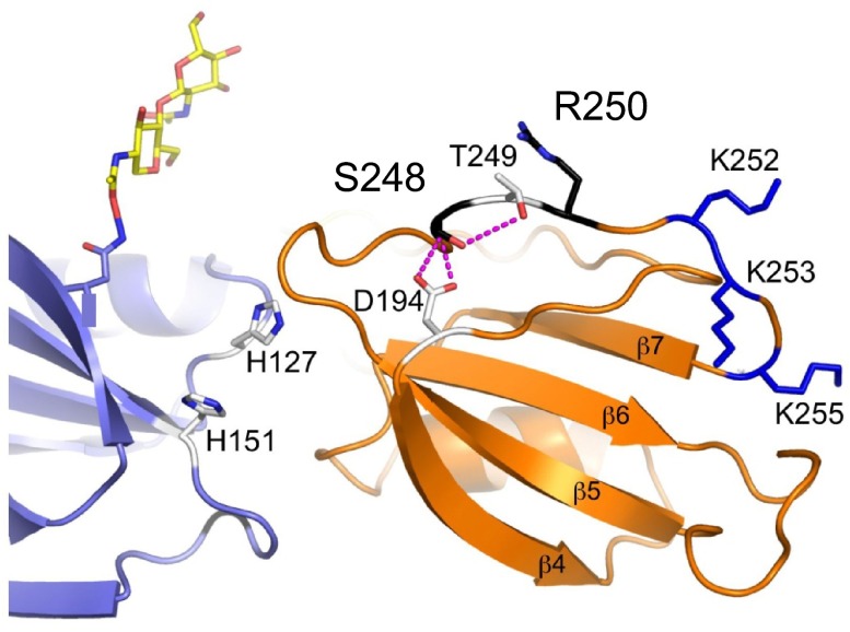
Figure 8.
Platelet-binding site on FXI. The locations of 2 residues (Ser248 and Arg250 in black), that probably form a GPIb platelet-binding site on the FXI A3 domain (orange), are shown relative to residues that form the heparin-binding site (Ly252, Lys253, and Lys255 in blue). Ser248 forms hydrogen bonds with Asp194 and Thr249, which are probably disrupted in the hereditary FXI mutation Ser248Gln. The adjacent A2 domain is shown in light blue. The position of an N-linked glycan moiety attached to residue Asn108 of the A2 domain is shown in yellow and red.

