

The last decade has seen a steady proliferation in the use of tissue-engineered cell culture systems (Deforest and Anseth, 2012), and these have been put to good use for studying neural axon growth and guidance (Li and Hoffman-Kim, 2008; Roy et al., 2013). These systems have been designed to more closely mimic the natural microenvironment of the developing or repairing nervous system and to enable spatiotemporal control over certain aspects of the microenvironment. The 3D nature of these culture systems provides a more physiologically-relevant microenvironment, while spatiotemporal control addresses aspects of tissue architecture and molecular presentation for quantitative investigation into how specific physical or molecular variables might influence axon growth. These capabilities should be tremendously important to the study of neural regeneration, since it is well known that developing and regenerating axons of both the central and peripheral nervous systems respond to particular attractive or repulsive cues presented in their microenvironments that may be sensed by the growth cones of these extending axons. Tissue-engineered model systems are well suited to the investigation into precise mechanisms of action of these cues, the elucidation of novel mechanisms, and for testing potential therapeutic strategies.
To date, biologists have yet to fully leverage the engineering developments of these model systems for neural regeneration research. This is important because molecular neuroscientists, who are trained for hypothesis-driven research, are arguably better suited to the utilization of these models than bioengineers (such as this author), who may be better suited to their design and validation. The main barriers to adoption of these tools are somewhat obvious: nonstandardized techniques and the need for specialized equipment, devices, and materials. But a more subtle barrier arises because engineers and molecular biologists approach problems differently, frequent different circles, and speak different scientific languages. These differences lead various fields of experts to ignore the advances or dismiss the methodologies of those in other fields, though they may be most relevant. Tissue-engineered culture models have come of age, and are poised to play an integral role in neural regeneration research. For this to occur, the technical and intellectual barriers to adoption of these models can and must be overcome.
Overcoming technical barriers: Tissue engineered culture systems often make use of custom equipment, methods, and materials, making adoption by non-specialists daunting. Of course collaboration is a way forward, and very fruitful research has and will continue to come about in this manner. But for truly widespread adoption, a certain level of standardization and availability of materials and methods is necessary to reduce the technical expertise required to use the techniques. This challenge is akin to the introduction of any new technology, like personal computers a few decades ago: while powerful, their practical use became much more widespread with user-friendly operating systems and plug-and-play peripherals. An analogous progression in biology has been the commercialization of “;kits” that make biological assays more user-friendly.
Fortunately, this trend toward reduced necessity for technical expertise is beginning in the realm of microfluidics and related MEMs devices for the study of axon growth (Millet and Gillette, 2012; Park et al., 2013). There are a number of relatively simple devices now commercially available, such as isolation chambers, useful for parsing cues that may act on the axon vs. the soma, or microfluidic gradient makers, which allow researchers to investigate axon guidance in response to quantifiable soluble gradients. The availability of these devices ease experiments that a decade ago would have been rather difficult or dependent on collaboration with engineers or physicists with access to clean-room microfabrication facilities. What is needed for tissue-engineered models to become more widely adopted is a similar trend toward user-friendly, plug-and-play designs.
Some research groups have been developing methods for microscale tissue engineering that may help make axon-growth culture models more user-friendly. For example, Molly Shoichet's research group demonstrated how a familiar confocal microscope can be used to study how gradients of guidance molecules influence axon growth (Yu et al., 2008). In my laboratory, we have been using a versatile digital projection photolithography scheme for forming micropatterns in hydrogels that is simple, rapid, can be used on a variety of regular cell culture materials, and eliminates the need for printing a new photomask or programming a scanning laser for each geometry desired. In this approach, one photocrosslinked hydrogel serves as a micro-mold to contain a second gel. That second gel can be used as a 3D supportive matrix for axon growth, and it may also be spatially modified for testing specific physical and molecular guidance cues (Curley and Moore, 2011; Horn-Ranney et al., 2013), as shown in Figure 1. Using these techniques, it is simple to generate geometries which mimic axonal pathfinding “choice points” (Catig et al., 2015).
Figure 1.
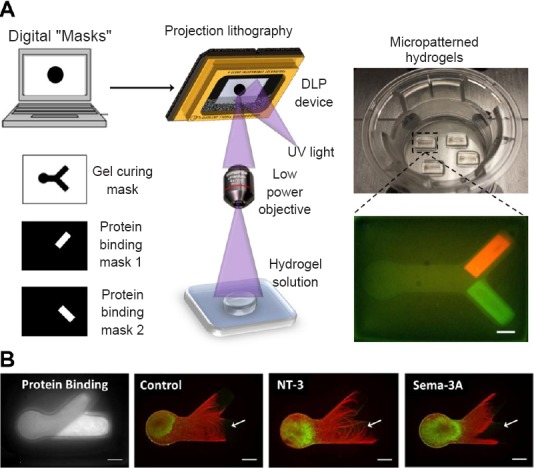
Digital projection photolithography for fabricating micropatterned 3D tissue culture matrices with bound proteins for axon growth and guidance studies.
(A) Black-and-white images corresponding to desired geometries (left) are uploaded via computer to a digital light projection device, which reflects UV light pixel-for-pixel through an objective lens onto a curable hydrogel (center). A series of digital “masks” can be used for curing the gels onto regular cell culture substrates (upper right), and for irradiating gels for binding proteins. Lower-right image represents two different fluorescent proteins covalently bound to the 3D gel in specific locations. (B) Fluorescent images show protein binding location as well as axon growth (red) from dorsal root ganglion neurons (green). A control protein led to equal axon growth in upper and lower forks, while NT-3 increased, and Sema-3A decreased, the amount of axon growth in the region of the bound protein, marked with arrows. All scale bars: 500 μm. Adapted from Moore and Curley, 2011; and Horn-Ranney et al., 2013, 2014. DLP: Digital projection lithography; NT-3: neurotrophin-3; Sema-3A: semaphorin 3A; UV: ultraviolet.
We validated our approach by demonstrating that a uniform concentration of semaphorin 3A bound to the matrix can repel dorsal root ganglion neurites, while bound neurotrophin-3 increases neurite growth (Horn-Ranney et al., 2014). We then used this approach to show that semaphorin 6A arrests axon growth from a subset of dorsal root ganglion neurons and that a concentration gradient of NGF can partially overcome this repulsion (Curley et al., 2014). A short time ago, such an experiment would have required a very complicated co-culture system using substrates lined with homogenized cell membranes from overexpressing cells. Models such as ours provide for placement of both soluble and immobilized guidance cues in gels with relative ease. This strategy represents a manner for the systematic and quantifiable investigation of how guidance cues may be either exploited or overcome to promote regeneration.
While the relative simplicity and flexibility of our model systems have perhaps reduced the barrier for others to adopt our methods, they still make use of custom-built optical apparatus and polymers which we synthesize in our own laboratory. These remaining barriers are not insurmountable. There are a number of commercially-available hydrogel products that can be used as homogeneous 3D culture matrices. However, to address tissue architecture or molecular anisotropy–so important to neural regeneration–methods of structural or molecular micropatterning are essential. The digital projection lithography method we employ, though not as precise as laser scanning, is easily adaptable with commercial attachments designed for optical microscopes, such as the Mosaic offered by Andor, or the Polygon by Mightex. Used with suitable low-power objectives and with software that enables uploading of images as digital photomasks, researchers could easily fabricate patterned cell cultures to their own specifications.
Some of the culture matrices on the market may be photocrosslinked to form patterned cultures. However, there still remain no commercially-available gels capable of binding biomolecules in response to light illumination, a capability that could advance our field if more widespread. What is needed is a standardization of the many variations of these culture materials that have been described. Kristi Anseth's laboratory has recently described a modular hydrogel design using orthogonal “click” chemistry, which would ostensibly accomplish just such a standardization (Azagarsamy and Anseth, 2013). Commercialization of this type of modular design, including mass production to reduce costs and development of kits to make them user-friendly, would greatly enhance the capabilities of our field.
Overcoming intellectual barriers: Once the technical advances of tissue-engineered models are more widely accessible, they will certainly gain growing acceptance in the field of neural regeneration. However, what has and will continue to slow the adoption of these methods is the reality that bioengineers, who largely develop the technologies, and molecular neuroscientists, who might employ them, tend to work in different circles, approach research differently, and speak different languages. This reality is probably not much different than for the many advances in biotechnology that have gone before. The difference today, however, is found in the increasing specialization in science, and the maturing of bioengineering as a sub-specialty.
Bioengineers now have access to dozens of journals dedicated to their own sub-specialty (and sub-sub-specialties), so it is simply easier to publish in these journals, which may not be appreciated or even noticed by most molecular neuroscientists. For example, all the author's papers cited in this perspective were published in biomaterials and biomedical engineering journals. The sheer rate of publications emerging from our own respective disciplines makes keeping abreast of other fields all the more challenging. With modern indexing services and software, and open access journals, advances in tissue engineered culture systems may eventually be discovered by the molecular neuroscience community and put to good use, so long as some are willing to overcome the technical barriers described above.
For these reasons, it is important that tissue engineers publish in relevant neuroscience journals, but this can be a difficult task because of the language barrier. Papers from engineering labs usually focus on demonstrating the feasibility of their design and validation of their approach. This scheme is not usually acceptable for publication in neuroscience journals. In molecular neuroscience, a much more mature discipline, an unwritten methodology has emerged in which testing a hypothesis typically requires one to demonstrate a particular molecular mechanism. Experiments are designed to demonstrate an effect, then to show that inhibiting the pathway upstream and/or downstream stops that effect, and further that blocking the inhibition restores the effect. The language barrier can be seen in contrasting results sections in engineering and biology journals. Engineers tend to write the subsection headings of their results in terms of what test we performed: “;Mechanical Analysis” or “;Binding Assay”; biologists, in terms of what they learned: “A is Necessary but Not Sufficient to Cause B”.
These intellectual barriers are also not insurmountable, but doing so may require more unnatural effort than overcoming the technical ones. First, language immersion is imperative: we must attend one another's seminars, read one another's journals, and frequent one another's conferences. Second, we must be aware of the differences in our approaches. In particular, engineers (this author chief among them), must learn to speak the language of molecular biology, if not fluently, then well enough to write papers that may be accepted in neuroscience journals. Likewise, neuroscientists would do well to explore the wealth of advanced culture techniques found in bioengineering journals. However, they need to learn to search the tissue engineering and biomaterials literature not for a specific effect, since feasibility and validation are usually the focus, but rather as examples of what could be done when applied to the molecular pathway in question. Finally, we must be deliberate in finding ways to foster cross-disciplinary research with the aim of solving particular clinical problems. This effort is seen in the formation of a few interdisciplinary research centers and can be fostered even more though purposeful symposia gathering experts of multiple disciplines in a single room.
A path forward: An area of research particularly ripe for implementation of this approach is counteracting inhibition of regeneration. For example, Jerry Silver's laboratory has pioneered the development of in vitro models for the study of growth cone inhibition in the glial scar (Tom et al., 2004). By overcoming the technical and intellectual barriers discussed in this perspective, more advanced model systems could readily be devised that provide a natural 3D environment with quantitative, precise placement of inhibitory cues. These model systems would be poised for the screening of potential stimulatory agents, which could also be presented with spatiotemporal precision, for the elucidation of promising treatment strategies. Such an endeavor would benefit from the seamless interaction between bioengineers and neuroscientists.
Tissue-engineered models are extremely powerful and translational biological tools, though remain largely untapped potential for neural regeneration research. We can overcome barriers to adoption of these models by condensing the many variations of the technology to simple, modular designs, by commercializing some of those designs as user-friendly kits, reducing the fear and intimidation in approaching new disciplines, and by learning to more effectively interact with our worthy colleagues in disparate fields.
I thank all my present and former students and colleagues whose work contributed to this article. I apologize to the many worthy investigators whose work inspired these thoughts but who were not named due to space constraints. Thanks to Dr. Taby Ahsan for helpful discussions and suggestions for the text. The work from my laboratory highlighted in this article has been funded by the Oliver Fund of Tulane University, the Louisiana Board of Regents (LEQSF[2009-11]-RD-A-18), the NIH (NS065374), and the NSF (CBET-1055990).
References
- Azagarsamy MA, Anseth KS. Bioorthogonal click chemistry: an indispensable tool to create multifaceted cell culture scaffolds. ACS Macro Lett. 2013;2:5–9. doi: 10.1021/mz300585q. [DOI] [PMC free article] [PubMed] [Google Scholar]
- Catig GC, Figueroa S, Moore MJ. Experimental and computational models of neurite extension at a choice point in response to controlled diffusive gradients. J Neural Eng. 2015;12:046012. doi: 10.1088/1741-2560/12/4/046012. [DOI] [PubMed] [Google Scholar]
- Curley JL, Moore MJ. Facile micropatterning of dual hydrogel systems for 3D models of neurite outgrowth. J Biomed Mater Res A. 2011;99:532–543. doi: 10.1002/jbm.a.33195. [DOI] [PMC free article] [PubMed] [Google Scholar]
- Curley JL, Catig GC, Horn-Ranney EL, Moore MJ. Sensory axon guidance with Semaphorin 6A and Nerve Growth Factor in a biomimetic choice point model. Biofabrication. 2014;6:034026. doi: 10.1088/1758-5082/6/3/035026. [DOI] [PMC free article] [PubMed] [Google Scholar]
- DeForest CA, Anseth KS. Advances in bioactive hydrogels to probe and direct cell fate. Annu Rev Chem Biomol Eng. 2012;3:421–444. doi: 10.1146/annurev-chembioeng-062011-080945. [DOI] [PubMed] [Google Scholar]
- Horn-Ranney EL, Khoshakhlagh P, Kaiga JW, Moore MJ. Light-reactive dextran gels with immobilized guidance cues for directed neurite growth in 3D models. Biomaterials Sci. 2014;2:1450–1459. doi: 10.1039/c4bm00043a. [DOI] [PubMed] [Google Scholar]
- Horn-Ranney EL, Curley JL, Catig GC, Huval RM, Moore MJ. Structural and molecular micropatterning of dual hydrogel constructs for neural growth models using photochemical strategies. Biomed Microdev. 2013;15:49–61. doi: 10.1007/s10544-012-9687-y. [DOI] [PMC free article] [PubMed] [Google Scholar]
- Li GN, Hoffman-Kim D. Tissue-engineered platforms of axon guidance. Tissue Eng B. 2008;14:33–51. doi: 10.1089/teb.2007.0181. [DOI] [PubMed] [Google Scholar]
- Millet LJ, Gillette MU. New perspectives on neuronal development via microfluidic environments. Trends Neurosci. 2012;35:752–761. doi: 10.1016/j.tins.2012.09.001. [DOI] [PMC free article] [PubMed] [Google Scholar]
- Park JW, Kim HJ, Kang MW, Jeon NL. Advances in microfluidics-based experimental methods for neuroscience research. Lab Chip. 2013;13:509–521. doi: 10.1039/c2lc41081h. [DOI] [PubMed] [Google Scholar]
- Roy J, Kennedy TE, Costantino S. Engineered cell culture substrates for axon guidance studies: moving beyond proof of concept. Lab Chip13. 2013:498–508. doi: 10.1039/c2lc41002h. [DOI] [PubMed] [Google Scholar]
- Tom VJ, Steinmetz MP, Miller JH, Doller CM, Silver J. Studies on the development and behavior of the dystrophic growth cone, the hallmark of regeneration failure, in an in vitro model of the glial scar and after spinal cord injury. J Neurosci. 2004;24:6531–6539. doi: 10.1523/JNEUROSCI.0994-04.2004. [DOI] [PMC free article] [PubMed] [Google Scholar]