Abstract
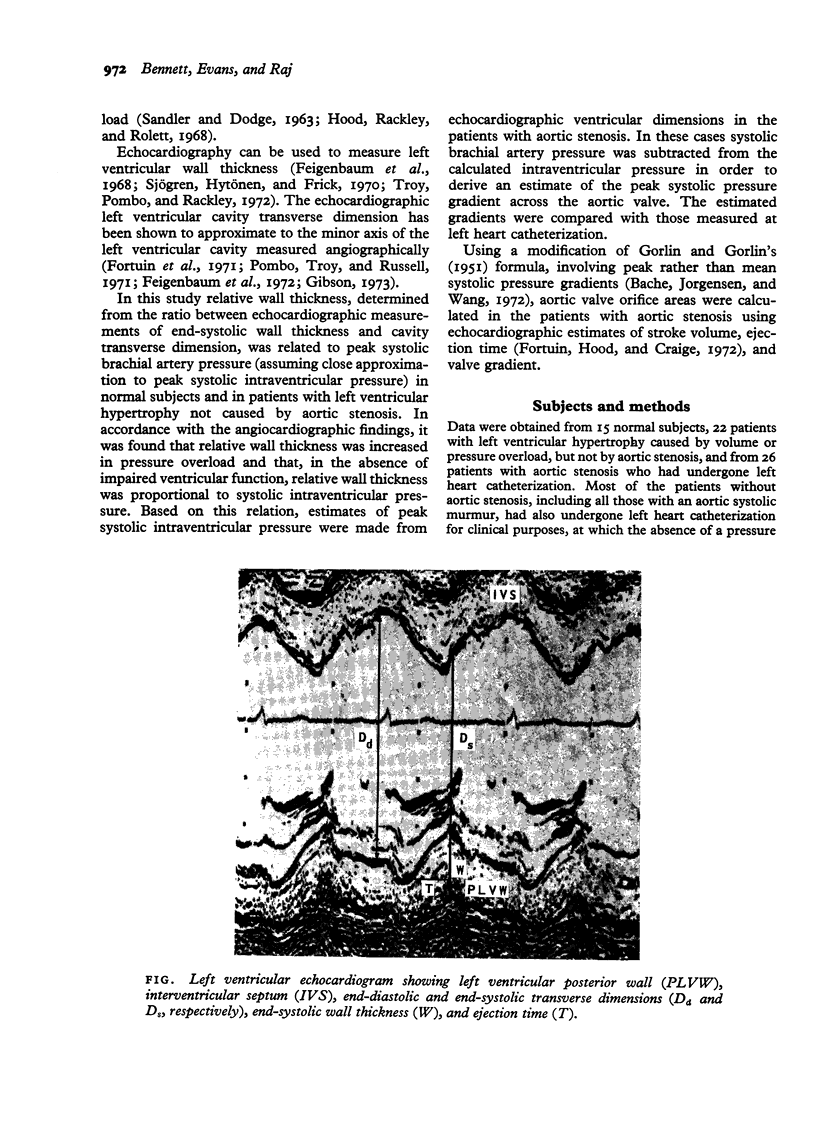
Left ventricular 'relative wall thickness', determined from the ratio between echocardiographic measurements of end-systolic wall thickness and cavity transverse dimension, was related to peak systolic intraventricular pressure in 15 normal subjects, in 15 patients with left ventricular volume or pressure overload without aortic stenosis, and in 23 patients with aortic stenosis. All these patients had a mean rate of circumferential fibre shortening greater than 1.0 circumference per second and were regarded as having good ventricular function. Relative wall thickness was found to be normal in cases of volume overload and to be increased in pressure overload, being proportional to the systolic intraventricular pressure. Values for the ratio of systolic intraventricular pressure to relative wall thickness in the normal subjects and patients without aortic stenosis were similar (mean 30 +/- 2.5). Based on this relation, estimates of peak systolic intraventricular pressure were made in the cases of aortic stenosis using the formula: systolic intraventricular pressure (kPa) equals 30 x wall thicknes divided by transverse dimension. Peak systolic aortic value gradients derived by subtracting brachial artery systolic pressure, measured by sphygmomanometer, from the echocardiographic estimates of intraventricular pressure compared favourably with the gradients measured at left heart catheterization (r equals 0.87, P less than 0.001). Aortic value orifice areas, derived from echocardiographic estimates of stroke volume, ejection time, and value gradient, ranged from 0.21 to 3.16 cm2 and appeared to correlate with the severity of aortic stenosis. All patients with aortic stenosis, with or without coexistent mild aortic regurgitation, who were recommended for aortic valve surgery, had estimated valve orifice areas of less than 0.8 cm2. A further 10 patients with pressure or volume overload had mean rates of circumferential fibre shortening of less than 1.0 circumference per second and were regarded as having poor ventricular function. In these cases values for relative wall thickness were lower than in those with good ventricular function and were not proportional to systolic intraventricular pressure. In patients with good left ventricular function systolic intraventricular pressure is proportional to, and can be estimated from, echocardiographic measurement of relative wall thickness.
Full text
PDF


Images in this article
Selected References
These references are in PubMed. This may not be the complete list of references from this article.
- Bache R. J., Jorgensen C. R., Wang Y. Simplified estimation of aortic valve area. Br Heart J. 1972 Apr;34(4):408–411. doi: 10.1136/hrt.34.4.408. [DOI] [PMC free article] [PubMed] [Google Scholar]
- Bennett D. H., Evans D. W., Raj M. V. Proceedings: Echocardiographic estimation of the sytolic pressure gradient in aortic stenosis. Br Heart J. 1975 May;37(5):557–557. [PubMed] [Google Scholar]
- Cooper R. H., O'Rourke R. A., Karliner J. S., Peterson K. L., Leopold G. R. Comparison of ultrasound and cineangiographic measurements of the mean rate of circumferential fiber shortening in man. Circulation. 1972 Nov;46(5):914–923. doi: 10.1161/01.cir.46.5.914. [DOI] [PubMed] [Google Scholar]
- Feigenbaum H., Popp R. L., Chip J. N., Haine C. L. Left ventricular wall thickness measured by ultrasound. Arch Intern Med. 1968 May;121(5):391–395. [PubMed] [Google Scholar]
- Feigenbaum H., Popp R. L., Wolfe S. B., Troy B. L., Pombo J. F., Haine C. L., Dodge H. T. Ultrasound measurements of the left ventricle. A correlative study with angiocardiography. Arch Intern Med. 1972 Mar;129(3):461–467. [PubMed] [Google Scholar]
- Fortuin N. J., Hood W. P., Jr, Craige E. Evaluation of left ventricular function by echocardiography. Circulation. 1972 Jul;46(1):26–35. doi: 10.1161/01.cir.46.1.26. [DOI] [PubMed] [Google Scholar]
- Fortun N. J., Hood W. P., Jr, Sherman M. E., Craige E. Determination of left ventricular volumes by ultrasound. Circulation. 1971 Oct;44(4):575–584. doi: 10.1161/01.cir.44.4.575. [DOI] [PubMed] [Google Scholar]
- GORLIN R., GORLIN S. G. Hydraulic formula for calculation of the area of the stenotic mitral valve, other cardiac valves, and central circulatory shunts. I. Am Heart J. 1951 Jan;41(1):1–29. doi: 10.1016/0002-8703(51)90002-6. [DOI] [PubMed] [Google Scholar]
- GORLIN R., McMILLAN I. K., MEDD W. E., MATTHEWS M. B., DALEY R. Dynamics of the circulation in aortic valvular disease. Am J Med. 1955 Jun;18(6):855–870. doi: 10.1016/0002-9343(55)90169-8. [DOI] [PubMed] [Google Scholar]
- Gibson D. G. Estimation of left ventricular size by echocardiography. Br Heart J. 1973 Feb;35(2):128–134. doi: 10.1136/hrt.35.2.128. [DOI] [PMC free article] [PubMed] [Google Scholar]
- Grant C., Greene D. G., Bunnell I. L. Left ventricular enlargement and hypertrophy. A clinical and angiocardiographic study. Am J Med. 1965 Dec;39(6):895–904. doi: 10.1016/0002-9343(65)90111-7. [DOI] [PubMed] [Google Scholar]
- Henry W. L., Clark C. E., Epstein S. E. Asymmetric septal hypertrophy. Echocardiographic identification of the pathognomonic anatomic abnormality of IHSS. Circulation. 1973 Feb;47(2):225–233. doi: 10.1161/01.cir.47.2.225. [DOI] [PubMed] [Google Scholar]
- Hermann H. J., Singh R., Dammann J. F. Evaluation of myocardial contractility in man. Am Heart J. 1969 Jun;77(6):755–766. doi: 10.1016/0002-8703(69)90409-8. [DOI] [PubMed] [Google Scholar]
- Hood W. P., Jr, Rackley C. E., Rolett E. L. Wall stress in the normal and hypertrophied human left ventricle. Am J Cardiol. 1968 Oct;22(4):550–558. doi: 10.1016/0002-9149(68)90161-6. [DOI] [PubMed] [Google Scholar]
- Hood W. P., Jr, Thomson W. J., Rackley C. E., Rolett E. L. Comparison of calculations of left ventricular wall stress in man from thin-walled and thick-walled ellipsoidal models. Circ Res. 1969 Apr;24(4):575–582. doi: 10.1161/01.res.24.4.575. [DOI] [PubMed] [Google Scholar]
- Kennedy J. W., Twiss R. D., Blackmon J. R., Dodge H. T. Quantitative angiocardiography. 3. Relationships of left ventricular pressure, volume, and mass in aortic valve disease. Circulation. 1968 Nov;38(5):838–845. doi: 10.1161/01.cir.38.5.838. [DOI] [PubMed] [Google Scholar]
- Kennedy J. W., Yarnall S. R., Murray J. A., Figley M. M. Quantitative angiocardiography. IV. Relationships of left atrial and ventricular pressure and volume in mitral valve disease. Circulation. 1970 May;41(5):817–824. doi: 10.1161/01.cir.41.5.817. [DOI] [PubMed] [Google Scholar]
- LEVINE N. D., ROCKOFF S. D., BRAUNWALD E. AN ANGIOCARDIOGRAPHIC ANALYSIS OF THE THICKNESS OF THE LEFT VENTRICULAR WALL AND CAVITY IN AORTIC STENOSIS AND OTHER VALVULAR LESIONS. HEMODYNAMIC-ANGIOGRAPHIC CORRELATIONS IN PATIENTS WITH OBSTRUCTION TO LEFT VENTRICULAR OUTFLOW. Circulation. 1963 Sep;28:339–345. doi: 10.1161/01.cir.28.3.339. [DOI] [PubMed] [Google Scholar]
- Ludbrook P., Karliner J. S., Peterson K., Leopold G., O'Rourke R. A. Comparison of ultrasound and cineangiographic measurements of left ventricular performance in patients with and without wall motion abnormalities. Br Heart J. 1973 Oct;35(10):1026–1032. doi: 10.1136/hrt.35.10.1026. [DOI] [PMC free article] [PubMed] [Google Scholar]
- Pombo J. F., Troy B. L., Russell R. O., Jr Left ventricular volumes and ejection fraction by echocardiography. Circulation. 1971 Apr;43(4):480–490. doi: 10.1161/01.cir.43.4.480. [DOI] [PubMed] [Google Scholar]
- Quinones M. A., Gaasch W. H., Alexander J. K. Echocardiographic assessment of left ventricular function with special reference to normalized velocities. Circulation. 1974 Jul;50(1):42–51. doi: 10.1161/01.cir.50.1.42. [DOI] [PubMed] [Google Scholar]
- SANDLER H., DODGE H. T. LEFT VENTRICULAR TENSION AND STRESS IN MAN. Circ Res. 1963 Aug;13:91–104. doi: 10.1161/01.res.13.2.91. [DOI] [PubMed] [Google Scholar]
- Sjögren A. L., Hytönen I., Frick M. H. Ultrasonic measurements of left ventricular wall thickness. Chest. 1970 Jan;57(1):37–40. doi: 10.1378/chest.57.1.37. [DOI] [PubMed] [Google Scholar]
- Troy B. L., Pombo J., Rackley C. E. Measurement of left ventricular wall thickness and mass by echocardiography. Circulation. 1972 Mar;45(3):602–611. doi: 10.1161/01.cir.45.3.602. [DOI] [PubMed] [Google Scholar]
- Wong A. Y., Rautaharju P. M. Stress distribution within the left ventricular wall approximated as a thick ellipsoidal shell. Am Heart J. 1968 May;75(5):649–662. doi: 10.1016/0002-8703(68)90325-6. [DOI] [PubMed] [Google Scholar]


