Abstract
Soft ticks, Argas vespertilionis, were collected from feces of bats in Japan. Cytopathic effect (CPE) was observed after inoculating the homogenates of ticks to Vero cells. Sequencing of RNA extracted from the cell supernatant was performed by next generation sequencer. The contigs had identity to segments of Bunyaviruses, Issyk-Kul virus. The identities of segment L, M and S were only 77, 76 and 79% to Issyk-Kul virus, respectively. Therefore, we named this novel virus Soft tick bunyavirus (STBV). In the phylogenetic tree, segment L of STBV was closely related to a cluster consisting of the genus Nairovirus of the family Bunyaviridae.
Keywords: novel Bunyavirus, soft tick
The ticks transmit various important diseases, such as Lyme disease, relapsing fever, rickettsiosis, viral hemorrhagic fever and tsutsugamushi disease (Scrub typhus). In 2013, severe fever with thrombocytopenia syndrome virus (SFTSV) was isolated from Japanese patients without a foreign tour experience [5]. Therefore, finding novel viruses from ticks is important for public sanitation to prevent an outbreak of further tick-borne infectious diseases.
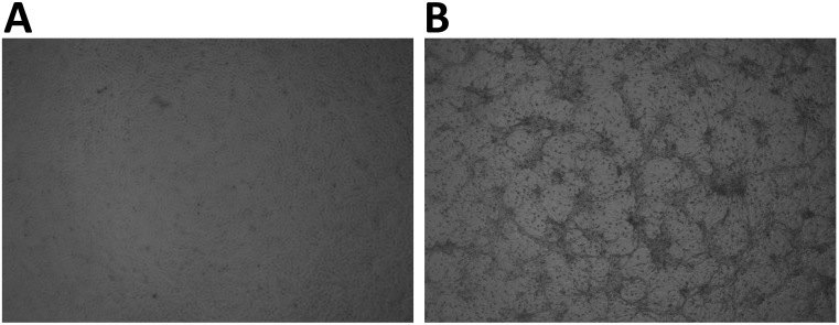
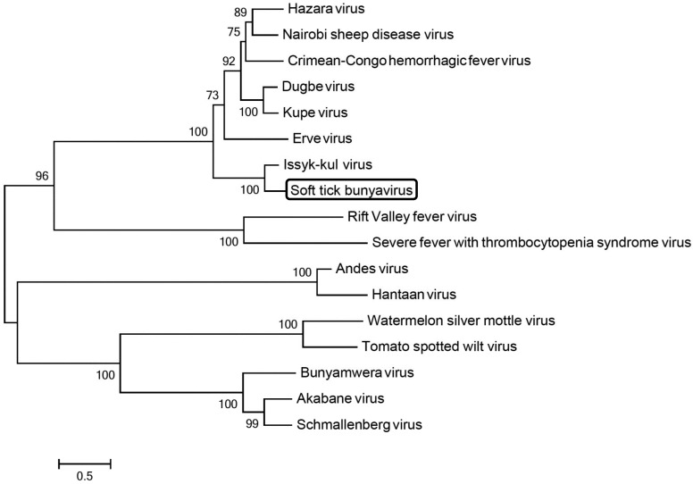
In this study, we tool the bat feces beneath bat colonies, and ticks in the feces were collected using microscopy. We also collected ticks invading the human habitat. Eighteen soft ticks, Argas vespertilionis, were collected in Japan. These ticks were homogenized in sucrose phosphate glucose solution. The homogenates were centrifuged, and supernatants were inoculated into Vero cells one by one. As shown in Fig. 1, cytopathic effect (CPE) was observed after the 2nd blind passage on 3 days post-inoculation. However, rickettsia and bacteria were not detected (data not shown). Total RNA was extracted from the cell supernatant using High Pure Viral Nucleic Acid kit (Roche, Basel, Switzerland), and a cDNA library for high-throughput sequencing was constructed using the NEBNext Ultra RNA Library Prep kit for Illumina (New England BioLabs, Ipswich, MA, U.S.A.) according to the manufacturer’s protocols. Sequencing was performed on MiSeq bench-top sequencer (Illumina, San Diego, CA, U.S.A.) using MiSeq Reagent Kit Nano v2 (300 cycles). The obtained 7,400,954 read sequences were assembled into 623 (˃200 mer) contigs using de novo assembly command in the CLC Genomics Workbench 6.5 (CLC bio, Aarhus, Denmark). Homology search in each contig was performed using the BLAST program in the National Center for Biotechnology Information (NCBI), and three of these contigs had identity to segments of Bunyaviruses (data not shown). To perform further analysis, the open reading frame (ORF) was extracted from those contigs using Find Open Reading Frames command in the CLC genomics Workbench and analyzed with the BLASTn with the BLAST program on the Internet site of NCBI. In the nucleotide sequence, the ORF of largest contig has homology to Issyk-Kul virus segment L (Accession No.KF892055.1), and the identity was only 77%. The other two contigs had 76 and 79% homology to Issyk-Kul virus segment M (Accession No. KF892056.1) and segment S (Accession No. KF892057.1). Thus, the virus is different from Issy-Kul virus. In this paper, we named this novel virus Soft tick bunyavirus (STBV), and the nucleotide sequence of S, M and L segments was deposited in GenBank (Accession No. LC027467, LC027466 and LC027465, respectively). The phylogenetic analysis with the nucleotide sequence of STBV segment L was performed by MEGA 6 software (Fig. 2). In the phylogenetic tree, STBV was closely related to a cluster consisting of the genus Nairovirus of the family Bunyaviridae including Crimean-Congo hemorrhagic fever virus (CCHFV), Hazara virus, Nairobi Sheep Disease Virus (NSDV), Kupe virus, Dugbe virus, Erve virus and Issyk-kul virus. Of these viruses, CCHFV and Hazara viruses are highly virulent pathogens to humans [1, 2], and NSDV is one of the most pathogenic agents of sheep and goats [4]. Recently, it has been reported that Issk-kul virus causes severe fever for humans and no deaths have been reported by this virus, but the high fever (39–41°C) caused by this virus lasted about eight days in the acute period, and the convalescent period was rather long (1–1.5 months) [3]. Therefore, STBV might be a potential pathogen to humans and animals, and further analysis is necessary to understand the characterization of this virus.
Fig. 1.
Cytopathic effect of the infectious agent on Vero cells. A: mock-infection, B: Vero cells inoculated by homogenized ticks.
Fig. 2.
Maximum likelihood tree based on nucleotide sequence of viruses of the family Bunyaviridae. The percentage of replicate trees in which the associated taxa are clustered together in the bootstrap test (1,000 replicates) was calculated. The length of the bar corresponds to the degree of sequence divergence. Phylogenetic analyses were conducted in MEGA 6. (Accession No.; CCHFV GU477492.1, Hazara virus DQ076419.1, NSDV EU697951.1, Kupe virus EU257628.1, Dugbe virus U15018.1, Erve virus JF911697.1, Issyk_Kul virus KF892055.1, Rift valley virus JF311376.1, SFTSV JQ684871.1, Hantaan virus JQ083393.1, Schmallenberg virus KC355457.1, Akabane virus AB190458.1 and Tomato spotted wilt virus KC261962.1)
Acknowledgments
This study was supported by the Research Project for Improving Food Safety and Animal Health of the Ministry of Agriculture, Forestry and Fisheries of Japan. This work was also supported by the grant of the Japanese Ministry of Health, Labour and Welfare.
REFERENCES
- 1.Al’khovskiĭ S. V., L’vov D. K., Shchelkanov M. I., Shchetinin A. M., Deriabin P. G., Samokhvalov E. I., Gitel’man A. K., Botikov A. G.2013. The taxonomy of the Issyk-Kul virus (ISKV, Bunyaviridae, Nairovirus), the etiologic agent of the Issyk-Kul fever isolated from bats (Vespertilionidae) and ticks Argas (Carios) vespertilionis (Latreille, 1796). Vopr. Virusol. 58: 11–15. [PubMed] [Google Scholar]
- 2.Dowall S. D., Findlay-Wilson S., Rayner E., Pearson G., Pickersgill J., Rule A., Merredew N., Smith H., Chamberlain J., Hewson R.2012. Hazara virus infection is lethal for adult type I interferon receptor-knockout mice and may act as a surrogate for infection with the human-pathogenic Crimean-Congo hemorrhagic fever virus. J. Gen. Virol. 93: 560–564. doi: 10.1099/vir.0.038455-0 [DOI] [PubMed] [Google Scholar]
- 3.Gavrilovskaya I. N.2001. Issyk-Kul virus disease pp. 231–234. In: Encyclopedia of Arthropod-Transmitted Infections of Man and Domesticated Animals (Service, M.W. ed.), CABI Publishing, New York. [Google Scholar]
- 4.Marczinke B. I., Nichol S. T.2002. Nairobi sheep disease virus, an important tick-borne pathogen of sheep and goats in Africa, is also present in Asia. Virology 303: 146–151. doi: 10.1006/viro.2002.1514 [DOI] [PubMed] [Google Scholar]
- 5.Takahashi T., Maeda K., Suzuki T., Ishido A., Shigeoka T., Tominaga T., Kamei T., Honda T., Ninomiya D., Sakai T., Senba T., Kaneyuki S., Sakaguchi S., Satoh A., Hosokawa T., Kawabe Y., Kurihara S., Izumikawa K., Kohno S., Azuma T., Suemori K., Yasukawa M., Mizutani T., Omatsu T., Katayama Y., Miyahara M., Ijuin M., Doi K., Okuda M., Umeki K., Saito T., Fukushima K., Nakajima K., Yoshikawa T., Tani H., Fukushi S., Fukuma A., Ogata M., Shimojima M., Nakajima N., Nagata N., Katano H., Fukumoto H., Sato Y., Hasegawa H., Yamagishi T., Oishi K., Kurane I., Morikawa S., Saijo M.2014. The first identification and retrospective study of severe fever with thrombocytopenia syndrome in Japan. J. Infect. Dis. 209: 816–827. doi: 10.1093/infdis/jit603 [DOI] [PMC free article] [PubMed] [Google Scholar]


