Abstract
VO2 and heart rate (HR) are widely used when determining appropriate training intensities for clinical, healthy, and athletic populations. It has been shown that if the % reserve (%R) is used, rather than % of max, HR and VO2 can be used interchangeably to accurately prescribe exercise intensities. Thus, heart rate reserve (HRR) can be prescribed if VO2 reserve (VO2R) is known. Therefore, the purpose of this study was to compare VO2 R and HRR during moderate intensity exercise (50%R). Physically active college students performed a maximal treadmill test to exhaustion. During which VO2 and HR were monitored to determine max values. Upon completion of the maximal test, calculations were made to determine the % grade expected to yield approximately 50% of the subjects VO2R. Subjects then returned to complete the submaximal test (50%R) at least two days later. The %VO2R and %HRR were calculated and compared to the predicted value as well as to each other. Statistical analysis revealed that VO2 at 50%R was significantly greater than the actual VO2 achieved, p < .001. Conversely, the mean predicted HR at 50%R was significantly less than the actual HR achieved, p < .001. In conclusion, this study indicated that VO2 could be more accurately predicted than HR during moderate intensity exercise. The weak correlation between VO2R and HRR indicates that caution should be used when relying on a HR to determine VO2.
Keywords: Exercise prescription, college students, submaximal testing
INTRODUCTION
Heart rate (HR) and VO2 are widely used to assess aerobic capacities and prescribe exercise programs to clinical, healthy, and athletic populations. However, it is important to be able to differentiate between % maximal oxygen uptake (%VO2max), % oxygen uptake reserve (%VO2R), % heart rate max (%HRmax), and % heart rate reserve (%HRR). This is due to the fact that VO2 and HR do not have absolute zeros, and their max values vary depending on the individual, thus %VO2max and %HRmax cannot be directly correlated. However, the term reserve describes the difference between the maximum and resting value for a given measurement providing more accurate predictive capabilities. Research has supported the finding that %VO2R and %HRR are more closely correlated than %VO2max and %HRmax (5). This association enables more precise exercise prescription.
This association between %VO2R and %HRR was first demonstrated in 1997 with the use of cycle ergometers and it was concluded that %VO2R and %HRR should be used when prescribing exercise intensities. (7). The same relationship was found a year later with the use of treadmills. When exercise intensities were prescribed using a percentage of VO2R, the same percentage of HRR was observed (8). Further research in this field has provided evidence that indicates this association to be present on a much larger scope. For example, this association was found to be present in elliptical exercise (4), as well as in individuals with substandard health, such as those suffering from heart disease (1). All of these studies looked at maximal tests, from which, regression lines were constructed to determine the association between %VO2R and %HRR. These experiments made the assumption that this regression held true throughout all intensity levels. However, the majority of research has not been conducted at moderate intensities to accurately determine the relationship between %VO2R and %HRR at these levels.
Currently exercise programs are prescribed using either %VO2R or %HRR and the American College of Sports Medicine (ACSM) provides guidelines of specific percentages for varying intensities (9). If earlier findings are correct then a subject exercising at a set %VO2R should exhibit the same %HRR, and vice-versa. However, recent research has been contradictory to earlier findings that a 1:1 ratio exists at all exercise intensities. It has been found that as exercise intensity increases, %VO2R and %HRR deviate from the expected 1:1 ratio (3). It is plausible to assume that this phenomenon exists during moderate exercise. Therefore, the purpose of this study was to determine if a 1:1 relationship exists between %HRR and %VO2R during moderate (50%) treadmill exercise.
METHODS
Participants
28 college students (11 male, 17 female, age 21.9 ± 1.6 years) participated in this study. The University’s Institutional Review Board approved this study and all participants were presented with an informed consent form as well as a Physical Activity Readiness Questionnaire (PAR-Q) prior to testing. If potential participants answered ‘yes’ to any of the questions on the PAR-Q, this resulted in automatic disqualification from the study. The informed consent highlighted the purpose of the study and what was expected of the participants.
Protocol
Subjects were required to perform two separate exercise bouts on two different days with at least 48 hours of rest between successive bouts. Weight (kg) was taken prior to each exercise bout using a Befour portable scale (Befour, Inc. Saukville, Wisconsin).
The first exercise bout was a treadmill VO2 max test to exhaustion. The protocol was the “12 kmh/7.5 mph VO2max Test” found in the COSMED Fitmate user manual, NH Edition (COSMED Srl - Italy) (Table 1). The procedure was programmed into a Quinton Q-Stress™ Stress Test System that was synced with a TM55 Treadmill (Quinton Instruments, Bothell, WA).
Table 1.
Run 12 kmh/7.5 mph VO2max test.
| Time (min) | Speed (mph) | Grade (%) |
|---|---|---|
| 1:00 | 4.4 | 0 |
| 2:00 | 5 | 0 |
| 3:00 | 5.6 | 0 |
| 4:00 | 6.2 | 0 |
| 5:00 | 6.9 | 0 |
| 6:00 | 7.5 | 0 |
| 7:00 | 7.5 | 2 |
| 8:00 | 7.5 | 4 |
| 9:00 | 7.5 | 6 |
| 10:00 | 7.5 | 8 |
| 11:00 | 7.5 | 10 |
| 12:00 | 7.5 | 12 |
| 13:00 | 7.5 | 14 |
| 14:00 | 7.5 | 16 |
| 15:00 | 7.5 | 18 |
| 16:00 | 7.5 | 20 |
| 17:00 | 7.5 | 22 |
| 18:00 | 7.5 | 24 |
| 19:00 | 7.5 | 25 |
In order to collect a minute by minute recording of HR, a Polar heart rate monitor (Polar Electro, Tempere, Finland) was lubricated using Spectra® 360 Electrode Gel (Scientific Digital Imaging, LLC. Oconomowoc, WI) and fitted to the subject. Minute-by-minute recordings of VO2 were collected via indirect calorimetry with the use of a metabolic cart (Parvo Medics True One 2400, Sandy UT, USA). To ensure complete analysis of expired gases a nosepiece was worn in addition to a mouthpiece that was held in place using a light headpiece.
The protocol was explained before the start of the test (Table 1). The subjects were encouraged to perform until absolute exhaustion at which point the participants were instructed to straddle the belt of the treadmill while it was brought to rest. At the completion of the test, VO2max and HRmax were collected from the PARVO system’s minute-by-minute printout and imported into Microsoft® Excel® for future analysis. An RER of > 1.1 was considered to indicate that a true max was achieved.
On a separate day, at least 48 hours following the initial test, subjects returned to the lab to complete an exercise bout on a treadmill at an intensity that was predicted to yield 50% of the participants VO2 reserve. This was calculated using the following equation: 50%VO2 = (VO2max – 3.5mL/kg/min) • 50% + 3.5mL/kg/min. Once 50%VO2 was calculated, exercise intensity was determined using a speed of 3 mph (80.4m/min) with varying degrees of incline, using the following equation: 50%VO2 = (.1mL/kg/min • 80.4m/min) + (80.4m/min • %grade • 1.8mL/kg/min) + (3.5mL/kg/min). The speed of 3 mph was kept constant to ensure that all participants would maintain a walking gait pattern. It has been shown that running requires two times the amount of oxygen as walking (11).
Prior to exercising, the participants resting heart rate was palpated for fifteen seconds and multiplied by four to determine beats per minute. The subjects would lie prone for ten minutes prior to palpation to ensure a true rested state. The subjects then warmed up for three minutes at a speed of 3 mph and 0% grade. Following the three-minute warm-up the grade was increased to the calculated value (value expected to yield 50% of reserve) for 5 minutes. VO2 and HR were monitored using the PARVO system. ACSM guidelines state that steady state is reached within 1–4 minutes during light exercise (10); consequently, a 5-minute exercise bout was used to ensure steady state was achieved. After the test, calculations were made to determine what percent of reserves (VO2R and HRR) had been reached. These values were then compared to the pre-test 50% calculation.
Statistical Analysis
While subjects performed the exercise bout at the predicted %50VO2R, VO2 and HR were obtained on a minute-by-minute basis. The mean VO2 (mL/kg/min) and HR (bpm) were then calculated for each subject between the fifth and eighth minute of exercise. Statistical analysis was carried out using IBM SPSS Statistics (Version 20). Paired t-tests were performed to determine the difference between the predicted and actual values for VO2 and HR at 50% of their reserves. Correlational analysis via Pearson’s r was used to compare the percent of VO2R and HRR achieved. Statistical significance was determined at p < 0.05, and Standard Error of Estimate (SEE) was also calculated.
RESULTS
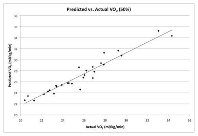
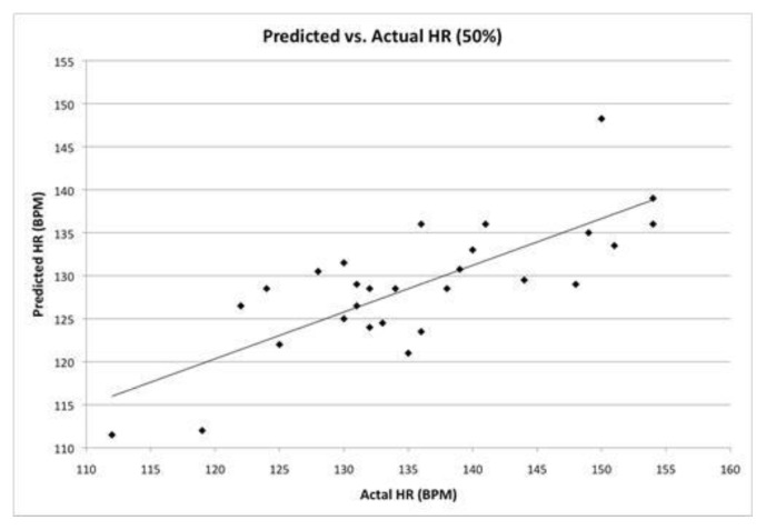
All results were reported as mean ± standard deviation. Statistical analysis revealed that the predicted 50%VO2 (27.1 ± 3.3) was significantly greater than the actual VO2 achieved (25.6 ± 3.3), t(27) = 8.2, p < .001 (Figure 1). It was also found that the mean predicted HR at 50% of the reserve (128.8 ± 7.5) was significantly less than the actual HR achieved (135.6 ± 10.6), t(27) = 5.4, p < .001 (Figure 2).
Figure 1.
Regression line of predicted and actual VO2 @ 50%R. Shows that a strong correlation existed (r = .96) and that predicted VO2 was significantly greater (27.1 ± 3.3) than actual VO2 achieved (25.6 ± 3.3), t(27) = 8.2, p < .001.
Figure 2.
Regression line of predicted and actual HR @ 50%R. Shows that a moderate correlation existed (r = .78) and that predicted HR was significantly less (128.8 ± 7.5) than actual HR achieved (135.6 ± 10.6), t(27) = 5.430, p < .001.
The correlation between measured and predicted VO2 at 50%R was r = .96, p < .001, with an SEE = .94 mL/kg/min. The correlation between measured and predicted HR at 50%R was r = .78, p < .001, with an SEE = 6.6 bpm.
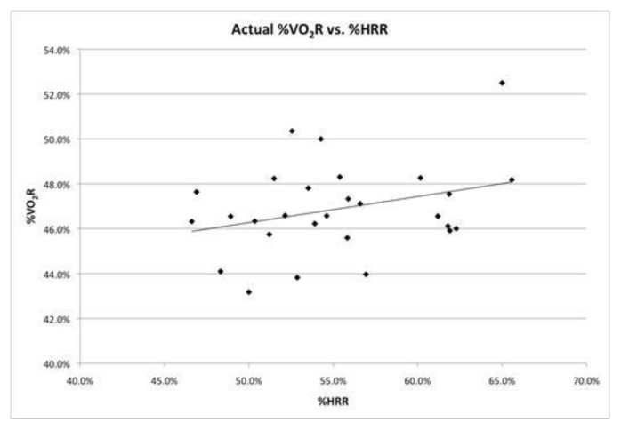
In addition to t-tests, Pearson’s r was used to analyze the correlation between %VO2R and %HRR during moderate intensity exercise. It was found that a statistically non-significant, weak positive, relationship existed between the two variables (r = .31, p = .105) (Figure 3).
Figure 3.
Regression line for %HRR at a given %VO2R. A statistically non-significant, weak positive, relationship existed between the two variables (r = .31, p = .105).
DISCUSSION
The primary purpose of this study was to determine the relationship between VO2R and HRR during moderate intensity exercise. Secondly, determining the predictability of VO2 and HR at moderate intensity exercise. It was hypothesized that VO2R and HRR would be directly correlated at moderate intensity exercise (50%R). It was also hypothesized that VO2 and HR could accurately be predicted. This study indicated that VO2 could be more accurately predicted than HR during moderate intensity exercise. Also, a weak positive correlation existed between VO2R and HRR. This indicated that it was less reliable to predict one variable given another (i.e., HR cannot be accurately predicted when the VO2 of a given exercise bout is known). On the basis of a 50%R (moderate intensity) prediction, mean %VO2R values were over-predicted (Actual %VO2R = 46.9% ± 2.0%) whereas mean %HRR values were under-predicted (Actual %HRR = 55.3% ± 5.4%).
Our results on the prediction of VO2 supported the findings of Cunha et al., which examined the association between VO2R and HRR as the intensity, and duration, of exercise were increased (28 physically active males). Cunha’s study concluded that predicted VO2 values were significantly greater than actual VO2 values. Additionally, it was found that as exercise intensity increased the strength of the relationship between %VO2R and %HRR weakened (3).
The results of our study were shown to overpredict VO2, however, the SEE value of .94 mL/kg/min suggests that accurate predictions may still be made in most practical circumstances. This finding supports the research conducted on 26 highly trained, healthy male cyclists by Lounana et al. They found that VO2 could be accurately predicted when using reserve values (5).
However, the results of this study that were dependent on HR data deviated from the majority of previous research findings. The majority of past research had found a direct correlation between %VO2R and %HRR (i.e., enabling accurate predictions of HR from VO2). Swain et al. investigated the relationship between %VO2R and %HRR in apparently healthy individuals (26 males, 24 women, 18–20 yrs) during treadmill exercise they found that the regression between VO2R and %HRR was r = 0.99 ± 0.002 (7) whereas our results found a weak correlation (r = .31).
These differences could have been the result of many different factors, the first of which is elevated levels of catecholamines in the blood circulating throughout the body. Catecholamines are hormones released by the body during exercise and are responsible for elevating blood pressure, breathing rate, and heart rate (6). Many participants in this study had exercised prior to performing the submaximal portion of the study. This would have resulted in elevated levels of these hormones. It was assumed that by lying prone for 10 minutes, an accurate resting HR, as well as an accurate ambulatory HR would be achieved. However, it is plausible that catecholamines were still circulating through the system, causing the participants resting HR to be elevated above that which was predicted.
Some other limitations consisted of making the assumption of 3.5mL/kg/min as the value for resting VO2 as well as inconsistency of HR measurements. The value of 3.5mL/kg/min was assumed for all participants as resting VO2 but upon further research it was found that resting VO2 has been shown to vary greatly from subject to subject. Cunha et al. found that their subjects measured resting VO2 values were significantly greater than the assumed value of 3.5mL/kg/min (4.3 ± 0.9 mL/kg/min, p < .001) (3). Conversely, in a study conducted by Byrne et al. that looked at two separate cohorts (593 women, 78 men, 18–74 yrs; 49 women 49 men, 38 ± 5 yr) resting VO2 values were found to be significantly lower than 3.5mL/kg/min (2.6 ± 0.4 mL/kg/min) (2).
When collecting exercising HR data, a HR monitor was used. However, resting HR’s were palpated, using the radial pulse, for fifteen seconds and multiplied by four. This method, while convenient and easy to use, has a large margin for error. The counting of one extra or one less beat would result in a difference of ± 4 beats per minute. By using a HR monitor to obtain both resting and exercising HR’s better consistency in readings would have been possible. A more meticulous approach to the collection of data could potentially indicate that HR can be accurately predicted using reserves. Furthermore, better collection of HR data could also indicate that a direct relationship between %VO2R and %HRR does exist. Such results would support the findings of Brawner and associates, Dalleck and Kravitz, Suain and Leutholtz, and Swain and colleagues (1,4,8,9). Lastly, it is possible that not all participants reached the desired exercise intensity, as the workload was a predictive calculation derived from their maximal test.
In conclusion, this study indicated that VO2 could be more accurately predicted than HR during moderate intensity exercise. The weak correlation between VO2R and HRR indicates that caution should be used when relying on a HR to determine VO2.
REFERENCES
- 1.Brawner CA, Keteyian SJ, Ehrman JK. The relationship of heart rate reserve to VO2 reserve in patients with heart disease. Med Sci Sports Exerc. 2002;34(3):418–422. doi: 10.1097/00005768-200203000-00006. [DOI] [PubMed] [Google Scholar]
- 2.Byrne NM, Hills AP, Hunter GR, Weinsier RL, Schutz Y. Metabolic equivalent: one size does not fit all. J Appl Physiol. 2005;99:1112–1119. doi: 10.1152/japplphysiol.00023.2004. [DOI] [PubMed] [Google Scholar]
- 3.Cunha FA, Midgley AW, Monteiro WD, Campos FK, Farinatti PTV. The relationship between oxygen uptake reserve and heart rate reserve is affected by intensity and duration during aerobic exercise at constant work rate. Appl Physiol Nutr Metab. 2011;36(6):839–847. doi: 10.1139/h11-100. [DOI] [PubMed] [Google Scholar]
- 4.Dalleck LC, Kravitz L. Relationship between % heart rate reserve and % VO2 reserve during elliptical crosstrainer exercise. J Sports Sci and Med. 2006;5:662–671. [PMC free article] [PubMed] [Google Scholar]
- 5.Lounana J, Campion F, Noakes TD, Medelli J. Relationship between %HRmax, %HR Reserve, %VO2max, and %VO2 Reserve in Elite Cyclists. Med Sci Sport Exer. 2007;39(2):350–357. doi: 10.1249/01.mss.0000246996.63976.5f. [DOI] [PubMed] [Google Scholar]
- 6.Schwarz NA, Spillane M, Bounty PL, Grandjean PW, Leutholtz B, Willoughby DS. Capsaicin and evodiamine ingestion does not augment energy expenditure and fat oxidation at rest or after moderately-intense exercise. Nutr Res. 2013;33:1034–1042. doi: 10.1016/j.nutres.2013.08.007. [DOI] [PubMed] [Google Scholar]
- 7.Swain DP, Leutholtz BC. Heart rate reserve is equivalent to % VO2Reserve, not to %VO2max. Med Sci Sports and Exer. 1997;29(3):410–414. doi: 10.1097/00005768-199703000-00018. [DOI] [PubMed] [Google Scholar]
- 8.Swain DP, Leutholtz BC, King ME, Haas LA, Branch DJ. Relationship between % heart rate reserve and % VO2 reserve in treadmill exercise. Med Sci Sports Exer. 1998;30(2):318–321. doi: 10.1097/00005768-199802000-00022. [DOI] [PubMed] [Google Scholar]
- 9.Thompson WR, Gordon NF, Pescatello LS. ACSM’s Guidelines for Exercise Testing and Prescription. 8th ed. Philadelphia (Pa): Lippincott Williams & Wilkins; 2010. p. 453. [Google Scholar]
- 10.Thompson WR, Gordon NF, Pescatello LS. ACSM’s Guidelines for Exercise Testing and Prescription. 8th ed. Philadelphia (Pa): Lippincott Williams & Wilkins; 2010. p. 49. [Google Scholar]
- 11.Thompson WR, Gordon NF, Pescatello LS. ACSM’s Guidelines for Exercise Testing and Prescription. 8th ed. Philadelphia (Pa): Lippincott Williams & Wilkins; 2010. p. 458. [Google Scholar]


