Abstract
Vibrio parahaemolyticus is an important seafood borne human pathogen worldwide due to it occurrence, prevalence and ability to cause gastrointestinal infections. This current study aim at investigating the incidence and prevalence of V. parahaemolyticus in seafood using systematic review-meta-analysis by exploring heterogeneity among primary studies. A comprehensive systematic review and meta-analysis of peer reviewed primary studies reported between 2003 and 2015 for the occurrence and prevalence of V. parahaemolyticus in seafood was conducted using “isolation”, “detection”, “prevalence”, “incidence”, “occurrence” or “enumeration” and V. parahaemolyticus as search algorithms in Web of Science (Science Direct) and ProQuest of electronic bibliographic databases. Data extracted from the primary studies were then analyzed with fixed effect meta-analysis model for effect rate to explore heterogeneity between the primary studies. Publication bias was evaluated using funnel plot. A total of 10,819 articles were retrieved from the data bases of which 48 studies met inclusion criteria. V. parahaemolyticus could only be isolated from 2761 (47.5 %) samples of 5811 seafood investigated. The result of this study shows that incidence of V. parahaemolyticus was more prevalent in oysters with overall prevalence rate of 63.4 % (95 % CI 0.592–0.674) than other seafood. Overall prevalence rate of clams was 52.9 % (95 % CI 0.490–0.568); fish 51.0 % (95 % CI 0.476–0.544); shrimps 48.3 % (95 % CI 0.454–0.512) and mussels, scallop and periwinkle: 28.0 % (95 % CI 0.255–0.307). High heterogeneity (p value <0.001; I2 = 95.291) was observed mussel compared to oysters (I2 = 91.024). It could be observed from this study that oysters harbor V. parahaemolyticus based on the prevalence rate than other seafood investigated. The occurrence and prevalence of V. parahaemolyticus is of public health importance, hence, more studies involving seafood such as mussels need to be investigated.
Keywords: Seafood safety and quality, Prevalence, Reservoir, V. parahaemolyticus, Shellfish
Background
Vibrio parahaemolyticus is a non-sucrose fermenting halophilic bacterium that grows between 10 and 44 °C and optimum temperature of 35–37 °C (Zamora-Pantoja et al. 2013; Wagley et al. 2009). The first outbreak of seafood borne disease due to consumption of V. parahaemolyticus contaminated sardine was reported in Japan in 1950 (Levin 2006). In this outbreak, 20 people were reported dead while over 270 people were likewise hospitalized. More outbreaks involving consumption of contaminated raw or undercooked seafood like oyster has been reported in United States (Iwamoto et al. 2010; McLaughlin et al. 2005; Drake et al. 2007), China (Liu et al. 2004), Taiwan (Chiou et al. 2000), Spain (Lozano-Leon et al. 2003), Italy (Ottaviani et al. 2008), Chile (Garcia et al. 2009), Peru (Gil et al. 2007) and (Leal et al. 2008) V. parahaemolyticus infection is characterized with vomiting, acute abdominal pain, abdominal pain, vomiting, watery or bloody diarrhea and gastroenteritis as result of production of thermostable direct hemolysin (TDH) and TDH-related hemolysin (TRH) toxins respectively (Jahangir Alam et al. 2002; Wagley et al. 2009) with an incubation period of 4–96 h (Levin 2006) however, non-pathogenic V. parahaemolyticus strains do not cause any infection. Several studies have been conducted globally regarding occurrence and prevalence of total or pathogenic V. parahaemolyticus in seafood yet there exist variability among the studies in terms of incidence and prevalence.
Meta-analysis is a quantitative statistical summarizing techniques aimed at extracting and combining scientific results from multiple primary studies that have investigated the same research question (Gonzales-Barron et al. 2013). Meta-analysis explains possible differences in outcomes of primary studies by extracting and encoding study characteristics such as research design features, data collection procedures, type of samples and year of study (DerSimonian and Laird 1986). This involves several steps like systematic review of literatures, data extraction of both qualitative and quantitative information from relevant primary studies, selection of effect size as described from each study, estimation of overall effect size of all the primary studies, assessment of heterogeneity of studies and presentation of meta-analysis using numerical (odd ratios, fixed effects size, p values, publication bias, meta regression, and random effect) and or graphical methods forest plot, funnel plot and others (Gonzales-Barron et al. 2013). Method of data generation differs from one study to another. Hence, researchers can either perform experiment to generate data or utilize available data from previous study (primary study) without experimental work (den Besten and Zwietering 2012). It was recently that food safety researchers stated conducting meta analytical studies as most meta-analytical study are conducted only in medical and social sciences (Gonzales Barron et al. 2008; Gonzales-Barron and Butler 2011; Patil et al. 2004). Meta-analytical studies could be carried out in food safety research in order to help answer various research questions involving prevalence pathogens in foods, treatment interventions, predictive modelling, microbial risk assessement, food safety knowledge, attitude and practices (Xavier et al. 2014).
Currently, no meta-analysis has been conducted on estimation of overall incidence, detection and prevalence of V. parahaemolyticus in seafood has been carried out in order to gain insight to source(s) of reservoir for these bacterial pathogens. This study therefore aim to systematically review and summarize primary studies describing incidence and prevalence of V. parahaemolyticus in seafood worldwide.
Methods
Definition
For the purpose of this study, incidence is defined as occurrence (presence) of V. parahaemolyticus in seafood samples analyzed in the primary studies while prevalence (p) is the number (n) of seafood that was positive for the presence of V. parahaemolyticus from the total sample (N). Primary studies imply all the studies carried out by other researchers used in this study. Population of study is the type of seafood investigated in each study. Seafood considered in this study are mollusks (oysters, clams, and mussels), finfish (salmon and tuna) and crustaceans (shrimp, crab, and lobster) (Iwamoto et al. 2010). In order to achieve the aim of this study, modified methods of Preferred Reporting Items for Systematic Reviews and Meta-Analyses—PRIMA (Moher et al. 2009) and (Gonzales-Barron and Butler 2011) were used. The steps consist of systematic review of literatures, data extraction of both qualitative and quantitative information from relevant primary studies, selection of effect size as described from each study, estimation of overall effect size of all the primary studies, assessment of heterogeneity of studies and meta-analysis representation of obtained result using numerical (odd ratios, fixed effects size, p values, publication bias, meta regression, and random effect) and or graphical methods forest plot, funnel plot and others).
Literature search, selection and relevance screening
This review was guided by a research question and problem statement. The research question was how prevalent is V. parahaemolyticus in seafood? While a problem statement describing the incidence and prevalence of V. parahaemolyticus in different seafood samples was formulated. Presence or absent of V. parahaemolyticus was considered as possible outcome of each primary study. Thereafter, a comprehensive literature search of electronic databases (ISI Web of science and ProQuest) and systematic review of available primary studies aimed at producing summary of relevant, quality and initial findings from such studies was carried out. The following search algorithms: “isolation” and V. parahaemolyticus, “detection” and V. parahaemolyticus, “prevalence” and V. parahaemolyticus, “incidence” and V. parahaemolyticus, “occurrence” and V. parahaemolyticus and “enumeration” and V. parahaemolyticus were used. Preliminary screening (Abstract-based relevance screening) of titles and abstracts of retrieved primary studies was carried out for eligibility and relevance to this study. Relevance of each article was screened using both inclusion and exclusion criteria. The inclusion criteria are: description of isolation method of V. parahaemolyticus from seafood using both conventional method (use of Thiosulphate Citrate Bile Salt agar—TCBS) and or molecular methods (Polymerase chain reaction—PCR). Full text and peer reviewed articles in English. The total number (population) of samples studied and number of samples that are positive for presence of V. parahaemolyticus clearly stated in the study. The exclusion criteria are: review articles, detection of V. parahaemolyticus in artificially contaminated samples, non-peer reviewed articles such as thesis, opinion articles, non-food related sources of V. parahaemolyticus such as clinical samples and conference abstract due to lack of access to full articles. Thereafter, full text screening of eligible primary studies were obtained from the databases. Articles that are not freely available were obtained via the service of the University of Tasmania’s library. Citations identified were retrieved and further checked for duplication using Endnote x7.1 software.
Data extraction and assessment of quality
Based on the inclusion and exclusion criteria, first author, year of publication or study, location, type of seafood studied, microbiological methods, number of sample positive for presence of V. parahaemolyticus were extracted.
Statistical analysis of extracted data
The pooled estimates of prevalence of V. parahaemolyticus in seafood were obtained by fixed effect meta-analysis model. The model was used to analyze combined extracted data while variation of incidence and prevalence of V. parahaemolyticus between the primary studies was evaluated using heterogeneity (I2). Heterogeneity of prevalence estimates between the studies was investigated using Q statistic (Bangar et al. 2014) and quantified by I2 Index (Higgins et al. 2003) as shown in below equations.
| 1 |
| 2 |
where df is the degree of freedom (N − 1), βw is the pooled estimate, βi is the estimate of individual primary study. Presence of bias in the publications was determined using funnel plots (odd of presence of V. parahaemolyticus in the samples) of standard error. Forest plots were however used to estimate the event rate at 95 % confidence intervals. Prevalence (p) and standard error (s.e.) were calculated by the following formulae: p = n/N and s.e. = √ p (1 − p)/N: where n = number of positive samples and N = number of samples (Tadesse and Tessema 2014). Modified method of (Greig et al. 2012) was used for the assessment of risk bias. Statistical analyses was carried out using Comprehensive Meta-Analysis (CMA) software. Statistical p values (p < 0.05) were considered as statistically significant.
Results and discussion
Literature search
The numbers of studies on V. parahaemolyticus has increased over the years. This current study is the first meta-analytical study to be carried out on incidence and prevalence of V. parahaemolyticus in seafood. Figure 1 shows results obtained from literature search. Literature search yielded 10,819 primary studies. However, when the source of articles was limited to peer review journals, 6876 articles were obtained. Further limiting of the subject to full text academic journals, V. parahaemolyticus, seafood and or shellfish, 149 articles were obtained. Abstract relevance screening of published articles reduced the study to 86 while only 63 articles remained after de-duplication. Hence, only few primary studies met the inclusion requirement of this meta-analysis. The primary studies considered in this meta-analysis described standard method for isolation and detection of V. parahaemolyticus from seafood samples. First author, year of publication or study, location of study, type of seafood studied, microbiological methods and number of sample positive for presence of V. parahaemolyticus were extracted from the following 48 primary studies: (Abd-Elghany and Sallam 2013; Amin and Salem 2012; Anjay et al. 2014; Bilung et al. 2005; Blanco-Abad et al. 2009; Chakraborty and Surendran 2008; Changchai and Saunjit 2014; Chao et al. 2009; Cook et al. 2002; Copin et al. 2012; Deepanjali et al. 2005; DePaola et al. 2003; Di Pinto et al. 2008, 2012; Dileep et al. 2003; Duan and Su 2005a, b; Eja et al. 2008; Fuenzalida et al. 2006, 2007; Han et al. 2007; Khouadja et al. 2013; Kirs et al. 2011; Koralage et al. 2012; Lee et al. 2008; Lu et al. 2006; Luan et al. 2008; Marlina et al. 2007; Miwa et al. 2006; Nakaguchi 2013; Nelapati and Krishnaiah 2010; Normanno et al. 2006; Ottaviani et al. 2005; Pal and Das 2010; Parveen et al. 2008; Paydar et al. 2013; Pereira et al. 2007; Raghunath et al. 2007; Ramos et al. 2014; Rizvi and Bej 2010; Robert-Pillot et al. 2014; Rosec et al. 2012; Schärer et al. 2011; Sobrinho Pde et al. 2011; Sobrinho et al. 2010; Sudha et al. 2012; Suffredini et al. 2014; Sun et al. 2012; Terzi et al. 2009; Vuddhakul et al. 2006; Xu et al. 2014; Yamamoto et al. 2008; Yang et al. 2008a, b; Yano et al. 2014; Zarei et al. 2012; Zhao et al. 2011; Zulkifli 2009). The outcome of this study revealed that oysters are more contaminated with this pathogen than other samples. It could be observed from this study that more studies have carried out on oyster than other samples. Oysters are eaten either raw or undercooked. This practice tend to increase the prevalence of outbreak of V. parahaemolyticus in oysters especially in countries like United States, China and Japan. There are limitations in meta-analysis study. Only studies that are published in English are used in this study. There could be possibility that positive results involving incidence of V. parahaemolyticus from other seafood are reported. This correlates with the publication bias observed in the study which involve publication of study with significant results. Additionally, primary research studies involving clinical samples were not included in this study
Fig. 1.
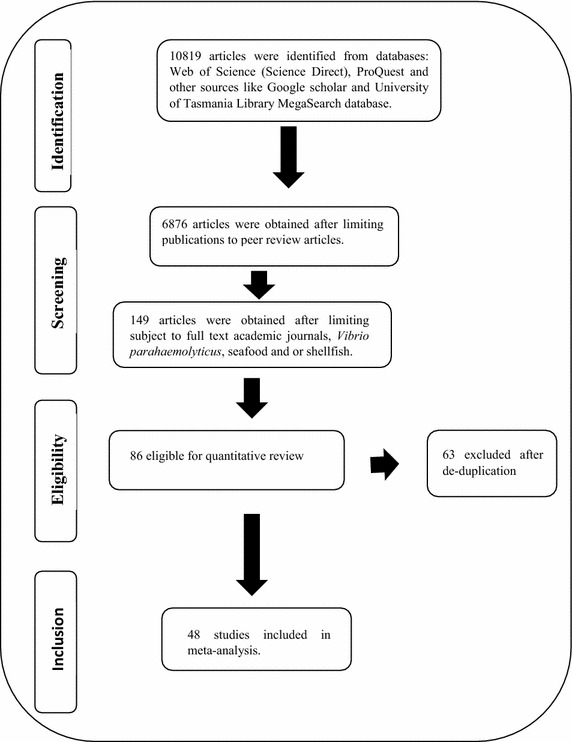
Flow diagram of selected studies included in fixed effect meta-analysis
Descriptive characteristics of eligible studies
As seen in Table 1, the studies were conducted and published between 2003 and 2015 from the following 24 countries: Brazil (3 studies); India (6 studies); Iran (1 study); United Kingdom (1 study); China (5 studies); Thailand (4 studies); Vietnam (1 study); Malaysia (3 studies); Indonesia (3 studies); Italy (5 studies); Japan (1 study); Chile (1 study); Egypt (2 studies); United States (3 studies); Turkey (1 study); France (3 studies); Spain (1 study); Mexico (1 study); Korea (1 study); Sri Lanka (1 study); Nigeria (1 study); Tunisia (1 study); New Zealand (1 study) and Switzerland (1 study). V. parahaemolyticus was isolated from 2761 (47.5 %) of 5811 mussel, scallop and periwinkle (1670) in 15 studies, oyster (951) in 17 studies, clam and cockle (830) in 18 studies,, shrimps, prawn and crab (1422) in 23 studies, fish, squid and cephalopod (998) in 20 studies of seafood investigated.
Table 1.
Descriptive characteristic of eligible studies in meta-analysis
| Sn | Sr | Ls | Yp | Ts | M | N | n | P (%) |
|---|---|---|---|---|---|---|---|---|
| 1 | Sobrinho Pde et al. (2011) | Brazil | 2011 | Oyster | TCBS/PCRm | 74 | 74 | 100 |
| 2 | Sudha et al. (2012) | India | 2012 | Finfish | TCBS/PCR | 182 | 82 | 45.1 |
| 3 | Zarei et al. (2012) | Iran | 2012 | Shrimps | TCBS/PCR | 300 | 146 | 43.9 |
| 4 | Wagley et al. (2009) | England | 2009 | Crabs | TCBS/PCR | 22 | 22 | 100 |
| 5 | Zhao et al. (2011) | Chinaa | 2011 | Oyster | TCBS/PCR | 80 | 39 | 48.8 |
| Clam | TCBS/PCR | 72 | 46 | 63.8 | ||||
| Scallop | TCBS/PCR | 70 | 42 | 60.0 | ||||
| Mussel | TCBS/PCR | 76 | 45 | 59.2 | ||||
| 6 | Nakaguchi (2013) | Thailand | 2013 | Cockle | TCBS/PCR | 109 | 76 | 69.4 |
| Mussel | TCBS/PCR | 73 | 54 | 74.5 | ||||
| Oyster | TCBS/PCR | 32 | 27 | 83.3 | ||||
| Clam | TCBS/PCR | 86 | 52 | 60.0 | ||||
| Vietnam | Fish | TCBS/PCR | 16 | 10 | 62.5 | |||
| Shrimp | TCBS/PCR | 18 | 13 | 73.2 | ||||
| Squid | TCBS/PCR | 7 | 2 | 28.6 | ||||
| Crab | TCBS/PCR | 5 | 2 | 40.0 | ||||
| Malaysia | Fish | TCBS/PCR | 11 | 6 | 54.5 | |||
| Squid | TCBS/PCR | 11 | 6 | 54.5 | ||||
| Indonesia | Shrimp | TCBS/PCR | 37 | 23 | 62.1 | |||
| Squid | TCBS/PCR | 29 | 4 | 13.8 | ||||
| 7 | Di Pinto et al. (2008) | Italy | 2008 | Mussel | TCBS/PCR | 144 | 47 | 32.6 |
| 8 | Yamamoto et al. (2008) | Thailandb | 2008 | Clams | MPNk/PCR | 32 | 32 | 100 |
| 9 | Miwa et al. (2006) | Japan | 2006 | Fish | MPN/PCR | 30 | 4 | 13.3 |
| Shrimp | MPN/PCR | 20 | 11 | 55.0 | ||||
| Cockle | MPN/PCR | 10 | 9 | 90 | ||||
| 10 | Fuenzalida et al. (2006) | Chile | 2006 | Mussel | TCBS/PCR | 35 | 9 | 25.7 |
| Clam | TCBS/PCR | 8 | 2 | 25 | ||||
| Oyster | TCBS/PCR | 5 | 1 | 20 | ||||
| 11 | Anjay et al. (2014) | India | 2014 | Fish | TCBS/PCR | 182 | 140 | 76.9 |
| Prawn | TCBS/PCR | 42 | 31 | 73.8 | ||||
| 12 | Abd-Elghany and Sallam (2013) | Egypt | 2013 | Shrimp | TCBS/PCR | 40 | 9 | 22.5 |
| Crab | TCBS/PCR | 40 | 8 | 20 | ||||
| Cockle | TCBS/PCR | 40 | 3 | 7.5 | ||||
| 13 | Changchai and Saunjit (2014) | Thailand | 2014 | Raw oystersl | MPN/PCR | 240 | 219 | 91 |
| 14 | Ramos et al. (2014) | Brazil | 2014 | Oyster | MPN/PCR | 60 | 29 | 48.3 |
| 15 | Chakraborty and Surendran (2008) | India | 2008 | Finfish | TCBS/MPN | 12 | 8 | 66.6 |
| Shellfish | TCBS/MPN | 25 | 21 | 84.0 | ||||
| Cephalopods | TCBS/MPN | 5 | 4 | 80 | ||||
| 16 | Bilung et al. (2005) | Malaysia | 2005 | Cockle | MPN/PCR | 100 | 62 | 62 |
| 17 | Rosec et al. (2012) | France | 2012 | Oyster | TCBS/C/PCR | 60 | 19 | 31.6 |
| Clams/mussel | TCBS/C/PCR | 9 | 1 | 11.1 | ||||
| 18 | Terzi et al. (2009) | Turkey | 2009 | Fish | TCBS/PCR | 30 | 9 | 30 |
| Mussel | TCBS/PCR | 60 | 35 | 58.3 | ||||
| 19 | Suffredini et al. (2014) | Italy | 2014 | Mussel | TCBS/PCR | 75 | 31 | 41.3 |
| Clams | TCBS/PCR | 51 | 22 | 43.1 | ||||
| 20 | Sun et al. (2012) | China | 2012 | Oyster | TCBS/LAMP | 10 | 2 | 20 |
| Clam | TCBS/LAMP | 16 | 2 | 12.5 | ||||
| 21 | Parveen et al. (2008) | US | 2008 | Oyster | TCBS/DCH/PCR | 33 | 22 | 67 |
| 22 | Di Pinto et al. (2012) | Italy | 2012 | Mussel | PCR/ELISA | 195 | 26 | 13.3 |
| 23 | Rizvi and Bej (2010) | Mexico | 2010 | Oyster | SYBR/PCR | 24 | 14 | 58.3 |
| 24 | Blanco-Abad et al. (2009) | Spain | 2009 | Mussel | TCBS/PCR | 48 | 5 | 10.4 |
| 25 | Marlina et al. (2007) | Indonesia | 2007 | Clam | RAPD/PCR | 35 | 13 | 37.1 |
| 26 | Luan et al. (2008) | China | 2008 | Shrimp | MPN/PCR | 80 | 66 | 82.5 |
| Crab | MPN/PCR | 15 | 14 | 93.3 | ||||
| Clam | MPN/PCR | 100 | 64 | 64 | ||||
| Fish | MPN/PCR | 10 | 10 | 100 | ||||
| Scallop | MPN/PCR | 20 | 11 | 55 | ||||
| 27 | Lu et al. (2006) | US | 2006 | Oyster | RAPD/PCR | 13 | 9 | 69 |
| Mussel | RAPD/PCR | 22 | 7 | 32 | ||||
| Clam | RAPD/PCR | 48 | 13 | 27 | ||||
| 28 | Robert-Pillot et al. (2014) | France | 2014 | Fish | RT/PCR | 27 | 5 | 18.5 |
| Mussel/Scallop | RT/PCR | 10 | 1 | 10 | ||||
| 29 | Zulkifli (2009) | Indonesia | 2009 | Cockle | C/PCR | 50 | 25 | 50 |
| 30 | Nelapati and Krishnaiah (2010) | India | 2010 | Fish | TCBS/PCR | 105 | 69 | 65.7 |
| 31 | Yano et al. (2014) | Thailand | 2014 | Shrimp | MPN/PCR | 16 | 6 | 37.5 |
| 32 | Duan and Su (2005a) | US | 2005 | Oyster | TCBS/PCR | 74 | 31 | 41.9 |
| 33 | Copin et al. (2012) | France | 2012 | Shrimp | MPN/PCR | 36 | 28 | 77.8 |
| 34 | Yang et al. (2008a) | China | 2008 | Fish | RADP/PCR | 197 | 58 | 29.7 |
| Crab | RADP/PCR | 49 | 22 | 44.9 | ||||
| Shrimp | RADP/PCR | 71 | 28 | 39.4 | ||||
| 35 | Ottaviani et al. (2005) | Italy | 2005 | Mussel | TCBS/PCR | 144 | 35 | 24.3 |
| 36 | Sobrinho et al. (2010) | Brazil | 2010 | Oyster | MPN/PCR | 123 | 122 | 99.2 |
| 37 | Xu et al. (2014) | China | 2014 | Shrimp | TCBS/PCR | 273 | 103 | 37.7 |
| 38 | Lee et al. (2008) | Korea | 2008 | Oyster | TCBS/PCR | 72 | 48 | 66.7 |
| 39 | Amin and Salem (2012) | Egypt | 2012 | Shrimp | TCBS/PCR | 20 | 4 | 20 |
| Crab | TCBS/PCR | 20 | 6 | 30 | ||||
| 40 | Koralage et al. (2012) | Sri Lanka | 2012 | Shrimp | TCBS/PCR | 170 | 155 | 91.2 |
| 41 | Schärer et al. (2011) | Switzerland | 2011 | Squid | TCBS/PCR | 2 | 2 | 100 |
| 42 | Paydar et al. (2013) | Malaysia | 2013 | Fish | TCBS/mPCR | 27 | 21 | 77.8 |
| Squid | TCBS/PCR | 7 | 4 | 57.1 | ||||
| Cockle | TCBS/PCR | 5 | 3 | 60 | ||||
| Shrimp | TCBS/PCR | 11 | 9 | 81.8 | ||||
| Clam | TCBS/PCR | 3 | 2 | 66.7 | ||||
| Prawn | TCBS/PCR | 7 | 5 | 71.4 | ||||
| Oyster | TCBS/PCR | 9 | 6 | 66.7 | ||||
| 43 | Dileep et al. (2003) | India | 2003 | Finfish | TCBS/PCR | 18 | 4 | 22.2 |
| Shrimp | TCBS/PCR | 10 | 3 | 30 | ||||
| 44 | Eja et al. (2008) | Nigeria | 2008 | Shrimp | TCBS/Biotyping | 120 | 26 | 21.7 |
| Clam | TCBS/Biotyping | 90 | 7 | 7.7 | ||||
| Periwinkle | TCBS/Biotyping | 98 | 9 | 9.2 | ||||
| 45 | Khouadja et al. (2013) | Tunisia | 2013 | Oyster | TCBS/PCR | 20 | 2 | 10.0 |
| Mussel | TCBS/PCR | 20 | 1 | 5.0 | ||||
| 46 | Kirs et al. (2011) | New Zealand | 2011 | Oyster | TCBS/RT/PCR | 58 | 55 | 94.8 |
| 47 | Normanno et al. (2006) | Italy | 2006 | Mussel | TCBS/API | 600 | 47 | 7.83 |
| 48 | Pal and Das (2010) | India | 2010 | Fish | TCBS/PCR | 90 | 60 | 66.7 |
i, shucked oyster; tb, Tillamook Bay; yb, Yaquina Bay; S, Selangor; pj, Padang and Jakarta; m, use of any molecular method like specie specific genes etc, k; mpn chrom agar; a, coastal province Jiangsu; China b, eastern coast of China. Sn = study number; Sr = study reference; Ls = location of study; Yp = year of publication; Ts = type of seafood; M = microbiological method(s); N = total sample; n = number of positive samples
Meta-analysis of prevalence of V. parahaemolyticus in mussel, scallop, and periwinkle
Meta-analysis of incidence and prevalence of V. parahaemolyticus in mussel, scallop, and periwinkle was carried out using data of 1670 samples from 15 studies. The results of estimates of prevalence are summarised in Table 2. The pooled prevalence estimate of V. parahaemolyticus was found to be 28.0 % (95 % CI 0.255–0.307) as shown in Table 2. The studies included in this meta-analysis were found to be of significant heterogeneity (Q = 297.293, df = 14, p < 0.001) between 15 studies. Heterogeneity quantified by I2 index was observed as 95.291 % as shown in the forest plot in Fig. 2. Squares represent effect estimates of individual studies with their 95 % confidence intervals of prevalence with size of squares proportional to the weight assigned to the study in the meta-analysis (Fig. 3).
Table 2.
Prevalence and meta-analysis statistics of V. parahaemolyticus in seafood investigated in the primary studies
| df | Sample | Effect size 95 % CI | Heterogeneity | Standard error | Variance | ||
|---|---|---|---|---|---|---|---|
| Q value | p value | I 2 | |||||
| 14 | Mussel, scallop, and periwinkle | 28.0 (0.255–0.307) | 297.293 | 0.0000 | 95.291 | 0.660 | 0.436 |
| 22 | Shrimp, prawn and crab | 48.3 (0.454–0.512) | 232.099 | 0.2590 | 90.521 | 0.484 | 0.2345 |
| 19 | Fish, squid and cephalopod | 51.0 (0.476–0.544) | 159.368 | 0.557 | 88.078 | 0.460 | 0.212 |
| 17 | Clam and cockle | 52.9 (0.490–0.568) | 132.490 | 0.145 | 87.169 | 0.429 | 0.184 |
| 16 | Oyster | 63.4 (0.592–0.674) | 178.260 | 0.0000 | 91.024 | 0.765 | 0.586 |
Q, Cochran’s test; I 2, inverse variance index; df, degree of freedom
Fig. 2.
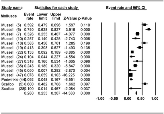
Forest plots of prevalence of V. parahaemolyticus in mussel, scallop and periwinkle for fixed effects meta-analyses. (Squares represent effect estimates of individual studies with their 95 % confidence intervals of prevalence with size of squares proportional to the weight assigned to the study in the meta-analysis)
Fig. 3.
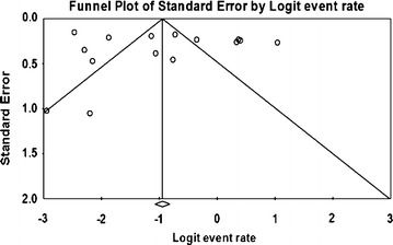
Funnel plot of prevalence of V. parahaemolyticus in mussel, scallop and periwinkle. Solid vertical line represents the summary prevalence rate derived from fixed-effect meta-analysis while the diagonal lines represent 95 % confidence interval
Meta-analysis of prevalence of V. parahaemolyticus in shrimp, prawn and crab
Meta-analysis of incidence and prevalence of V. parahaemolyticus in shrimp, prawn and crab was carried out using data of 1422 samples from 24 studies. The pooled prevalence estimate of V. parahaemolyticus was found to be 48.3 % (95 % CI 0.454–0.512). The primary studies included in this meta-analysis were found to be of significant heterogeneity (Q = 232.099, df = 22, p > 0.001) between 24 studies. Heterogeneity quantified by I2 index was observed as 90.521 % as shown in the forest plot in Fig. 4. Squares represent effect estimates of individual studies with their 95 % confidence intervals of prevalence with size of squares proportional to the weight assigned to the study in the meta-analysis (Fig. 5).
Fig. 4.
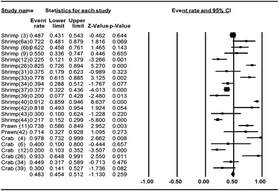
Forest plots of prevalence of V. parahaemolyticus in shrimp, prawn and crab for fixed effects meta-analyses. (Squares represent effect estimates of individual studies with their 95 % confidence intervals of prevalence with size of squares proportional to the weight assigned to the study in the meta-analysis)
Fig. 5.
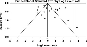
Funnel plot of prevalence of V. parahaemolyticus in shrimp, prawn and crab. Solid vertical line represents the summary prevalence rate derived from fixed-effect meta-analysis while the diagonal lines represent 95 % confidence interval
Meta-analysis of prevalence of V. parahaemolyticus in fish, squid and cephalopod
Meta-analysis of incidence and prevalence of V. parahaemolyticus in fish, squid and cephalopod was carried out using data of 998 samples from 20 studies. The pooled prevalence estimate of V. parahaemolyticus was found to be 51.0 % (95 % CI 0.476–0.544). The studies included in this meta-analysis were has found to be significant heterogeneity (Q = 159.368, df = 19, p > 0.001) between 20 studies. Heterogeneity quantified by I2 index was observed as 88.078 % as shown in the forest plot in Fig. 6. Squares represent effect estimates of individual studies with their 95 % confidence intervals of prevalence with size of squares proportional to the weight assigned to the study in the meta-analysis (Fig. 7).
Fig. 6.
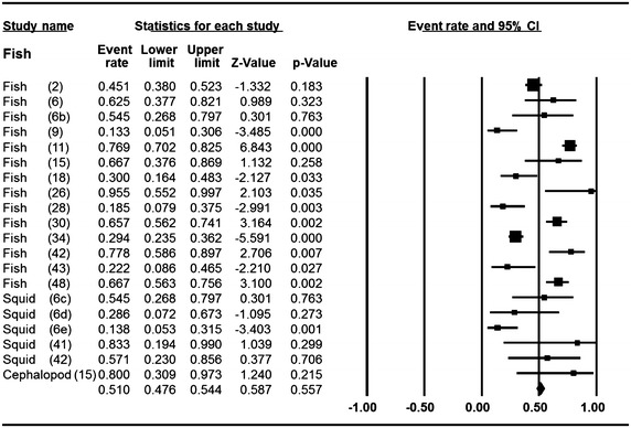
Forest plots of prevalence of V. parahaemolyticus in fish, squid and cephalopod for fixed effects meta-analyses. (Squares represent effect estimates of individual studies with their 95 % confidence intervals of prevalence with size of squares proportional to the weight assigned to the study in the meta-analysis)
Fig. 7.
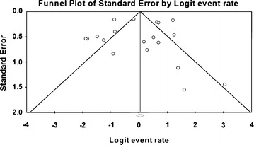
Funnel plot of prevalence of V. parahaemolyticus in fish, squid and cephalopod. Solid vertical line represents the summary prevalence rate derived from fixed-effect meta-analysis while the diagonal lines represent 95 % confidence interval
Meta-analysis of prevalence of V. parahaemolyticus in clam and cockle
Meta-analysis of incidence and prevalence of V. parahaemolyticus in clam and cockle was carried out using data of 830 samples from 18 studies. The pooled prevalence estimate of V. parahaemolyticus was found to be 52.9 % (95 % CI 0.490–0.568). The studies included in this meta-analysis were has found to be significant heterogeneity (Q = 132.490, df = 17, p > 0.001) between 18 studies. Heterogeneity quantified by I2 index was observed as 87.169 % as shown in the forest plot in Fig. 8. Squares represent effect estimates of individual studies with their 95 % confidence intervals of prevalence with size of squares proportional to the weight assigned to the study in the meta-analysis (Fig. 9).
Fig. 8.
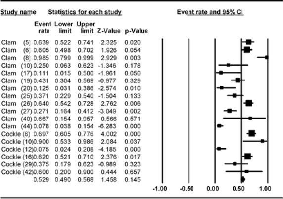
Forest plots of prevalence of V. parahaemolyticus in clam and cockle for fixed effects meta-analyses. (Squares represent effect estimates of individual studies with their 95 % confidence intervals of prevalence with size of squares proportional to the weight assigned to the study in the meta-analysis)
Fig. 9.
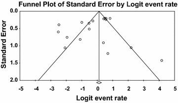
Funnel plot of prevalence of V. parahaemolyticus in clam and cockle. Solid vertical line represents the summary prevalence rate derived from fixed-effect meta-analysis while the diagonal lines represent 95 % confidence interval
Meta-analysis of prevalence of V. parahaemolyticus in oyster
Meta-analysis of incidence and prevalence of V. parahaemolyticus in oyster was carried out using data of 951 samples from 17 studies. The pooled prevalence estimate of V. parahaemolyticus was found to be 63.40 % (95 % CI 0.592–0.674). The studies included in this meta-analysis were has found to be significant heterogeneity (Q = 178.260, df = 16, p < 0.001) between 17 studies. Heterogeneity quantified by I2 index was observed as 91.024 % as shown in the forest plot in Fig. 10. Squares represent effect estimates of individual studies with their 95 % confidence intervals of prevalence with size of squares proportional to the weight assigned to the study in the meta-analysis (Fig. 11).
Fig. 10.
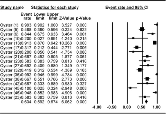
Forest plots of prevalence of V. parahaemolyticus in oyster for fixed effects meta-analyses. (Squares represent effect estimates of individual studies with their 95 % confidence intervals of prevalence with size of squares proportional to the weight assigned to the study in the meta-analysis)
Fig. 11.
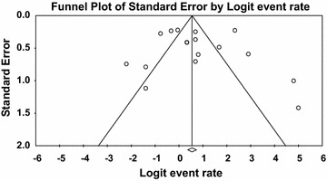
Funnel plot of prevalence of V. parahaemolyticus in oyster. Solid vertical line represents the summary prevalence rate derived from fixed-effect meta-analysis while the diagonal lines represent 95 % confidence interval
Publication bias among the primary studies
Both publication bias and quality of primary studies are limiting factors in any meta-analytical study (Noble Jr. 2006). In meta-analysis, publication bias is usually graphically assessed using funnel plot (Soon et al. 2012; Gonzales-Barron and Butler 2011). This was obtained by plotting of study size (usually standard error or precision) on the vertical axis as a function of effect size on the horizontal axis. In this current study, publication bias could be observed among the primary studies due to asymmetric nature of the plots. Solid vertical line in the funnel plots represents the summary of prevalence rate derived from fixed-effect meta-analysis while the diagonal lines represent 95 % confidence interval. Studies with large samples appeared toward the top of the graph, and tend to cluster near the mean effect size while studies with smaller samples appeared toward the bottom of the graph. It should be noted that sampling variation in effect size estimates in the studies with smaller seafood samples affects the plots.
Conclusion
In conclusion, higher prevalence rate of V. parahaemolyticus was observed in oysters than other seafood investigated. The occurrence and prevalence of V. parahaemolyticus is of public health importance, hence, more studies involving seafood such as mussels need to be investigated. Additionally, the study is a trial to develop a new data analysis tool. There is need to investigate prevalence of this pathogen in other seafood and also intervention strategies to reduce V. parahaemolyticus in seafood.
Acknowledgements
The University of Tasmania is appreciated for provision of Tasmania Graduate Research Scholarship (TGRS) and University of Tasmania Full Tuition Scholarship.
Competing interests
The author declares no competing interest.
References
- Abd-Elghany SM, Sallam KI. Occurrence and molecular identification of Vibrio parahaemolyticus in retail shellfish in Mansoura, Egypt. Food Control. 2013;33:399–405. doi: 10.1016/j.foodcont.2013.03.024. [DOI] [Google Scholar]
- Amin RA, Salem AM. Specific detection of pathogenic Vibrio species in shellfish by using multiplex polymerase chain reaction. Glob Vet. 2012;8:525–531. [Google Scholar]
- Anjay SC, Das A, Kumar P Kaushik, Kurmi B. Occurrence of Vibrio parahaemolyticus in marine fish and shellfish. Indian J Geo-Mar Sci. 2014;43:887–890. [Google Scholar]
- Bangar YC, Singh B, Dohare AK, Verma MR. A systematic review and meta-analysis of prevalence of subclinical mastitis in dairy cows in India. Trop Anim Health Prod. 2014;47:291–297. doi: 10.1007/s11250-014-0718-y. [DOI] [PubMed] [Google Scholar]
- Bilung LM, Radu S, Bahaman AR, Rahim RA, Napis S, Ling MW, Tanil GB, Nishibuchi M. Detection of Vibrio parahaemolyticus in cockle (Anadara granosa) by PCR. FEMS Microbiol Lett. 2005;252:85–88. doi: 10.1016/j.femsle.2005.08.053. [DOI] [PubMed] [Google Scholar]
- Blanco-Abad V, Ansede-Bermejo J, Rodriguez-Castro A, Martinez-Urtaza J. Evaluation of different procedures for the optimized detection of Vibrio parahaemolyticus in mussels and environmental samples. Int J Food Microbiol. 2009;129:229–236. doi: 10.1016/j.ijfoodmicro.2008.11.028. [DOI] [PubMed] [Google Scholar]
- Chakraborty R, Surendran PK. Occurrence and distribution of virulent strains of Vibrio parahaemolyticus in seafoods marketed from Cochin (India) World J Microbiol Biotechnol. 2008;24:1929–1935. doi: 10.1007/s11274-008-9698-2. [DOI] [Google Scholar]
- Changchai N, Saunjit S. Occurrence of Vibrio parahaemolyticus and Vibrio vulnificus in retail raw oysters from the eastern coast of Thailand. Southeast Asian J Trop Med Public Health. 2014;45:662–669. [PubMed] [Google Scholar]
- Chao G, Jiao X, Zhou X, Yang Z, Huang J, Zhou L, Qian X. Distribution, prevalence, molecular typing, and virulence of Vibrio parahaemolyticus isolated from different sources in coastal province Jiangsu, China. Food Control. 2009;20:907–912. doi: 10.1016/j.foodcont.2009.01.004. [DOI] [Google Scholar]
- Chiou C-S, Hsu S-Y, Chiu S-I, Wang T-K, Chao C-S. Vibrio parahaemolyticus serovar O3:K6 as cause of unusually high incidence of food-borne disease outbreaks in Taiwan from 1996 to 1999. J Clin Microbiol. 2000;38:4621–4625. doi: 10.1128/jcm.38.12.4621-4625.2000. [DOI] [PMC free article] [PubMed] [Google Scholar]
- Cook DW, Bowers JC, DePaola A. Density of total and pathogenic (tdh+) Vibrio parahaemolyticus in Atlantic and Gulf Coast molluscan shellfish at harvest. J Food Prot. 2002;65:1873–1880. doi: 10.4315/0362-028x-65.12.1873. [DOI] [PubMed] [Google Scholar]
- Copin S, Robert-Pillot A, Malle P, Quilici ML, Gay M. Evaluation of most-probable-number–PCR method with internal amplification control for the counting of total and pathogenic Vibrio parahaemolyticus in frozen shrimps. J Food Prot. 2012;75:150–153. doi: 10.4315/0362-028X.JFP-11-165. [DOI] [PubMed] [Google Scholar]
- Deepanjali A, Kumar HS, Karunasagar I. Seasonal variation in abundance of total and pathogenic Vibrio parahaemolyticus bacteria in oysters along the southwest coast of India. Appl Environ Microbiol. 2005;71:3575–3580. doi: 10.1128/AEM.71.7.3575-3580.2005. [DOI] [PMC free article] [PubMed] [Google Scholar]
- den Besten HMW, Zwietering MH. Meta-analysis for quantitative microbiological risk assessments and benchmarking data. Trends Food Sci Technol. 2012;25:34–39. doi: 10.1016/j.tifs.2011.12.004. [DOI] [Google Scholar]
- DePaola A, Nordstrom JL, Bowers JC, Wells JG, Cook DW. Seasonal abundance of total and pathogenic Vibrio parahaemolyticus in Alabama oysters. Appl Environ Microbiol. 2003;69:1521–1526. doi: 10.1128/AEM.69.3.1521-1526.2003. [DOI] [PMC free article] [PubMed] [Google Scholar]
- DerSimonian R, Laird N. Meta-analysis in clinical trials. Control Clin Trials. 1986;7:177–188. doi: 10.1016/0197-2456(86)90046-2. [DOI] [PubMed] [Google Scholar]
- Di Pinto A, Ciccarese G, De Corato R, Novello L, Terio V. Detection of pathogenic Vibrio parahaemolyticus in southern Italian shellfish. Food Control. 2008;19:1037–1041. doi: 10.1016/j.foodcont.2007.10.013. [DOI] [Google Scholar]
- Di Pinto A, Terio V, Di Pinto P, Colao V, Tantillo G. Detection of Vibrio parahaemolyticus in shellfish using polymerase chain reaction-enzyme-linked immunosorbent assay. Lett Appl Microbiol. 2012;54:494–498. doi: 10.1111/j.1472-765X.2012.03231.x. [DOI] [PubMed] [Google Scholar]
- Dileep V, Kumar H, Kumar Y, Nishibuchi M, Karunasagar I, Karunasagar I. Application of polymerase chain reaction for detection of Vibrio parahaemolyticus associated with tropical seafoods and coastal environment. Lett Appl Microbiol. 2003;36:423–427. doi: 10.1046/j.1472-765X.2003.01333.x. [DOI] [PubMed] [Google Scholar]
- Drake SL, DePaola A, Jaykus L-A. An overview of Vibrio vulnificus and Vibrio parahaemolyticus. Compr Rev Food Sci Food Saf. 2007;6:120–144. doi: 10.1111/j.1541-4337.2007.00022.x. [DOI] [Google Scholar]
- Duan J, Su Y-C. Comparison of a chromogenic medium with Thiosulfate-citrate-bile salts-sucrose agar for detecting Vibrio parahaemolyticus. J Food Sci. 2005;70:M125–M128. doi: 10.1111/j.1365-2621.2005.tb07102.x. [DOI] [Google Scholar]
- Duan J, Su Y-C. Occurrence of Vibrio parahaemolyticus in two Oregon oyster-growing bays. J Food Sci. 2005;70:M58–M63. doi: 10.1111/j.1365-2621.2005.tb09047.x. [DOI] [Google Scholar]
- Eja M, Abriba C, Etok C, Ikpeme E, Arikpo G, Enyi-Idoh K, Ofor U. Seasonal occurrence of Vibrios in water and shellfish obtained from the Great Kwa river estuary, Calabar, Nigeria. Bull Environ Contam Toxicol. 2008;81:245–248. doi: 10.1007/s00128-008-9482-x. [DOI] [PubMed] [Google Scholar]
- Fuenzalida L, Hernández C, Toro J, Rioseco ML, Romero J, Espejo RT. Vibrio parahaemolyticus in shellfish and clinical samples during two large epidemics of diarrhoea in southern Chile. Environ Microbiol. 2006;8:675–683. doi: 10.1111/j.1462-2920.2005.00946.x. [DOI] [PubMed] [Google Scholar]
- Fuenzalida L, Armijo L, Zabala B, Hernández C, Rioseco ML, Riquelme C, Espejo RT. Vibrio parahaemolyticus strains isolated during investigation of the summer 2006 seafood related diarrhea outbreaks in two regions of Chile. Int J Food Microbiol. 2007;117:270–275. doi: 10.1016/j.ijfoodmicro.2007.03.011. [DOI] [PubMed] [Google Scholar]
- Garcia K, Torres R, Uribe P, Hernandez C, Rioseco ML, Romero J, Espejo RT. Dynamics of clinical and environmental Vibrio parahaemolyticus strains during seafood-related summer diarrhea outbreaks in southern Chile. Appl Environ Microbiol. 2009;75:7482–7487. doi: 10.1128/AEM.01662-09. [DOI] [PMC free article] [PubMed] [Google Scholar]
- Gil AI, Miranda H, Lanata CF, Prada A, Hall ER, Barreno CM, Nusrin S, Bhuiyan NA, Sack DA, Nair GB. O3:K6 serotype of Vibrio parahaemolyticus identical to the global pandemic clone associated with diarrhea in Peru. Int J Infect Dis. 2007;11:324–328. doi: 10.1016/j.ijid.2006.08.003. [DOI] [PubMed] [Google Scholar]
- Gonzales Barron U, Bergin D, Butler F. A meta-analysis study of the effect of chilling on prevalence of Salmonella on pig carcasses. J Food Prot. 2008;71:1330–1337. doi: 10.4315/0362-028x-71.7.1330. [DOI] [PubMed] [Google Scholar]
- Gonzales-Barron U, Butler F. The use of meta-analytical tools in risk assessment for food safety. Food Microbiol. 2011;28:823–827. doi: 10.1016/j.fm.2010.04.007. [DOI] [PubMed] [Google Scholar]
- Gonzales-Barron U, Cadavez V, Sheridan JJ, Butler F. Modelling the effect of chilling on the occurrence of Salmonella on pig carcasses at study, abattoir and batch levels by meta-analysis. Int J Food Microbiol. 2013;163:101–113. doi: 10.1016/j.ijfoodmicro.2013.03.006. [DOI] [PubMed] [Google Scholar]
- Greig JD, Waddell L, Wilhelm B, Wilkins W, Bucher O, Parker S, Rajić A. The efficacy of interventions applied during primary processing on contamination of beef carcasses with Escherichia coli: a systematic review-meta-analysis of the published research. Food Control. 2012;27:385–397. doi: 10.1016/j.foodcont.2012.03.019. [DOI] [Google Scholar]
- Han F, Walker RD, Janes ME, Prinyawiwatkul W, Ge B. Antimicrobial susceptibilities of Vibrio parahaemolyticus and Vibrio vulnificus isolates from Louisiana Gulf and retail raw oysters. Appl Environ Microbiol. 2007;73:7096–7098. doi: 10.1128/AEM.01116-07. [DOI] [PMC free article] [PubMed] [Google Scholar]
- Higgins JP, Thompson SG, Deeks JJ, Altman DG. Measuring inconsistency in meta-analyses. BMJ. 2003;327:557–560. doi: 10.1136/bmj.327.7414.557. [DOI] [PMC free article] [PubMed] [Google Scholar]
- Iwamoto M, Ayers T, Mahon BE, Swerdlow DL. Epidemiology of seafood-associated infections in the United States. Clin Microbiol Rev. 2010;23:399–411. doi: 10.1128/CMR.00059-09. [DOI] [PMC free article] [PubMed] [Google Scholar]
- Jahangir Alam M, Tomochika K-I, Miyoshi S-I, Shinoda S. Environmental investigation of potentially pathogenic Vibrio parahaemolyticus in the Seto-Inland Sea, Japan. FEMS Microbiol Lett. 2002;208:83–87. doi: 10.1111/j.1574-6968.2002.tb11064.x. [DOI] [PubMed] [Google Scholar]
- Khouadja S, Suffredini E, Spagnoletti M, Croci L, Colombo MM, Amina B. Presence of pathogenic Vibrio Parahaemolyticus in waters and seafood from the Tunisian Sea. World J Microbiol Biotechnol. 2013;29:1341–1348. doi: 10.1007/s11274-013-1297-1. [DOI] [PubMed] [Google Scholar]
- Kirs M, Depaola A, Fyfe R, Jones JL, Krantz J, Van Laanen A, Cotton D, Castle M. A survey of oysters (Crassostrea gigas) in New Zealand for Vibrio parahaemolyticus and Vibrio vulnificus. Int J Food Microbiol. 2011;147:149–153. doi: 10.1016/j.ijfoodmicro.2011.03.012. [DOI] [PubMed] [Google Scholar]
- Koralage MSG, Alter T, Pichpol D, Strauch E, Zessin K-H, Huehn S. Prevalence and molecular characteristics of Vibrio spp. isolated from preharvest shrimp of the North Western Province of Sri Lanka. J Food Prot. 2012;75:1846–1850. doi: 10.4315/0362-028X.JFP-12-115. [DOI] [PubMed] [Google Scholar]
- Leal NC, da Silva SC, Cavalcanti VO, Figueiroa AC, Nunes VV, Miralles IS, Hofer E. Vibrio parahaemolyticus serovar O3:K6 gastroenteritis in northeast Brazil. J Appl Microbiol. 2008;105:691–697. doi: 10.1111/j.1365-2672.2008.03782.x. [DOI] [PubMed] [Google Scholar]
- Lee J-K, Jung D-W, Eom S-Y, Oh S-W, Kim Y, Kwak H-S, Kim Y-H. Occurrence of Vibrio parahaemolyticus in oysters from Korean retail outlets. Food Control. 2008;19:990–994. doi: 10.1016/j.foodcont.2007.10.006. [DOI] [Google Scholar]
- Levin RE. Vibrio parahaemolyticus, a notably lethal human pathogen derived from seafood: a review of its pathogenicity, characteristics, subspecies characterization, and molecular methods of detection. Food Biotechnol. 2006;20:93–128. doi: 10.1080/08905430500524275. [DOI] [Google Scholar]
- Liu X, Chen Y, Wang X, Ji R. [Foodborne disease outbreaks in China from 1992 to 2001 national foodborne disease surveillance system]. Wei sheng yan jiu= J Hyg Res. 2004;33:725–727. [PubMed] [Google Scholar]
- Lozano-Leon A, Torres J, Osorio CR, Martinez-Urtaza J. Identification of tdh-positive Vibrio parahaemolyticus from an outbreak associated with raw oyster consumption in Spain. FEMS Microbiol Lett. 2003;226:281–284. doi: 10.1016/S0378-1097(03)00604-9. [DOI] [PubMed] [Google Scholar]
- Lu S, Liu B, Cao J, Zhou B, Levin R. Incidence and enumeration of Vibrio parahaemolyticus in shellfish from two retail sources and the genetic diversity of isolates as determined by RAPD-PCR analysis. Food Biotechnol. 2006;20:193–209. doi: 10.1080/08905430600709644. [DOI] [Google Scholar]
- Luan X, Chen J, Liu Y, Li Y, Jia J, Liu R, Zhang XH. Rapid quantitative detection of Vibrio parahaemolyticus in seafood by MPN-PCR. Curr Microbiol. 2008;57:218–221. doi: 10.1007/s00284-008-9177-x. [DOI] [PubMed] [Google Scholar]
- Marlina, Radu S, Kqueen CY, Napis S, Zakaria Z, Mutalib SA, Nishibuchi M. Detection of tdh and trh genes in Vibrio parahaemolyticus isolated from Corbicula moltkiana prime in West Sumatera, Indonesia. Southeast Asian J Trop Med Public Health. 2007;38:349–355. [PubMed] [Google Scholar]
- McLaughlin JB, DePaola A, Bopp CA, Martinek KA, Napolilli NP, Allison CG, Murray SL, Thompson EC, Bird MM, Middaugh JP. Outbreak of Vibrio parahaemolyticus gastroenteritis associated with Alaskan oysters. N Engl J Med. 2005;353:1463–1470. doi: 10.1056/NEJMoa051594. [DOI] [PubMed] [Google Scholar]
- Miwa N, Kashiwagi M, Kawamori F, Masuda T, Sano Y, Hiroi M, Kurashige H. Levels of Vibrio parahaemolyticus and thermostable direct hemolysin gene-positive organisms in retail seafood determined by the most probable number-polymerase chain reaction (MPN-PCR) method. J Food Hyg Soc Jpn. 2006;47:41–45. doi: 10.3358/shokueishi.47.41. [DOI] [PubMed] [Google Scholar]
- Moher D, Liberati A, Tetzlaff J, Altman DG. Preferred reporting items for systematic reviews and meta-analyses: the PRISMA statement. Ann Intern Med. 2009;151:264–269. doi: 10.7326/0003-4819-151-4-200908180-00135. [DOI] [PubMed] [Google Scholar]
- Nakaguchi Y. Contamination by Vibrio parahaemolyticus and Its virulent strains in seafood marketed in Thailand, Vietnam, Malaysia, and Indonesia. Trop Med Health. 2013;41:95–102. doi: 10.2149/tmh.2011-06. [DOI] [PMC free article] [PubMed] [Google Scholar]
- Nelapati S, Krishnaiah N. Detection of total and pathogenic Vibrio parahaemolyticus by Polymerase chain reaction using toxR, tdh and trh genes. Veterinary World. 2010;3:268–271. [Google Scholar]
- Noble JH., Jr Meta-analysis: methods, strengths, weaknesses, and political uses. J Lab Clin Med. 2006;147:7–20. doi: 10.1016/j.lab.2005.08.006. [DOI] [PubMed] [Google Scholar]
- Normanno G, Parisi A, Addante N, Quaglia NC, Dambrosio A, Montagna C, Chiocco D. Vibrio parahaemolyticus, Vibrio vulnificus and microorganisms of fecal origin in mussels (Mytilus galloprovincialis) sold in the Puglia region (Italy) Int J Food Microbiol. 2006;106:219–222. doi: 10.1016/j.ijfoodmicro.2005.05.020. [DOI] [PubMed] [Google Scholar]
- Ottaviani D, Santarelli S, Bacchiocchi S, Masini L, Ghittino C, Bacchiocchi I. Presence of pathogenic Vibrio parahaemolyticus strains in mussels from the Adriatic Sea, Italy. Food Microbiol. 2005;22:585–590. doi: 10.1016/j.fm.2005.01.005. [DOI] [Google Scholar]
- Ottaviani D, Leoni F, Rocchegiani E, Santarelli S, Canonico C, Masini L, DiTrani V, Carraturo A. First clinical report of pandemic Vibrio parahaemolyticus O3:K6 infection in Italy. J Clin Microbiol. 2008;46:2144–2145. doi: 10.1128/JCM.00683-08. [DOI] [PMC free article] [PubMed] [Google Scholar]
- Pal D, Das N. Isolation, identification and molecular characterization of Vibrio parahaemolyticus from fish samples in Kolkata. Eur Rev Med Pharmacol Sci. 2010;14:545–549. [PubMed] [Google Scholar]
- Parveen S, Hettiarachchi KA, Bowers JC, Jones JL, Tamplin ML, McKay R, Beatty W, Brohawn K, Dasilva LV, Depaola A. Seasonal distribution of total and pathogenic Vibrio parahaemolyticus in Chesapeake Bay oysters and waters. Int J Food Microbiol. 2008;128:354–361. doi: 10.1016/j.ijfoodmicro.2008.09.019. [DOI] [PubMed] [Google Scholar]
- Patil SR, Morales R, Cates S, Anderson D, Kendall D. An application of meta-analysis in food safety consumer research to evaluate consumer behaviors and practices. J Food Prot. 2004;67:2587–2595. doi: 10.4315/0362-028x-67.11.2587. [DOI] [PubMed] [Google Scholar]
- Paydar M, Teh CSJ, Thong KL. Prevalence and characterisation of potentially virulent Vibrio parahaemolyticus in seafood in Malaysia using conventional methods, PCR and REP-PCR. Food Control. 2013;32:13–18. doi: 10.1016/j.foodcont.2012.11.034. [DOI] [Google Scholar]
- Pereira CS, Possas Cde A, Viana CM, Rodrigues Ddos P. Characteristics of Vibrio parahaemolyticus isolated from mussels (Perna perna) commercialized at Niteroi, Rio de Janeiro. Rev Soc Bras Med Trop. 2007;40:56–59. doi: 10.1590/S0037-86822007000100011. [DOI] [PubMed] [Google Scholar]
- Raghunath P, Pradeep B, Karunasagar I, Karunasagar I. Rapid detection and enumeration of trh-carrying Vibrio parahaemolyticus with the alkaline phosphatase-labelled oligonucleotide probe. Environ Microbiol. 2007;9:266–270. doi: 10.1111/j.1462-2920.2006.01145.x. [DOI] [PubMed] [Google Scholar]
- Ramos RJ, Miotto LA, Miotto M, Silveira Junior N, Cirolini A, Silva HS, Rodrigues Ddos P, Vieira CR. Occurrence of potentially pathogenic Vibrio in oysters (Crassostrea gigas) and waters from bivalve mollusk cultivations in the South Bay of Santa Catarina. Rev Soc Bras Med Trop. 2014;47:327–333. doi: 10.1590/0037-8682-0069-2014. [DOI] [PubMed] [Google Scholar]
- Rizvi AV, Bej AK. Multiplexed real-time PCR amplification of tlh, tdh and trh genes in Vibrio parahaemolyticus and its rapid detection in shellfish and Gulf of Mexico water. Antonie Van Leeuwenhoek. 2010;98:279–290. doi: 10.1007/s10482-010-9436-2. [DOI] [PubMed] [Google Scholar]
- Robert-Pillot A, Copin S, Himber C, Gay M, Quilici ML. Occurrence of the three major Vibrio species pathogenic for human in seafood products consumed in France using real-time PCR. Int J Food Microbiol. 2014;189:75–81. doi: 10.1016/j.ijfoodmicro.2014.07.014. [DOI] [PubMed] [Google Scholar]
- Rosec JP, Causse V, Cruz B, Rauzier J, Carnat L. The international standard ISO/TS 21872-1 to study the occurence of total and pathogenic Vibrio parahaemolyticus and Vibrio cholerae in seafood: ITS improvement by use of a chromogenic medium and PCR. Int J Food Microbiol. 2012;157:189–194. doi: 10.1016/j.ijfoodmicro.2012.04.026. [DOI] [PubMed] [Google Scholar]
- Schärer K, Savioz S, Cernela N, Saegesser G, Stephan R. Occurrence of Vibrio spp. in fish and shellfish collected from the Swiss market. J Food Prot. 2011;74:1345–1347. doi: 10.4315/0362-028X.JFP-11-001. [DOI] [PubMed] [Google Scholar]
- Sobrinho Pde S, Destro MT, Franco BD, Landgraf M. Occurrence and distribution of Vibrio parahaemolyticus in retail oysters in Sao Paulo State, Brazil. Food Microbiol. 2011;28:137–140. doi: 10.1016/j.fm.2010.09.006. [DOI] [PubMed] [Google Scholar]
- Sobrinho PdSC, Destro MT, Franco BDGM, Landgraf M. Correlation between environmental factors and prevalence of Vibrio parahaemolyticus in oysters harvested in the southern coastal area of Sao Paulo State, Brazil. Appl Environ Microbiol. 2010;76:1290–1293. doi: 10.1128/AEM.00861-09. [DOI] [PMC free article] [PubMed] [Google Scholar]
- Soon JM, Baines R, Seaman P. Meta-analysis of food safety training on hand hygiene knowledge and attitudes among food handlers. J Food Prot. 2012;75:793–804. doi: 10.4315/0362-028X.JFP-11-502. [DOI] [PubMed] [Google Scholar]
- Sudha S, Divya PS, Francis B, Hatha AA. Prevalence and distribution of Vibrio parahaemolyticus in finfish from Cochin (south India) Vet Ital. 2012;48:269–281. [PubMed] [Google Scholar]
- Suffredini E, Mioni R, Mazzette R, Bordin P, Serratore P, Fois F, Piano A, Cozzi L, Croci L. Detection and quantification of Vibrio parahaemolyticus in shellfish from Italian production areas. Int J Food Microbiol. 2014;184:14–20. doi: 10.1016/j.ijfoodmicro.2014.04.016. [DOI] [PubMed] [Google Scholar]
- Sun X, Xu Q, Pan Y, Lan W, Zhao Y, Wu VH. A loop-mediated isothermal amplification method for rapid detection of Vibrio parahaemolyticus in seafood. Ann Microbiol. 2012;62:263–271. doi: 10.1007/s13213-011-0255-0. [DOI] [Google Scholar]
- Tadesse G, Tessema TS. A meta-analysis of the prevalence of Salmonella in food animals in Ethiopia. BMC Microbiol. 2014;14:270. doi: 10.1186/s12866-014-0270-y. [DOI] [PMC free article] [PubMed] [Google Scholar]
- Terzi G, Buyuktanir O, Yurdusev N. Detection of the tdh and trh genes in Vibrio parahaemolyticus isolates in fish and mussels from Middle Black Sea Coast of Turkey. Lett Appl Microbiol. 2009;49:757–763. doi: 10.1111/j.1472-765X.2009.02736.x. [DOI] [PubMed] [Google Scholar]
- Vuddhakul V, Soboon S, Sunghiran W, Kaewpiboon S, Chowdhury A, Ishibashi M, Nakaguchi Y, Nishibuchi M. Distribution of virulent and pandemic strains of Vibrio parahaemolyticus in three molluscan shellfish species (Meretrix meretrix, Perna viridis, and Anadara granosa) and their association with foodborne disease in southern Thailand. J Food Prot. 2006;69:2615–2620. doi: 10.4315/0362-028x-69.11.2615. [DOI] [PubMed] [Google Scholar]
- Wagley S, Koofhethile K, Rangdale R. Prevalence and potential pathogenicity of Vibrio parahaemolyticus in Chinese Mitten Crabs (Eriocheir sinensis) Harvested from the River Thames Estuary, England. J Food Prot. 2009;72:60–66. doi: 10.4315/0362-028x-72.1.60. [DOI] [PubMed] [Google Scholar]
- Xavier C, Gonzales-Barron U, Paula V, Estevinho L, Cadavez V. Meta-analysis of the incidence of foodborne pathogens in Portuguese meats and their products. Food Res Int. 2014;55:311–323. doi: 10.1016/j.foodres.2013.11.024. [DOI] [Google Scholar]
- Xu X, Wu Q, Zhang J, Cheng J, Zhang S, Wu K. Prevalence, pathogenicity, and serotypes of Vibrio parahaemolyticus in shrimp from Chinese retail markets. Food Control. 2014;46:81–85. doi: 10.1016/j.foodcont.2014.04.042. [DOI] [Google Scholar]
- Yamamoto A, Iwahori JI, Vuddhakul V, Charernjiratragul W, Vose D, Osaka K, Shigematsu M, Toyofuku H, Yamamoto S, Nishibuchi M, Kasuga F. Quantitative modeling for risk assessment of Vibrio parahaemolyticus in bloody clams in southern Thailand. Int J Food Microbiol. 2008;124:70–78. doi: 10.1016/j.ijfoodmicro.2008.02.021. [DOI] [PubMed] [Google Scholar]
- Yang Z-Q, Jiao X-A, Zhou X-H, Cao G-X, Fang W-M, Gu R-X. Isolation and molecular characterization of Vibrio parahaemolyticus from fresh, low-temperature preserved, dried, and salted seafood products in two coastal areas of eastern China. Int J Food Microbiol. 2008;125:279–285. doi: 10.1016/j.ijfoodmicro.2008.04.007. [DOI] [PubMed] [Google Scholar]
- Yano Y, Hamano K, Satomi M, Tsutsui I, Ban M, Aue-umneoy D. Prevalence and antimicrobial susceptibility of Vibrio species related to food safety isolated from shrimp cultured at inland ponds in Thailand. Food Control. 2014;38:30–36. doi: 10.1016/j.foodcont.2013.09.019. [DOI] [Google Scholar]
- Zamora-Pantoja DR, Quiñones-Ramírez EI, Fernández FJ, Vazquez-Salinas C. Virulence factors involved in the pathogenesis of Vibrio parahaemolyticus. Rev Med Microbiol. 2013;24:41–47. doi: 10.1097/MRM.0b013e32835a1f02. [DOI] [Google Scholar]
- Zarei M, Borujeni MP, Jamnejad A, Khezrzadeh M. Seasonal prevalence of Vibrio species in retail shrimps with an emphasis on Vibrio parahaemolyticus. Food Control. 2012;25:107–109. doi: 10.1016/j.foodcont.2011.10.024. [DOI] [Google Scholar]
- Zhao F, Zhou D-Q, Cao H-H, Ma L-P, Jiang Y-H. Distribution, serological and molecular characterization of Vibrio parahaemolyticus from shellfish in the eastern coast of China. Food Control. 2011;22:1095–1100. doi: 10.1016/j.foodcont.2010.12.017. [DOI] [Google Scholar]
- Zulkifli Y. Identification of Vibrio parahaemolyticus isolates by PCR targeted to the toxR gene and detection of virulence genes. Serdang: Faculty of Food Science and Technology, Universiti Putra Malaysia; 2009. [Google Scholar]


