

Abstract
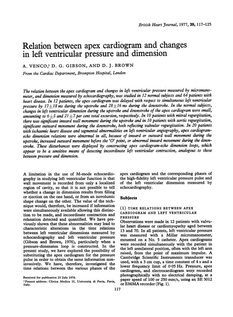
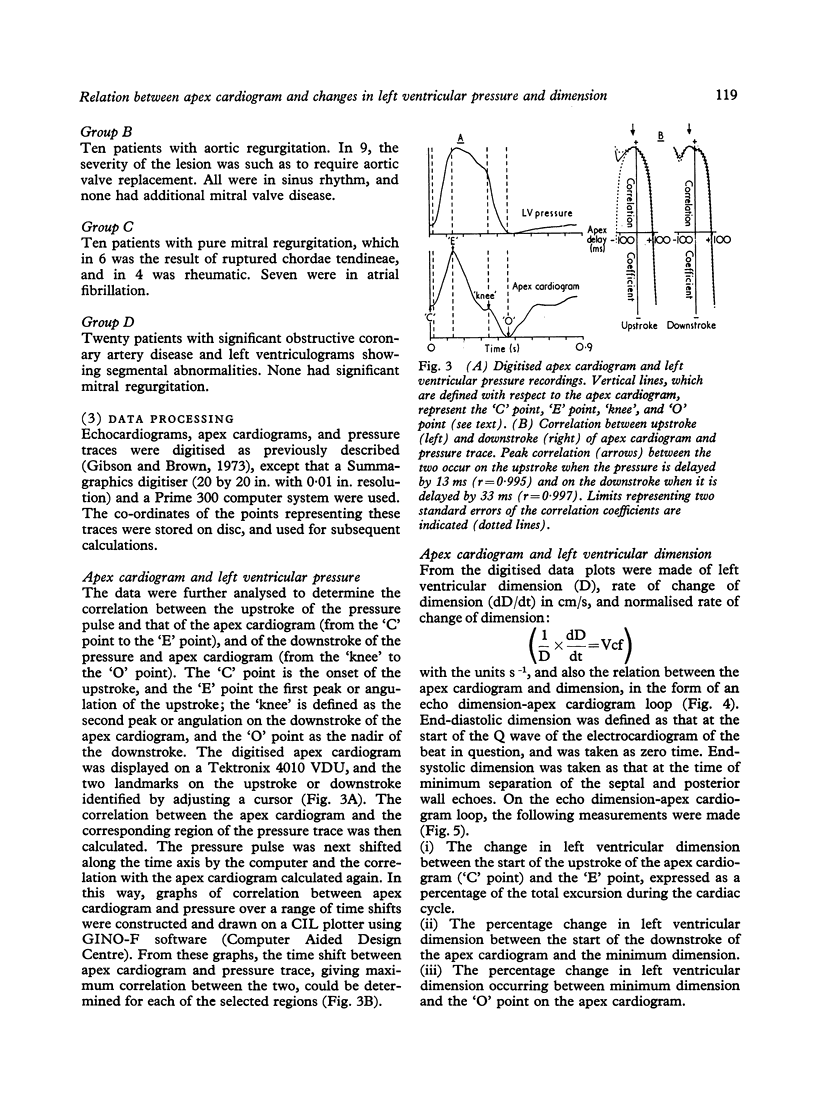
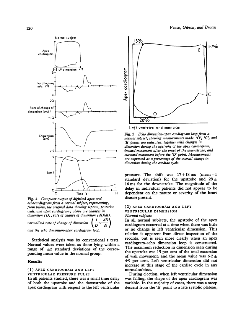
The relation between the apex cardiogram and changes in left ventricular pressure measured by micromanometer, and dimension measured by echocardiography, was studied in 12 normal subjects and 64 patients with heart disease. In 12 patients, the apex cardiogram was delayed with respect to simultaneous left ventricular pressure by 17 +/- 18 ms during the upstroke and 28 +/- 16 ms during the downstroke. In the normal subjects, changes in left ventricular dimension during the upstroke and downstroke of the apex cardiogram were small, amounting to 6 +/- 5 and 21 +/- 7 per cent total excursion, respectively. In 10 patients with mitral regurgitation, there was significant inward wall movement during the upstroke and in 10 patients with aortic regurgitation, significant outward movement during the downstroke, both reflecting valvular regurgitation. In 20 patients with ischaemic heart disease and segmental abnormalities on left ventricular angiography, apex cardiogram-echo dimension relations were abnormal in all, because of inward or outward wall movement during the upstroke, increased outward movement before the 'O' point, or abnormal inward movement during the downstroke. These disturbances were displayed by constructing apex cardiogram-echo dimension loops, which appear to be a sensitive means of detecting incoordinate left ventricular contraction, analogous to those between pressure and dimension.
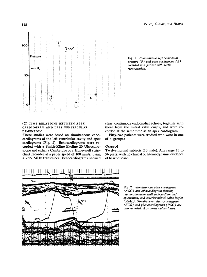
Full text
PDF
Images in this article
Selected References
These references are in PubMed. This may not be the complete list of references from this article.
- Altieri P. I., Wilt S. M., Leighton R. F. Left ventricular wall motion during the isovolumic relaxation period. Circulation. 1973 Sep;48(3):499–505. doi: 10.1161/01.cir.48.3.499. [DOI] [PubMed] [Google Scholar]
- Gibson D. G., Brown D. Measurement of instantaneous left ventricular dimension and filling rate in man, using echocardiography. Br Heart J. 1973 Nov;35(11):1141–1149. doi: 10.1136/hrt.35.11.1141. [DOI] [PMC free article] [PubMed] [Google Scholar]
- Gibson D. G., Prewitt T. A., Brown D. J. Analysis of left ventricular wall movement during isovolumic relaxation and its relation to coronary artery disease. Br Heart J. 1976 Oct;38(10):1010–1019. doi: 10.1136/hrt.38.10.1010. [DOI] [PMC free article] [PubMed] [Google Scholar]
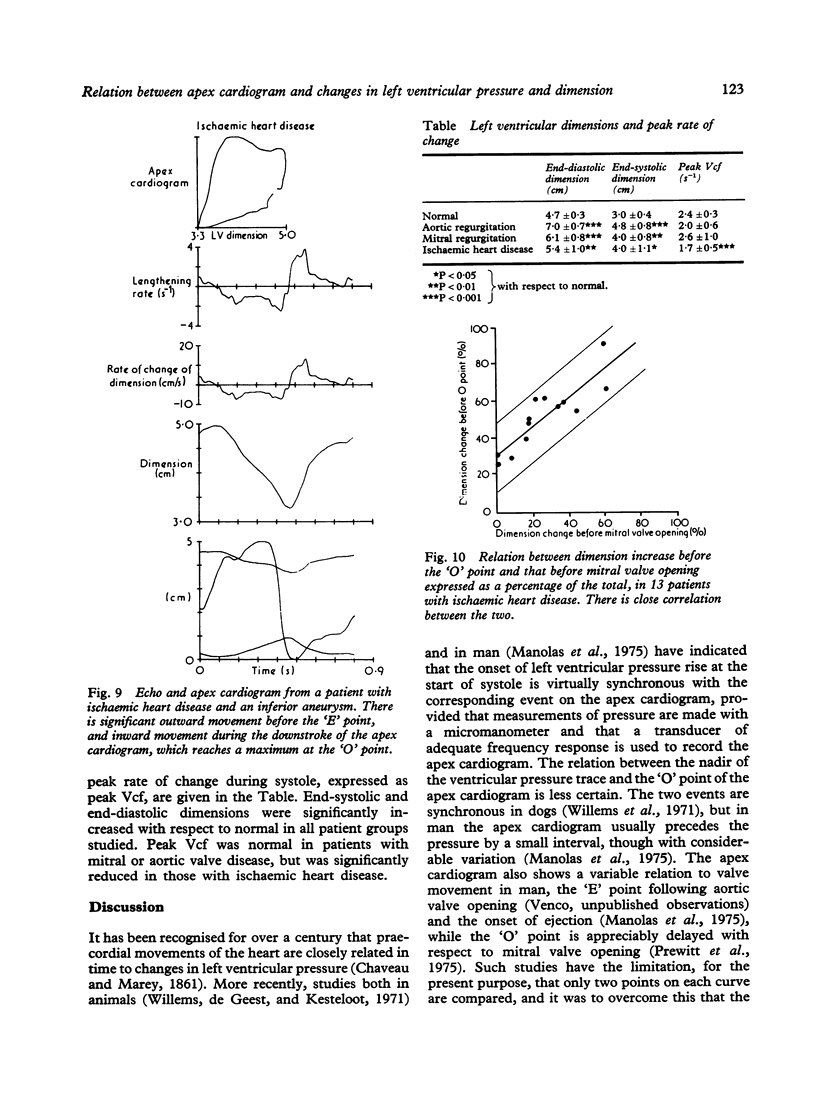
- Hamby R. I., Aintablian A., Tabrah F., Reddy K., Wisoff G. Late systolic bulging of left ventricle in patients with angina pectoris. A form of asynchronous contraction. Chest. 1974 Feb;65(2):169–175. doi: 10.1378/chest.65.2.169. [DOI] [PubMed] [Google Scholar]
- Karliner J. S., Bouchard R. J., Gault J. H. Dimensional changes of the human left ventricle prior to aortic valve opening. A cineangiographic study in patients with and without left heart disease. Circulation. 1971 Sep;44(3):312–322. doi: 10.1161/01.cir.44.3.312. [DOI] [PubMed] [Google Scholar]
- Manolas J., Rutishauser W., Wirz P., Arbenz U. Time relation between apex cardiogram and left ventricular events using simultaneous high-fidelity tracings in man. Br Heart J. 1975 Dec;37(12):1263–1267. doi: 10.1136/hrt.37.12.1263. [DOI] [PMC free article] [PubMed] [Google Scholar]
- Prewitt T., Gibson D., Brown D., Sutton G. The 'rapid filling wave' of the apex cardiogram. Its relation to echocardiographic and cineangiographic measurements of ventricular filling. Br Heart J. 1975 Dec;37(12):1256–1262. doi: 10.1136/hrt.37.12.1256. [DOI] [PMC free article] [PubMed] [Google Scholar]
- Rackley C. E., Behar V. S., Whalen R. E., McIntosh H. D. Biplane cineangiographic determinations of left ventricular function: pressure-volume relationships. Am Heart J. 1967 Dec;74(6):766–779. doi: 10.1016/0002-8703(67)90096-8. [DOI] [PubMed] [Google Scholar]
- Ruttley M. S., Adams D. F., Cohn P. F., Abrams H. L. Shape and volume changes during "isovolumetric relaxation" in normal and asynergic ventricles. Circulation. 1974 Aug;50(2):306–316. doi: 10.1161/01.cir.50.2.306. [DOI] [PubMed] [Google Scholar]
- Upton M. T., Gibson D. G., Brown D. J. Echocardiographic assessment of abnormal left ventricular relaxation in man. Br Heart J. 1976 Oct;38(10):1001–1009. doi: 10.1136/hrt.38.10.1001. [DOI] [PMC free article] [PubMed] [Google Scholar]
- Willems J. L., De Geest H., Kesteloot H. On the value of apex cardiography for timing intracardiac events. Am J Cardiol. 1971 Jul;28(1):59–66. doi: 10.1016/0002-9149(71)90035-x. [DOI] [PubMed] [Google Scholar]
- Wilson C. S., Krueger S., Forker A. D., Weaver W. F. Correlation between segmental early relaxation of the left ventricular wall and coronary occlusive disease. Am Heart J. 1975 Apr;89(4):474–479. doi: 10.1016/0002-8703(75)90154-4. [DOI] [PubMed] [Google Scholar]