

Abstract
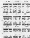
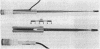
Although echocardiography is ideally suited for repetitive use on a patient for evaluation of left ventricular function, the value of this application is minimised by the uncertainty as to whether changes in left ventricular dimensions observed on a patient at different times or by different observers are real or result from the ultrasonic beam penetrating the left ventricle at different angles. Accordingly, an instrument was designed and constructed in our laboratory to improve the reproducibility of echocardiographic measurements of left ventricular dimensions. The instrument represents an orthogonal reference frame by means of which the spatial orientation of the ultrasonic beam relative to the chest is determined and reproduced in subsequent studies, while the point of entrance of the beam is marked on the chest wall. Using this instrument, left ventricular echograms were initially recorded on a group (I) of 23 subjects with or without heart disease and the study was repeated 8 hours to 49 (mean 7) days later by the same observer and also, in 16 cases, by an independent observer. The average values from 2 to 6 (mean 4) heart cycles were used for the left ventricular end-diastolic dimension (Dd), end-systolic (Ds) dimension, and their difference (delta D). Differences in all three variables between studies were random and statistically insignificant, never exceeding 3-5 mm for Dd or Ds, and 4 mm for deltaD. For comparison, left ventricular internal dimensions were also obtained in a seprate group (II) of 14 subjects by the standard method of using the mitral valve as an internal landmark, without the benefit of this instrument. All 14 subjects had the initial study repeated within 8 hours to 11 (mean 3-8) days later by the same and also by an independent observer. Though in the group as a whole there was no significant difference in left ventricular dimensions between studies, individual variations reached 11 mm for Dd, 9 mm for Ds, and 9 mm for deltaD, and the degree of scatter was significantly larger than in group I. This initial experience indicates that the use of this instrument improves the reproducibility and enhances the reliability of estimates of echocardiographic left ventricular dimensions and function on a patient examined at different times by the same or independent observers.
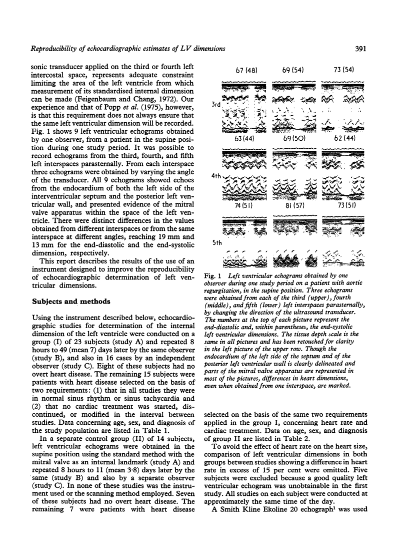
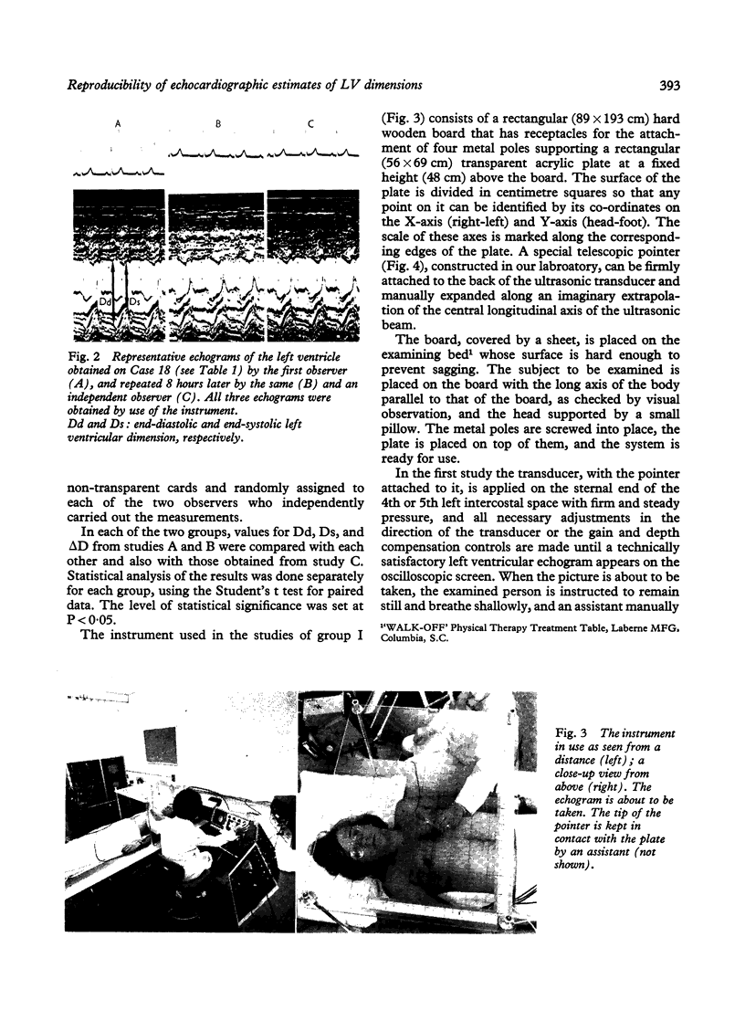
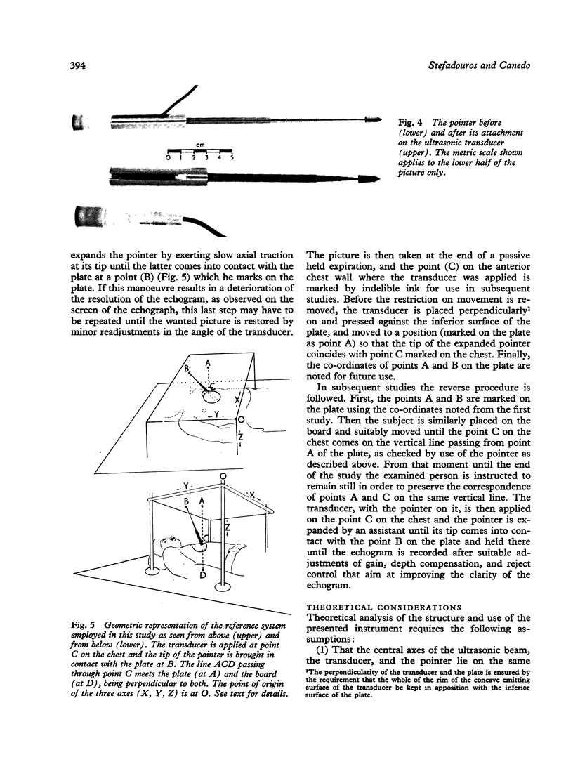
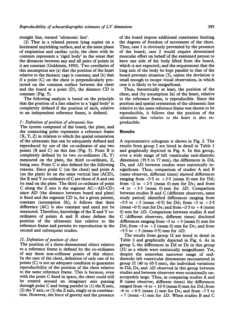
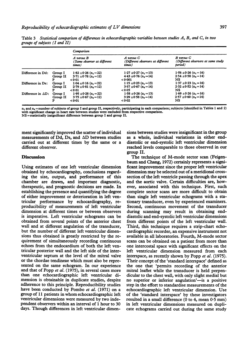
Full text
PDF
Images in this article
Selected References
These references are in PubMed. This may not be the complete list of references from this article.
- Cooper R. H., O'Rourke R. A., Karliner J. S., Peterson K. L., Leopold G. R. Comparison of ultrasound and cineangiographic measurements of the mean rate of circumferential fiber shortening in man. Circulation. 1972 Nov;46(5):914–923. doi: 10.1161/01.cir.46.5.914. [DOI] [PubMed] [Google Scholar]
- Feigenbaum H., Stone J. M., Lee D. A., Nasser W. K., Chang S. Identification of ultrasound echoes from the left ventricle by use of intracardiac injections of indocyanine green. Circulation. 1970 Apr;41(4):615–621. doi: 10.1161/01.cir.41.4.615. [DOI] [PubMed] [Google Scholar]
- Fortuin N. J., Hood W. P., Jr, Craige E. Evaluation of left ventricular function by echocardiography. Circulation. 1972 Jul;46(1):26–35. doi: 10.1161/01.cir.46.1.26. [DOI] [PubMed] [Google Scholar]
- Fortun N. J., Hood W. P., Jr, Sherman M. E., Craige E. Determination of left ventricular volumes by ultrasound. Circulation. 1971 Oct;44(4):575–584. doi: 10.1161/01.cir.44.4.575. [DOI] [PubMed] [Google Scholar]
- Linhart J. W., Mintz G. S., Segal B. L., Kawai N., Kotler M. N. Left ventricular volume measurement by echocardiography: fact or fiction? Am J Cardiol. 1975 Jul;36(1):114–118. doi: 10.1016/0002-9149(75)90877-2. [DOI] [PubMed] [Google Scholar]
- Paraskos J. A., Grossman W., Saltz S., Dalen J. E., Dexter L. A noninvasive technique for the determination of velocity of circumferential fiber shortening in man. Circ Res. 1971 Dec;29(6):610–615. doi: 10.1161/01.res.29.6.610. [DOI] [PubMed] [Google Scholar]
- Pombo J. F., Troy B. L., Russell R. O., Jr Left ventricular volumes and ejection fraction by echocardiography. Circulation. 1971 Apr;43(4):480–490. doi: 10.1161/01.cir.43.4.480. [DOI] [PubMed] [Google Scholar]
- Popp R. L., Filly K., Brown O. R., Harrison D. C. Effect of transducer placement on echocardiographic measurement of left ventricular dimensions. Am J Cardiol. 1975 Apr;35(4):537–540. doi: 10.1016/0002-9149(75)90837-1. [DOI] [PubMed] [Google Scholar]
- Popp R. L., Harrison D. C. Ultrasonic cardiac echography for determining stroke volume and valvular regurgitation. Circulation. 1970 Mar;41(3):493–502. doi: 10.1161/01.cir.41.3.493. [DOI] [PubMed] [Google Scholar]
- Popp R. L., Wolfe S. B., Hirata T., Feigenbaum H. Estimation of right and left ventricular size by ultrasound. A study of the echoes from the interventricular septum. Am J Cardiol. 1969 Oct;24(4):523–530. doi: 10.1016/0002-9149(69)90495-0. [DOI] [PubMed] [Google Scholar]